- Target
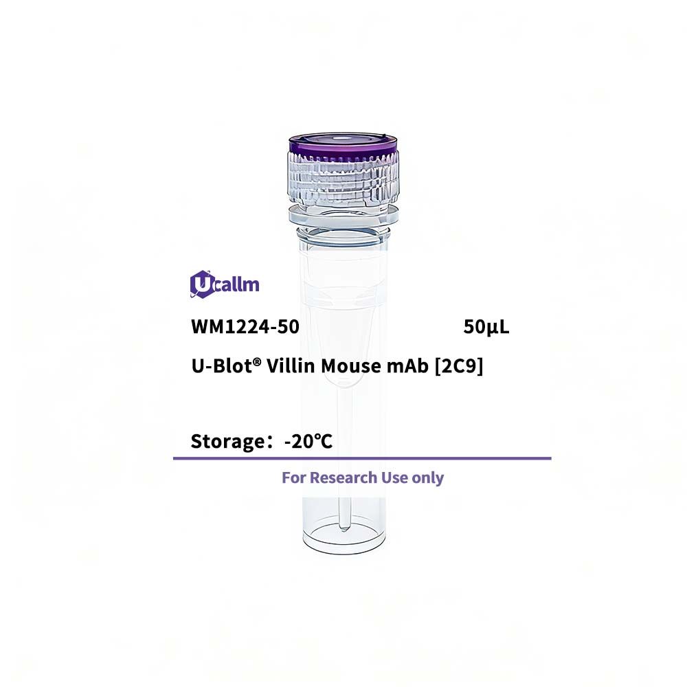
- Villin
- Reactivity
- Human,Mouse,Rat
- Application
- WB,IHC
- MW(Calculated)
- --
- MW(Observed)
- 93kD
- Host Species
- Mouse
- Isotype
- --
- Conjugate/Modification
- Unmodified
- Modification site
- --
- Recommended Dilution Ratio
- WB 1:500-2000;IHC 1:50-200
- Form
- Liquid in PBS containing 50% glycerol, and 0.02% sodium azide.
- Source
- --
- Purification
- The antibody was affinity-purified from mouse ascites by affinity-chromatography using epitope-specific immunogen.
- Purity
- --
- storageCondition
- -15°C to -25°C/1 year(Do not lower than -25°C)
- Concentration
- 1 mg/ml
- Clonality
- Monoclonal
- Clone No.
- 2C9
- Immunogen
- Synthesized peptide derived from human Villin
- Sequence
- --
- Specificity
- This antibody detects endogenous levels of Villin at Human, Mouse,Rat
- Gene Name
- VIL1 VIL
- Protein Name
- Villin-1
- Other Name
- Villin-1
- Organism-1
- Human
- Gene ID-1
- 7429
- SwissProt-1
- P09327
- Organism-2
- Mouse
- Gene ID-2
- 22349
- SwissProt-2
- Q62468
- Organism-3
- --
- Gene ID-3
- --
- SwissProt-3
- --
- Organism-4
- --
- gene ID-4
- --
- SwissProt-4
- --
- Cellular Localization
- Cytoplasm, cytoskeleton. Cell projection, lamellipodium. Cell projection, ruffle. Cell projection, microvillus. Cell projection, filopodium tip . Cell projection, filopodium . Relocalized in the tip of cellular protrusions and filipodial extensions upon infection with S.flexneri in primary intestinal epithelial cells (IEC) and in the tail-like structures forming the actin comets of S.flexneri. Redistributed to the leading edge of hepatocyte growth factor (HGF)-induced lamellipodia (By similarity). Rapidly redistributed to ruffles and lamellipodia structures in response to autotaxin, lysophosphatidic acid (LPA) and epidermal growth factor (EGF) treatment. .