
info@ucallmlabs.com
Three-dimensional models can more closely recapitulate the spatial architecture of real tissues and can further integrate tumor cells, stromal cells, and immune cells, making them useful for studying tumor–immune interactions and evaluating immunotherapies. For immune cell research, an important value of 3D models is their ability to mimic the complex microenvironment encountered by immune cells after entering tumor tissues. These models can be further used to investigate immune cell migration, infiltration, recognition, killing, activation, and exhaustion processes, as well as to evaluate immune checkpoint inhibitors, CAR-T cells, NK cells, and other immunotherapeutic strategies.
Organoid Research Topic
Learn about our organoid culture consumables and reagents
Learn about our organoid services
Organoids
Organoids are self-organizing three-dimensional cellular structures generated from stem cells, progenitor cells, or patient-derived tissue cells under 3D culture conditions. Compared with traditional two-dimensional cell lines, organoids can partially preserve the cellular composition, spatial organization, and certain physiological and pathological functions of the tissues from which they are derived. Therefore, they have become important models for studying human tissue development, disease mechanisms, and tumor biology. Particularly in cancer research, patient-derived tumor organoids can be established from patient tumor tissues and maintain some original genetic and phenotypic characteristics of the tumors in vitro, providing significant value for personalized research.
Tumor organoids can be used to study tumor cell proliferation, differentiation, invasion, drug responses, and tumor heterogeneity. Compared with traditional tumor cell lines, organoids better preserve key biological features of patient-derived tumors and can serve as patient-specific models for drug screening and precision medicine research. For example, tumor organoids can be established from different patients and individually treated with different drugs or therapeutic combinations to compare treatment sensitivities among patient tumors.
Therefore, current organoid research is gradually evolving from simple “tumor organoids” toward more complex tumor immune organoids and organoid-based tumor microenvironment models that incorporate or reconstruct additional microenvironmental components. By introducing immune cells, fibroblasts, vascular-associated cells, or other microenvironmental elements, these models enable further investigation of dynamic interactions between tumor cells and the immune system, while improving their ability to mimic real tumor tissues.
Learn about our clinical organoid culture products
Learn about our clinical organoid services
Data display:
Tumor Spheroids
Tumor spheroids are another widely used type of three-dimensional tumor model, typically generated by allowing tumor cells to aggregate into three-dimensional cell clusters under non-adhesive or low-adhesion conditions. Compared with organoids, tumor spheroids generally have simpler structures; however, they are easier to establish, less expensive, and require shorter experimental periods. Therefore, they are particularly suitable for studying tumor cell growth, cell–cell interactions, and drug responses. Spheroids are also one of the most widely applied systems among current 3D cancer models.
As the number of cells increases, tumor spheroids can develop gradients of oxygen, nutrients, and metabolic products, and may contain proliferative regions, quiescent regions, and hypoxic or even necrotic areas. These spatial features partially resemble those found within solid tumors, making spheroids useful for studying tumor cell survival states and therapeutic responses under different microenvironmental conditions.
In immune cell research, tumor–immune spheroid models can be further established by co-culturing tumor cells with T cells, NK cells, or other immune cells. Through this approach, researchers can investigate immune cell migration toward tumor spheroids, infiltration into the spheroid structure, and cytotoxic effects against tumor cells. For example, three-dimensional imaging and live-cell imaging can be used to observe immune cell aggregation around spheroids, penetration into the interior, and cytotoxic interactions after direct contact with tumor cells. Therefore, tumor spheroids provide a relatively simple and controllable system for studying immune-cell infiltration and cytotoxicity.
Learn about our ultra-low attachment cell culture plates
Learn about our media, fetal bovine serum (FBS), and cryopreservation solutions
Data display:
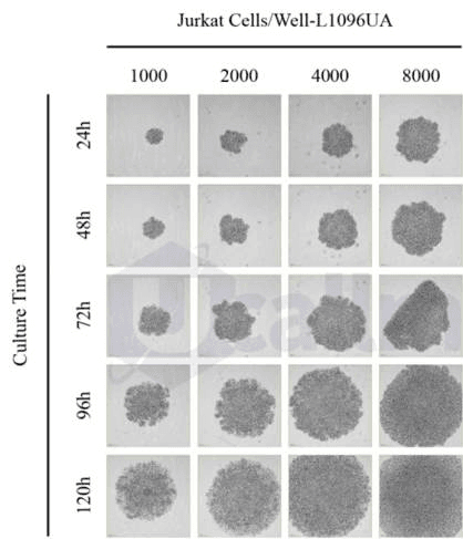
Jurkat cultured on Ucallm® Ultra-Low Attachment Surface forms tumor spheroids. Jurkat T cells were planted in 96-well tissue culture plates and ultra-low attachment plates at concentrations of 500, 1000, 2000, 4000, and 8000 cells per well. Imaging was conducted at 24, 48, 72, 96, and 120 hours after seeding. Scale bars represent 200 μm.
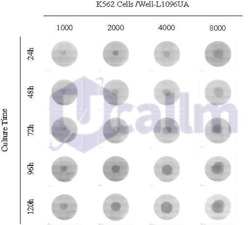
K562 cells cultured on Ucallm® Ultra-Low Attachment Surface forms tumor spheroids. K562 cells were planted in 96-well tissue culture plates and ultra-low attachment plates at concentrations of 500, 1000, 2000, 4000, and 8000 cells per well. Live-cell imaging was conducted at 24, 48, 72, 96, and 120 hours after seeding. Scale bars represent 200 μm.
Immune-Organoid Co-culture
Immune-organoid co-culture (immune cell–organoid co-culture) is a three-dimensional in vitro model established by introducing immune cells into tumor organoids, thereby creating a system that more closely resembles the real tumor immune microenvironment. The core concept is to combine the three-dimensional tumor architecture provided by organoids with the functional properties of immune cells, allowing researchers to investigate interactions between immune cells and tumor cells within a patient-derived tumor context.
Immune-organoid co-culture is particularly suitable for studying tumor–immune cell interactions. For example, it can be used to determine whether T cells can recognize and attack patient-derived tumor cells, as well as to analyze immune cell infiltration, spatial distribution, and cytotoxic effects after entering organoids. Through approaches including live-cell imaging, immunofluorescence, flow cytometry, single-cell sequencing, and cytokine analysis, immune cell activation, exhaustion, and functional changes can be further characterized.
Immune cell–organoid co-culture systems have been applied in the evaluation of immune checkpoint inhibitors (ICIs), CAR-T therapy research, T-cell-mediated tumor killing, NK-cell cytotoxicity studies, and personalized immunotherapy screening. For example, combining patient-derived tumor organoids with autologous or allogeneic immune cells enables in vitro evaluation of the effects of different immunotherapeutic strategies on tumors from specific patients. Therefore, these patient-specific organoid–immune co-culture systems are considered promising platforms for the development of personalized immunotherapy screening approaches.
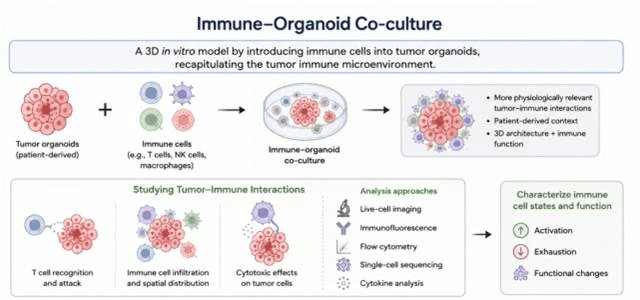
Schematic of immune-organoid co-culture for studying tumor–immune interactions. Immune-organoid co-culture is a 3D in vitro model that incorporates immune cells into patient-derived tumor organoids to mimic the tumor immune microenvironment. It preserves patient context, 3D architecture and immune function to recapitulate tumor–immune interactions. Researchers use live imaging, immunofluorescence, flow cytometry, single-cell sequencing and cytokine assays to examine immune infiltration, tumor cytotoxicity, immune activation, exhaustion and functional changes.
Learn about our clinical organoid services
Data Presentation: