
info@ucallmlabs.com
Cardiovascular disease (CVD) ranks as the global leading cause of mortality and imposes substantial economic pressure on healthcare systems worldwide. Conventional biomedical studies predominantly adopt cell lines for disease modeling and drug screening, yet such in vitro models fail to recapitulate the full structural complexity of native in vivo tissues. To address these limitations, organoid culture systems have been established to restore pivotal biological features, including the physiological cellular microenvironment and intercellular crosstalk. The rapid advancement of organoid engineering and biomimetic models has innovated fundamental biological research, human pathogenic mechanism exploration, and drug response assessment, significantly accelerating the development of novel therapeutic strategies for diverse diseases.
Cardiac organoids are structurally organized miniature cardiac constructs self-assembled by cardiac progenitor cells, cardiomyocytes, endothelial cells and fibroblasts within a three-dimensional microenvironment. They faithfully mimic the biological properties of endogenous human cardiac tissues and recapitulate the histogenetic processes, physiological functions and temporal developmental characteristics of the human heart. Owing to these superior biomimetic features, cardiac organoids have emerged as a powerful platform for cardiovascular disease modeling, high-throughput drug screening and toxicological evaluation, serving as an indispensable core tool for basic cardiac functional research and the development of personalized therapeutic regimens.
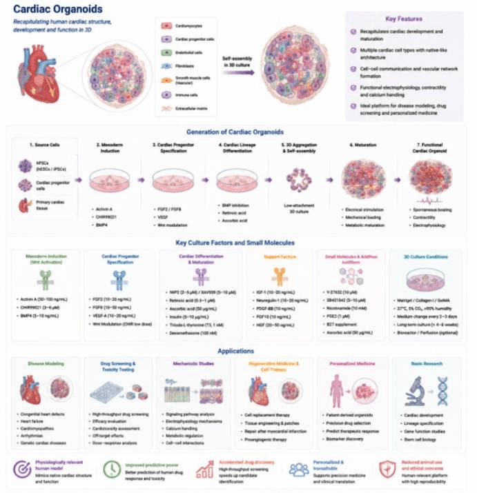
Generation, Culture, and Applications of Human Cardiac Organoids.
Common Reagents for Cardiac Organoid Culture
Recommended Markers for Cardiac Organoids
Data display.
(A) Organoid culture status (bright-field morphology)
(B) Multicolor immunofluorescence staining
(C) Post-translational modification proteomics
(D) Epigenetic profiling (ChIP-seq / ATAC-seq)
(E) Gene editing validation (CRISPR)