
info@ucallmlabs.com
```
```
Alkaliptosis
1.1 Mechanism Overview
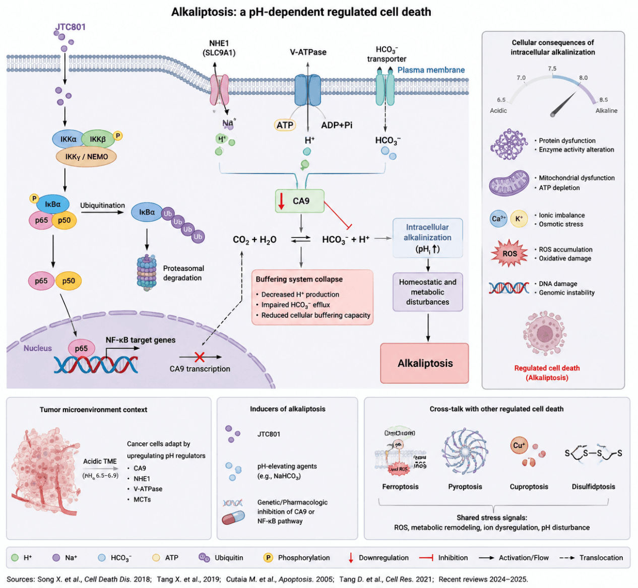
Alkaliptosis is a novel form of regulated cell death driven by intracellular alkalinization, first proposed by Song et al. in pancreatic cancer research in 2018. Studies have revealed that the small molecule JTC801 induces a cell death modality distinct from apoptosis, necroptosis and ferroptosis, characterized by sustained elevation of intracellular pH and disrupted acid‑base homeostasis. Mechanistic investigations demonstrate that JTC801 activates the IKK/NF‑κB signaling pathway to promote nuclear translocation of the transcription factor NF‑κB p65 (RELA), which subsequently suppresses the expression of carbonic anhydrase 9 (CA9). As a critical regulator maintaining acid‑base balance in tumor cells, CA9 catalyzes the conversion of CO₂ and H₂O into HCO₃⁻ and H⁺, participating in intra‑ and extracellular pH modulation. Inhibition of CA9 impairs cellular pH buffering capacity, hinders H⁺ extrusion and triggers intracellular HCO₃⁻ accumulation, leading to persistent intracellular alkalinization. Elevated pH further disrupts metabolic homeostasis, ionic equilibrium and protein function, initiating irreversible cellular damage and alkaliptosis.
Beyond the NF‑κB/CA9 axis, emerging evidence links alkaliptosis to plasma membrane transport systems, metabolic reprogramming and the tumor microenvironment. Na⁺/H⁺ exchanger 1 (NHE1/SLC9A1), vacuolar H⁺‑ATPase (V‑ATPase) and bicarbonate transporters collectively sustain cellular pH homeostasis, and their dysregulation may determine cellular sensitivity to alkalinizing stimuli. Furthermore, tumor cells generate an acidic microenvironment via the Warburg effect and rely heavily on pH regulators such as CA9 for survival. Therefore, targeting pH homeostasis represents a promising strategy to trigger alkaliptosis and enhance anti‑tumor efficacy. Current data indicate that alkaliptosis can function as an independent death modality and engage in crosstalk with other regulated cell death pathways including ferroptosis, pyroptosis and cuproptosis, jointly governing tumor cell fate.
Alkaliptosis Model Diagram. JTC801 inhibits CA9 expression by activating the IKK/NF‑κB signaling pathway, disrupting the CO₂/HCO₃⁻/H⁺ buffering system and inducing persistent intracellular alkalinization. This subsequently triggers metabolic disturbance, mitochondrial damage, ROS accumulation and ionic imbalance, ultimately initiating a novel regulated cell death distinct from classical cell death modalities, namely alkaliptosis.
1.2 Target List
| Mechanism Module | Target | Full Name | Biological Function | Mechanism in Alkaliptosis | Research Value / Detection Index |
|---|---|---|---|---|---|
| Core NF‑κB Signaling Pathway | NF‑κB p65 (RELA) | Nuclear factor kappa B subunit p65 (RELA) | Transcription factor regulating inflammatory and stress‑response genes | Key upstream regulator; NF‑κB activation suppresses CA9 expression and promotes intracellular alkalinization | Core signaling activation marker |
| p50 (NFKB1) | Nuclear factor kappa B subunit 1 | Forms canonical NF‑κB heterodimer with p65 | Participates in NF‑κB transcriptional regulation during alkaliptosis | Indicator of canonical NF‑κB activation | |
| IKKα (CHUK) | Inhibitor of nuclear factor kappa‑B kinase subunit alpha | Serine/threonine kinase initiating NF‑κB activation | Phosphorylates IκB proteins and promotes NF‑κB pathway activation | Upstream NF‑κB activation marker | |
| IKKβ (IKBKB) | Inhibitor of nuclear factor kappa‑B kinase subunit beta | Kinase responsible for IκBα phosphorylation and degradation | Promotes release and nuclear translocation of NF‑κB | NF‑κB pathway activation indicator | |
| IκBα (NFKBIA) | NF‑κB inhibitor alpha | Inhibitory protein retaining NF‑κB in cytoplasm | Reduced expression indicates NF‑κB activation | Negative regulator of NF‑κB signaling | |
| Core Execution Axis | CA9 | Carbonic anhydrase IX | Carbonic anhydrase regulating intracellular pH balance | NF‑κB‑mediated CA9 suppression decreases CO₂/HCO₃⁻ buffering capacity, resulting in intracellular alkalinization | Most critical marker of alkaliptosis |
| pH Homeostasis Regulation | NHE1 / SLC9A1 | Sodium/hydrogen exchanger 1 | Exchanges intracellular H⁺ with extracellular Na⁺ to regulate pH | Modulates intracellular alkalization through H⁺ extrusion | pH regulation marker |
| V‑ATPase | Vacuolar‑type H⁺‑ATPase | ATP‑dependent proton pump maintaining proton gradients | Regulates proton transport and intracellular pH adaptation | H⁺ transport regulation marker | |
| SLC4 family (HCO₃⁻ transporters) | Solute carrier family 4 bicarbonate transporters | Maintains bicarbonate buffering system | Regulates intracellular bicarbonate balance during alkalinization stress | pH buffering capacity indicator | |
| MCT1/MCT4 (SLC16A1/SLC16A3) | Monocarboxylate transporter 1/4 | Transports lactate and H⁺ across membranes | Participates in tumor cell pH adaptation | Tumor pH regulation marker | |
| pH Detection Assays | Intracellular pH (pHi) | Intracellular hydrogen ion concentration / intracellular pH | Direct measurement of intracellular acid‑base status | Increased pHi represents intracellular alkalinization, the defining feature of alkaliptosis | Direct confirmation of alkaliptosis occurrence |
| HCO₃⁻ level | Bicarbonate concentration | Major intracellular buffering component | Altered bicarbonate levels reflect disruption of pH buffering | Evaluation of buffering system changes | |
| Mitochondrial Injury | COX IV | Cytochrome c oxidase subunit IV | Component of mitochondrial respiratory Complex IV | Alkaliptosis‑associated stress impairs mitochondrial respiratory function | Mitochondrial quality control marker |
| ATP5A | ATP synthase F1 subunit alpha | Catalyzes ATP synthesis during oxidative phosphorylation | Mitochondrial dysfunction reduces ATP production capacity | Energy metabolism indicator | |
| TOM20 | Translocase of outer mitochondrial membrane 20 | Maintains mitochondrial protein import and structure | Reflects mitochondrial structural damage | Mitochondrial integrity marker | |
| JC‑1 | Mitochondrial membrane potential fluorescent probe | Detects mitochondrial membrane potential (ΔΨm) | Loss of ΔΨm indicates mitochondrial dysfunction | Mitochondrial membrane potential detection | |
| Oxidative Stress | ROS | Reactive oxygen species | Reactive molecules reflecting oxidative stress | Intracellular alkalinization induces oxidative stress accumulation | Oxidative stress evaluation |
| MitoSOX | Mitochondrial superoxide indicator | Detects mitochondrial superoxide production | Indicates mitochondrial ROS elevation during alkaliptosis | Mitochondrial oxidative stress marker | |
| SOD2 | Superoxide dismutase 2 (MnSOD) | Antioxidant enzyme converting superoxide to hydrogen peroxide | Reflects mitochondrial antioxidant response | Oxidative defense marker | |
| GPX4 | Glutathione peroxidase 4 | Removes lipid peroxides and protects against ferroptosis | Used to exclude or compare ferroptotic mechanisms | Ferroptosis exclusion marker | |
| Ion Homeostasis | Ca²⁺ | Calcium ion | Essential second messenger regulating cellular functions | Alkalinization disrupts Ca²⁺ homeostasis and contributes to cell stress | Calcium imbalance detection |
| K⁺ channel‑related proteins | Potassium channel‑related proteins | Maintain intracellular potassium balance | Altered K⁺ transport contributes to ionic homeostasis disturbance | Ion imbalance evaluation |
1.3 Application Scheme
| Marker | Significance |
|---|---|
| p65/RELA | NF‑κB activation |
| IκBα | NF‑κB liberation |
| CA9 | Core execution molecule of alkaliptosis |
| pHi (BCECF‑AM) | Intracellular alkalinization phenotype |
| Annexin V/PI | Cell death outcome |
1.4 References
- Song X, et al. JTC801 Induces pH‑dependent Death Specifically in Cancer Cells and Slows Growth of Tumors in Mice. Gastroenterology. 2018;154(5):1480‑1493.e8.
- Chen F, Kang R, Liu J, Tang D. Mechanisms of alkaliptosis. Frontiers in Cell and Developmental Biology. 2023;11:1213995