
info@ucallmlabs.com
Autoimmune diseases are a group of disorders caused by abnormal immune responses against self-antigens, resulting in persistent inflammation and tissue damage. Their development usually involves coordinated interactions between innate and adaptive immune systems, including immune cells such as T cells, B cells, macrophages, dendritic cells, and neutrophils, as well as various immune mediators including cytokines, chemokines, and autoantibodies.
Learn more about our autoimmune disease organoid models.
1.1 Rheumatoid Arthritis
Rheumatoid arthritis (RA) is an autoimmune disease characterized primarily by chronic synovial inflammation and progressive joint destruction. Its immunopathological processes involve multiple cell types, including CD4⁺ T cells, B cells, macrophages, dendritic cells, and fibroblast-like synoviocytes (FLS).
In RA, antigen-presenting cells (APCs) activate CD4⁺ T cells, followed by the involvement of Th1 and Th17 T-cell subsets in inflammatory responses. B cells produce autoantibodies such as rheumatoid factor (RF) and anti-citrullinated protein antibodies (ACPAs). Meanwhile, macrophages and other inflammatory cells produce inflammatory mediators including TNF-α, IL-1β, IL-6, and IL-17, which promote synovial cell activation, inflammatory cell recruitment, and osteoclast differentiation, ultimately leading to cartilage destruction and bone erosion.
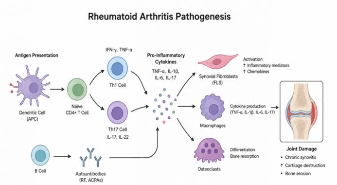
Immunopathogenesis of rheumatoid arthritis. Autoantigen presentation by dendritic cells activates CD4⁺ T cells and promotes Th1/Th17 polarization. Th1 and Th17 cells release multiple proinflammatory cytokines. B cells produce autoantibodies including RF and ACPAs. Cytokines activate synovial fibroblasts and macrophages, and induce osteoclast formation. This inflammatory circuit triggers chronic synovitis, cartilage degradation and bone erosion, causing progressive joint injury in rheumatoid arthritis.
Common Research Markers
| Research focus | Representative markers |
| T-cell activation | CD3, CD4, CD25, CD69 |
| Th17 response | IL-17A, RORγt |
| B cells | CD19, CD20 |
| Macrophages | CD68, CD163 |
| Inflammatory cytokines | TNF-α, IL-1β, IL-6 |
| Autoantibodies | RF, ACPA |
| Bone destruction | RANKL, RANK, TRAP |
1.2 Systemic Lupus Erythematosus
Systemic lupus erythematosus (SLE) is a systemic autoimmune disease that can affect multiple organ systems. Its major characteristics include autoantibody production, immune complex formation, and chronic inflammation. In SLE, nuclear-derived self-antigens can be recognized by dendritic cells and other antigen-presenting cells, promoting the activation of B cells and T cells. B cells produce autoantibodies such as antinuclear antibodies (ANA) and anti-double-stranded DNA antibodies (anti-dsDNA antibodies), leading to the formation of immune complexes. Meanwhile, plasmacytoid dendritic cells (pDCs) produce large amounts of type I interferons, particularly IFN-α, resulting in a prominent interferon signature. Deposition of immune complexes and persistent inflammatory signaling can activate the complement system and multiple inflammatory immune cells, ultimately causing tissue damage in the kidneys, skin, joints, and other organs.
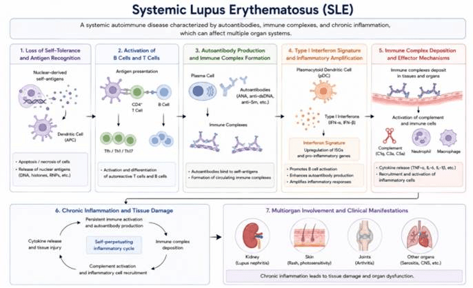
Immunopathogenesis of systemic lupus erythematosus. SLE is a multisystem autoimmune disease. Cell death releases nuclear autoantigens, breaking self-tolerance. Dendritic cells present autoantigens to activate autoreactive T and B cells. Plasma cells generate autoantibodies that form immune complexes. pDC-derived type I interferons create an interferon signature and amplify autoimmunity. Tissue-deposited immune complexes activate complement and inflammatory cells, establishing a self-sustaining inflammatory cycle. Chronic inflammation leads to multi-organ damage affecting kidney, skin, joints and other organs.
Common Research Markers
| Research focus | Representative markers |
| B cells | CD19, CD20, CD27 |
| Plasma cells | CD138 |
| T cells | CD3, CD4, CD8 |
| pDCs | CD123, BDCA2/CD303 |
| Type I IFN response | IFN-α, IFN-β, ISG15, MX1 |
| Autoantibodies | ANA, anti-dsDNA |
| Complement | C3, C4 |
| Inflammation | IL-6, TNF-α, IL-1β |
1.3 Multiple Sclerosis
Multiple sclerosis (MS) is an autoimmune disease characterized primarily by inflammation in the central nervous system (CNS) and myelin damage. Its immunopathological processes involve peripheral immune cell activation, disruption of the blood–brain barrier, and persistent inflammation within the CNS. In MS, autoreactive CD4⁺ T cells, particularly Th1 and Th17 cells, contribute to the recognition of CNS-associated antigens and promote inflammatory responses. Activated T cells and B cells can cross the blood–brain barrier (BBB) and infiltrate the central nervous system (CNS). Subsequently, macrophages, microglia, and other immune cells participate in inflammatory responses by producing cytokines, chemokines, and reactive oxygen species (ROS), thereby promoting oligodendrocyte damage, demyelination, and axonal injury.
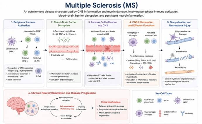
Immunopathogenesis of multiple sclerosis. MS is an autoimmune disorder targeting CNS myelin. Dendritic cells activate autoreactive Th1/Th17 cells and B cells in the periphery. Proinflammatory cytokines disrupt the blood‑brain barrier, allowing immune cell infiltration into the CNS. Activated immune cells and microglia release inflammatory mediators that injure oligodendrocytes, causing demyelination and axonal damage. Antigen release and epitope spreading sustain a self‑amplifying neuroinflammatory cycle, resulting in relapsing or progressive neurological deficits.
Common Research Markers
| Research focus | Representative markers |
| T cells | CD3, CD4, CD8 |
| Th1 | IFN-γ, T-bet |
| Th17 | IL-17A, RORγt |
| B cells | CD19, CD20 |
| Microglia/macrophages | Iba1, CD68 |
| Inflammatory signaling | NF-κB, STAT3 |
| Cytokines | IFN-γ, IL-17A, TNF-α, IL-6 |
| Myelin injury | MBP, MOG, PLP1 |
| Axonal injury | Neurofilament |
1.4 Inflammatory Bowel Disease
Inflammatory bowel disease (IBD) mainly includes Crohn’s disease (CD) and ulcerative colitis (UC). Its development is closely associated with abnormal activation of the intestinal immune system, intestinal barrier dysfunction, and gut microbiota dysbiosis. Following disruption of the intestinal epithelial barrier, microbial products can more easily enter the mucosal immune system and activate macrophages, dendritic cells, and other innate immune cells. Subsequently, T cells, particularly Th1 and Th17 cells, contribute to sustained inflammatory responses. Large amounts of inflammatory cytokines, including TNF-α, IL-1β, IL-6, IL-12, IL-17, and IL-23, further promote immune cell recruitment and activation, leading to the establishment of a persistent inflammatory loop.
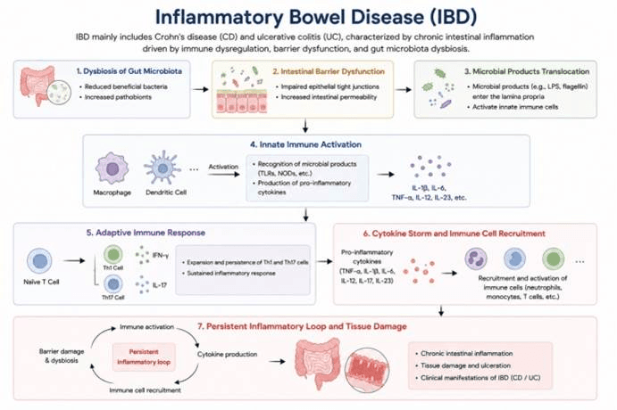
Immunopathogenesis of inflammatory bowel disease. IBD includes Crohn’s disease and ulcerative colitis. Gut microbiota dysbiosis impairs intestinal tight junctions and increases mucosal permeability, enabling translocation of microbial products into the lamina propria. Macrophages and dendritic cells recognize microbial ligands and secrete proinflammatory cytokines, which promote Th1/Th17 differentiation. Robust cytokine production recruits inflammatory immune cells and forms a perpetual inflammatory cycle. Chronic mucosal inflammation causes tissue injury and ulceration, producing the clinical phenotypes of IBD.
Common Research Markers
| Research focus | Representative markers |
| Macrophages | CD68, CD163 |
| Dendritic cells | CD11c, HLA-DR |
| T cells | CD3, CD4, CD8 |
| Th1 | IFN-γ, T-bet |
| Th17 | IL-17A, RORγt |
| Cytokines | TNF-α, IL-1β, IL-6, IL-12, IL-23 |
| Chemokines | CXCL8/IL-8, CCL2 |
| Barrier function | ZO-1, Occludin, Claudin-1 |
| Tissue injury | MPO, LDH |
Learn more about our autoimmune disease organoid models
Data display