
info@ucallmlabs.com
```
```
Calcicoptosis
1.1 Mechanism Overview
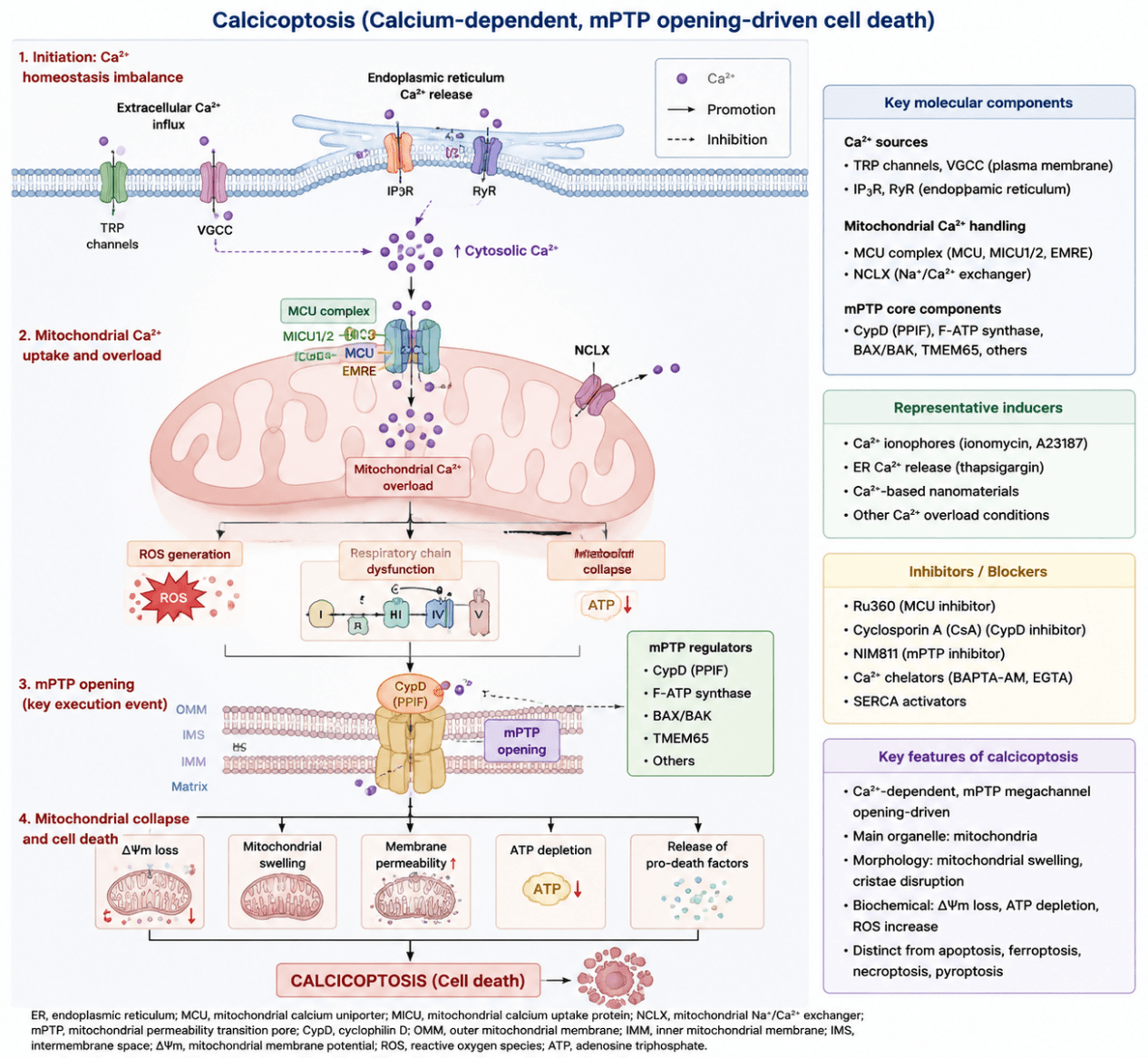
Calcicoptosis is a novel type of regulated cell death driven by disrupted intracellular calcium (Ca²⁺) homeostasis. Its hallmark is sustained opening of the mitochondrial permeability transition pore (mPTP) triggered by mitochondrial Ca²⁺ overload, which ultimately leads to mitochondrial functional collapse and cell death. Unlike conventional cell death modalities such as apoptosis and ferroptosis that rely on specific protein‑mediated execution cascades, calcicoptosis is predominantly governed by disturbed intracellular Ca²⁺ dynamics and impaired mitochondrial energy metabolism, representing a mitochondria‑centered cell death process.
Persistent mitochondrial Ca²⁺ overload acts as the critical initiating event of calcicoptosis. Excessive Ca²⁺ uptake by mitochondria boosts the production of reactive oxygen species (ROS), disrupts electron transport chain activity, and impairs oxidative phosphorylation together with ATP synthesis. Meanwhile, elevated Ca²⁺ facilitates aberrant opening of the mPTP, elevating the permeability of mitochondrial inner and outer membranes. This results in the loss of mitochondrial membrane potential (ΔΨm), matrix osmotic imbalance and mitochondrial swelling. Eventually, structural destruction of mitochondria and energy metabolism collapse drive irreversible cell death. Morphologically and functionally, calcicoptosis is characterized by prominent mitochondrial swelling, disrupted inner membrane architecture, compromised respiratory chain function and ATP depletion. In contrast to apoptosis, typical chromatin condensation and caspase cascade activation are absent during calcicoptosis.
Calcicoptosis Mechanism Diagram. Stimuli disrupt Ca²⁺ homeostasis and trigger MCU‑mediated mitochondrial Ca²⁺ overload. Subsequently, ROS accumulation and CypD‑dependent mPTP opening lead to loss of mitochondrial membrane potential, ATP depletion and structural damage, ultimately initiating calcicoptosis.
1.2 Target List
| Mechanism Module | Target (Gene/Protein/Marker) | Full Name | Biological Function | Mechanism in Calcicoptosis | Research Value / Detection Index |
|---|---|---|---|---|---|
| Disrupted Ca²⁺ Homeostasis | Fluo‑4 AM | Fluo‑4 acetoxymethyl ester calcium fluorescent probe | Fluorescent indicator for intracellular Ca²⁺ measurement | Detects abnormal elevation of cytoplasmic Ca²⁺ concentration during Ca²⁺ overload | Direct detection of intracellular Ca²⁺ accumulation |
| Rhod‑2 AM | Rhod‑2 acetoxymethyl ester mitochondrial calcium probe | Fluorescent probe for mitochondrial Ca²⁺ measurement | Detects Ca²⁺ transfer from cytoplasm into mitochondria | Verification of mitochondrial Ca²⁺ overload | |
| IP3R1 | Inositol 1,4,5‑trisphosphate receptor type 1 | Ca²⁺ release channel controlling ER calcium release | Abnormal activation increases intracellular Ca²⁺ release | Indicator of Ca²⁺ source dysregulation | |
| RyR | Ryanodine receptor | Intracellular Ca²⁺ release channel | Excessive Ca²⁺ release contributes to calcium overload | Marker of Ca²⁺ transport abnormality | |
| TRPM7 | Transient receptor potential melastatin 7 | Cation channel regulating Ca²⁺ and Mg²⁺ influx | Promotes extracellular Ca²⁺ entry and disrupts Ca²⁺ homeostasis | Indicator of Ca²⁺ influx activation | |
| MCU‑Mediated Ca²⁺ Uptake | MCU | Mitochondrial calcium uniporter | Main channel mediating mitochondrial Ca²⁺ uptake | Excessive activation increases mitochondrial Ca²⁺ accumulation and triggers calcicoptosis | Core marker of mitochondrial Ca²⁺ overload |
| MICU1 | Mitochondrial calcium uptake 1 | Calcium sensor regulating MCU channel activity | Controls mitochondrial Ca²⁺ uptake threshold | Regulatory component of MCU complex | |
| MICU2 | Mitochondrial calcium uptake 2 | Regulatory subunit of MCU complex | Modulates MCU‑dependent Ca²⁺ entry | Evaluation of MCU regulation | |
| EMRE | Essential MCU regulator | Accessory component required for MCU channel function | Maintains MCU complex stability and Ca²⁺ transport activity | MCU complex functional marker | |
| NCLX | Sodium/calcium lithium exchanger | Mediates mitochondrial Ca²⁺ efflux | Reduced activity impairs mitochondrial Ca²⁺ clearance | Indicator of mitochondrial Ca²⁺ removal capacity | |
| Mitochondrial Injury Triggered by Ca²⁺ Overload | COX4 | Cytochrome c oxidase subunit 4 | Component of respiratory Complex IV involved in oxidative phosphorylation | Ca²⁺ overload damages respiratory chain function | Mitochondrial respiratory injury marker |
| ATP5A | ATP synthase F1 subunit alpha | Catalyzes mitochondrial ATP synthesis | Mitochondrial dysfunction reduces ATP production | Energy metabolism marker | |
| NDUFS1 | NADH dehydrogenase [ubiquinone] Fe‑S protein 1 | Core subunit of respiratory Complex I | Ca²⁺ overload impairs electron transport activity | Complex I functional marker | |
| JC‑1 | Mitochondrial membrane potential fluorescent probe | Detects mitochondrial membrane potential (ΔΨm) | Loss of ΔΨm indicates mitochondrial depolarization | Detection of mitochondrial dysfunction | |
| TMRM / TMRE | Tetramethylrhodamine methyl ester / ethyl ester | Fluorescent indicators of mitochondrial membrane potential | Detect mitochondrial membrane potential collapse | ΔΨm measurement | |
| ATP level | Adenosine triphosphate level | Reflects cellular energy status | Mitochondrial injury causes ATP depletion | Energy crisis evaluation | |
| ROS Amplification Cascade | MitoSOX | Mitochondrial superoxide indicator | Detects mitochondrial superoxide production | Ca²⁺ overload enhances mitochondrial ROS generation | Detection of mtROS accumulation |
| DCFH‑DA | 2',7'‑Dichlorodihydrofluorescein diacetate | General ROS fluorescent probe | Detects intracellular oxidative stress increase | ROS measurement | |
| 4‑HNE | 4‑Hydroxynonenal | Lipid peroxidation product | Reflects oxidative damage caused by ROS | Lipid oxidative injury marker | |
| MDA | Malondialdehyde | End product of lipid peroxidation | Indicates oxidative membrane damage | Oxidative stress marker | |
| mPTP Opening | CypD / PPIF | Cyclophilin D / Peptidyl‑prolyl cis‑trans isomerase F | Core regulator of mitochondrial permeability transition pore | Ca²⁺ overload activates mPTP opening and promotes mitochondrial damage | Core execution regulator of calcicoptosis |
| F‑ATP synthase | F‑type ATP synthase | ATP synthesis complex involved in mitochondrial function | Participates in mPTP structural regulation | mPTP assembly indicator | |
| VDAC1 | Voltage‑dependent anion channel 1 | Controls mitochondrial metabolite exchange | Participates in mitochondrial permeability regulation | mPTP structural component | |
| ANT (SLC25A4) | Adenine nucleotide translocator 1 | Exchanges ATP/ADP across mitochondrial membrane | Regulates mitochondrial permeability transition sensitivity | mPTP regulation marker | |
| Terminal Cell Death Event | TOM20 | Translocase of outer mitochondrial membrane 20 | Maintains mitochondrial protein import and structure | Mitochondrial structural disruption alters TOM20 integrity | Mitochondrial integrity marker |
| PI staining | Propidium iodide staining | Detects loss of membrane integrity | Increased permeability indicates cell death | Cell death validation | |
| LDH release | Lactate dehydrogenase release | Marker of plasma membrane rupture | Indicates terminal membrane damage and cell death | Necrotic cell death confirmation |
1.3 Application Scheme
| Purpose | Marker |
|---|---|
| Elevated Ca²⁺ levels | Fluo‑4 AM / Rhod‑2 AM |
| Mitochondrial Ca²⁺ uptake | MCU |
| MCU regulation | MICU1 |
| mPTP opening | CypD (PPIF) |
| Mitochondrial injury | COX4 / ATP5A |
| Reduced mitochondrial membrane potential | JC‑1 |
| Increased ROS production | MitoSOX |
| ATP depletion | ATP assay |
| Exclusion of apoptosis | Cleaved‑Caspase3 |
1.4 References
- Hu H, Chen Z, Li Y, et al. Metal‑dependent regulated cell death: Molecular architecture and translational frontiers.iMeta, 2026, DOI: 10.1002/imt2.70141