
info@ucallmlabs.com
```
```
Cuproptosis
1.1 Mechanism Overview
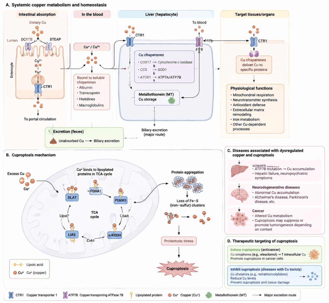
Cuproptosis is a form of metal‑dependent regulated cell death. Its core mechanism relies on abnormal intracellular accumulation of copper ions (Cu⁺) and the activity of the mitochondrial tricarboxylic acid (TCA) cycle. Excessive copper ions are transported into mitochondria and participate in redox reactions. By binding to lipoylated mitochondrial metabolic enzymes, particularly the lipoylated protein DLAT within the pyruvate dehydrogenase complex (PDH), copper triggers abnormal aggregation of lipoylated proteins, induces loss of iron‑sulfur (Fe‑S) cluster proteins and mitochondrial proteotoxic stress, and ultimately disrupts proteostasis, impairs mitochondrial function and initiates cell death.
The copper carrier FDX1 acts as a pivotal regulator of cuproptosis, facilitating the reduction of Cu²⁺ to Cu⁺ and modulating protein lipoylation. LIAS, LIPT1, DLD and DLAT participate in the generation and metabolic regulation of lipoylated proteins, whereas ATP7A, ATP7B and SLC31A1 (CTR1) mediate cellular copper uptake and efflux. Essentially, cuproptosis represents a novel cell death modality driven by the cascade of "copper accumulation – aberrant protein lipoylation – mitochondrial metabolic disturbance – proteotoxic stress". Its execution strongly depends on mitochondrial respiration and TCA cycle activity, rendering it highly valuable for research on tumor metabolic reprogramming, anti‑tumor therapy, metabolic disorders and mitochondrial dysfunction.
Cuproptosis Mechanism Diagram. Copper enters cells via CTR1 and participates in mitochondrial function under the regulation of copper chaperones. Excess Cu⁺ translocates into mitochondria, binds to lipoylated proteins in the TCA cycle, induces protein aggregation and disruption of Fe‑S clusters, causes proteotoxic stress, and ultimately triggers cuproptosis. Dysregulated copper homeostasis is involved in liver diseases, neurodegenerative disorders and cancers, and modulation of cuproptosis emerges as a novel therapeutic strategy.
1.2 Target List
Core Targets of Cuproptosis
| Mechanism Module | Target (Gene/Protein) | Full Name | Biological Function | Mechanism in Cuproptosis | Research Value / Detection Index |
|---|---|---|---|---|---|
| Core Regulatory Axis of Cuproptosis | FDX1 | Ferredoxin 1 | Participates in electron transfer and metabolic regulation | A key regulator of cuproptosis; promotes Cu²⁺ reduction to Cu⁺, facilitates lipoylated protein formation, and mediates copper toxicity | Core marker of cuproptosis |
| LIAS | Lipoic acid synthetase | Catalyzes lipoic acid synthesis | Promotes lipoylation of TCA cycle proteins and generates Cu⁺‑binding targets | Determines cuproptosis sensitivity | |
| Protein Lipoylation | LIPT1 | Lipoyltransferase 1 | Transfers lipoic acid to lysine residues of target proteins | Regulates the lipoylation levels of DLAT, DLST, and other mitochondrial proteins | Regulatory node of cuproptosis |
| Copper‑dependent Lipoylated Protein Aggregation | DLAT | Dihydrolipoamide S‑acetyltransferase | E2 subunit of the pyruvate dehydrogenase (PDH) complex | Cu⁺ directly binds to lipoylated DLAT, inducing protein aggregation and acting as the primary execution target of cuproptosis | Classical detection protein for cuproptosis |
| Pyruvate Metabolism Regulation | PDHA1 | Pyruvate dehydrogenase E1 alpha 1 | Catalyzes the conversion of pyruvate to acetyl‑CoA | Regulates pyruvate entry into the TCA cycle and influences cuproptosis sensitivity | Indicator of mitochondrial metabolic status |
| PDHB | Pyruvate dehydrogenase E1 beta subunit | Component of the PDH complex | Participates in the metabolic network of lipoylated proteins | Auxiliary detection indicator | |
| Mitochondrial Electron Transfer | DLD | Dihydrolipoamide dehydrogenase | Mediates electron transfer within the PDH and α‑ketoglutarate dehydrogenase (α‑KGDH) complexes | Contributes to the formation of Cu‑sensitive lipoylated protein complexes | Indicator of mitochondrial metabolism |
| Lipoylated TCA Cycle Target Proteins | DLST | Dihydrolipoamide succinyltransferase | E2 subunit of the α‑ketoglutarate dehydrogenase (α‑KGDH) complex | Cu⁺ binding to lipoylated DLST induces protein aggregation and leads to TCA cycle dysfunction | Indicator of cuproptosis execution |
Key Downstream Damage Targets
| Mechanism Module | Target (Gene/Protein) | Full Name | Biological Function | Mechanism in Cuproptosis | Research Value / Detection Index |
|---|---|---|---|---|---|
| Fe‑S Cluster Assembly and Homeostasis | ISCU | Iron‑sulfur cluster scaffold protein | Core scaffold protein for Fe‑S cluster assembly | Cuproptosis induces Fe‑S protein loss and disrupts Fe‑S cluster stability | Indicator of Fe‑S damage |
| NFS1 | Cysteine desulfurase | Supplies sulfur required for Fe‑S cluster synthesis | Regulates Fe‑S cluster biogenesis capacity and mitochondrial Fe‑S metabolism | Mitochondrial iron‑sulfur metabolic marker | |
| FXN | Frataxin | Regulates Fe‑S cluster maturation and mitochondrial iron utilization | Impaired Fe‑S cluster maturation contributes to mitochondrial dysfunction | Indicator of Fe‑S homeostasis | |
| TCA Cycle Fe‑S Protein Dysfunction | ACO2 | Aconitase 2 | Catalyzes the conversion of citrate to isocitrate in the TCA cycle | Disruption of Fe‑S clusters inactivates ACO2 and impairs TCA cycle activity | Functional verification marker for cuproptosis |
| Mitochondrial Complex I Dysfunction | NDUFS1 | NADH dehydrogenase Fe‑S protein 1 | Fe‑S‑containing subunit of mitochondrial respiratory Complex I | Loss of Fe‑S clusters impairs Complex I activity and oxidative phosphorylation | Mitochondrial function detection |
| NDUFS2 | NADH dehydrogenase Fe‑S protein 2 | Participates in electron transfer within Complex I | Copper toxicity destabilizes Complex I and disrupts respiratory chain function | Respiratory chain damage marker | |
| Mitochondrial Complex IV Dysfunction | COX17 | Cytochrome c oxidase copper chaperone | Delivers copper ions to cytochrome c oxidase for Complex IV assembly | Altered copper metabolism affects COX maturation and mitochondrial respiration | Copper transport and mitochondrial marker |
| Oxidative Phosphorylation Dysfunction | COX4 | Cytochrome c oxidase subunit IV | Maintains cytochrome c oxidase activity and oxidative phosphorylation | Cuproptosis suppresses mitochondrial respiration and impairs Complex IV function | Widely used mitochondrial WB marker |
| Mitochondrial Energy Metabolism Failure | ATP5A | ATP synthase F1 subunit alpha | Catalyzes ATP synthesis through oxidative phosphorylation | Mitochondrial dysfunction causes ATP depletion | Evaluation of mitochondrial function |
1.3 Application Scheme
| Purpose | Target Panel |
|---|---|
| Determine whether cuproptosis occurs | FDX1 + LIAS + DLAT |
| Detect copper accumulation | CTR1 + ATP7B |
| Evaluate TCA cycle damage | DLST + DLD + ACO2 |
| Assess mitochondrial injury | COX4 + ATP5A + NDUFS1 |
| Exclude other forms of cell death | GPX4/SLC7A11 (ferroptosis), Caspase3/PARP (apoptosis) |
1.4 References
- Tsvetkov P, Coy S, Petrova B, et al. Copper induces cell death by targeting lipoylated TCA cycle proteins. Science. 2022;375(6586):1254‑1261. doi:10.1126/science.abf0529.
- Zhang L, Deng R, Guo R, et al. Recent progress of methods for cuproptosis detection. Front Mol Biosci. 2024;11:1460987. doi:10.3389/fmolb.2024.1460987.
- Bian Z, Fan R, Xie L. A novel cuproptosis‑related gene signature for predicting prognosis and immune landscape in cancer. Front Oncol. 2022;12:954940. doi:10.3389/fonc.2022.954940.
- Zhao C, Zhang Y, Zeng Y, et al. Elesclomol induces cuproptosis by targeting mitochondrial copper homeostasis and FDX1‑dependent pathways. Cell Death Dis. 2022;13:995. doi:10.1038/s41419‑022‑05359‑7.
- Xiong Y, Xiao C, Li Z, et al. Cuproptosis: a new form of programmed cell death and its implications in cancer therapy. Cell Mol Immunol. 2022;19:1301‑1303. doi:10.1038/s41423‑022‑00912‑4.