
info@ucallmlabs.com
```
```
Ferroptosis
1.1 Mechanism Overview
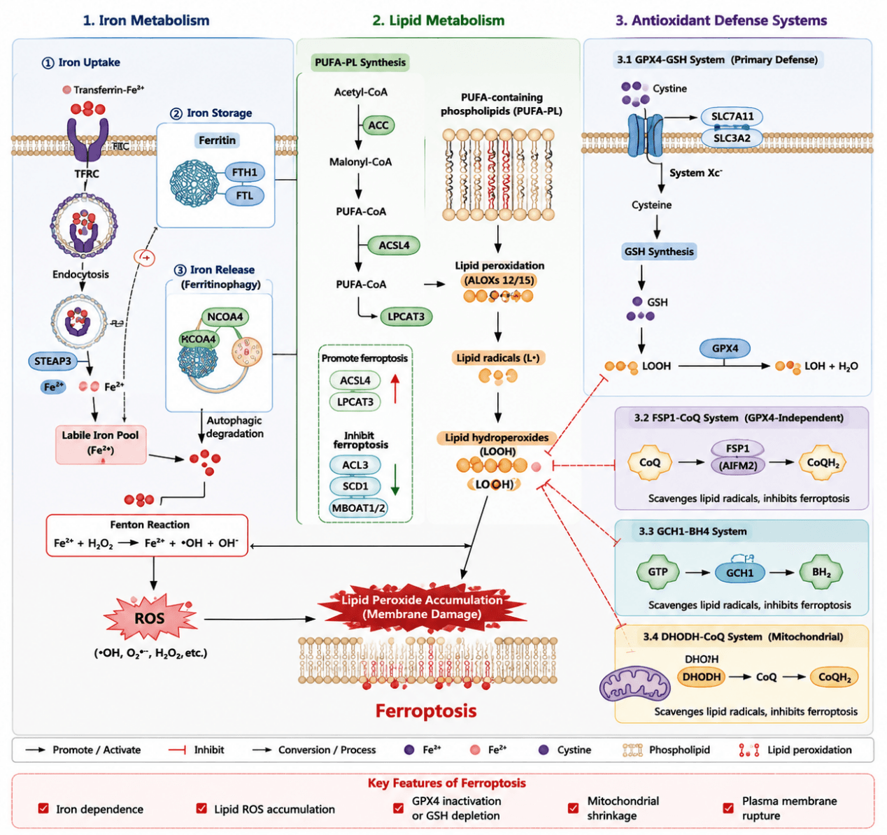
Ferroptosis is a form of regulated cell death driven by iron‑dependent lipid peroxidation. Its initiation is jointly determined by disrupted iron metabolism, remodeled lipid metabolism and the collapse of antioxidant defense systems. Cells take up iron ions via transferrin receptor (TFRC) and store iron in ferritin. NCOA4‑mediated ferritinophagy expands the labile iron pool (LIP). Subsequently, Fe²⁺ generates abundant reactive oxygen species (ROS) through the Fenton reaction and facilitates the formation of lipid radicals. Meanwhile, ACSL4 and LPCAT3 mediate the biosynthesis of polyunsaturated fatty acid‑containing phospholipids (PUFA‑PLs), enriching cell membranes with oxidation‑prone lipids. Lipid peroxidation occurs under the action of ROS and lipoxygenases (ALOX), leading to accumulation of lipid peroxides (lipid‑OOH) and damage to membrane structures, ultimately triggering ferroptosis.
To maintain redox homeostasis, cells rely on the antioxidant defense axis centered on system Xc⁻ (SLC7A11/SLC3A2)–glutathione (GSH)–GPX4 to eliminate lipid peroxides. In addition, GPX4‑independent pathways including the FSP1–CoQ axis, GCH1–BH4 axis and mitochondrial DHODH–CoQ axis suppress ferroptosis by scavenging lipid radicals. Accordingly, ferroptosis essentially arises from the imbalance among elevated iron load, intensified PUFA lipid peroxidation and impaired antioxidant capacity. TFRC/NCOA4 govern iron supply; ACSL4/LPCAT3 determine cellular susceptibility to lipid oxidation; GPX4, FSP1, GCH1 and DHODH constitute the core anti‑ferroptotic defense network. Collectively, these factors dictate cellular sensitivity to ferroptosis.
Ferroptosis Mechanism Diagram: Ferroptosis is triggered by iron metabolic imbalance‑induced Fe²⁺ accumulation, which generates ROS via the Fenton reaction and facilitates ACSL4/LPCAT3‑mediated peroxidation of PUFA‑containing phospholipids. When antioxidant systems including GPX4–GSH, FSP1–CoQ, GCH1–BH4 and DHODH–CoQ fail to scavenge lipid peroxides, oxidative damage emerges on cell membranes, ultimately inducing iron‑dependent programmed cell death.
1.2 Target List
| Mechanism Module | Target (Gene/Protein) | Full Name | Main Function | Effect on Ferroptosis |
|---|---|---|---|---|
| Iron Uptake | TFRC (TfR1) | Transferrin receptor 1 | Mediates cellular uptake of transferrin‑bound iron | ↑ Promote ferroptosis |
| TF | Transferrin | Delivers Fe³⁺ into cells | ↑ Promote ferroptosis | |
| SLC11A2 (DMT1) | Divalent metal transporter 1 | Transmembrane transport of Fe²⁺ | ↑ Promote ferroptosis | |
| Iron Storage | FTH1 | Ferritin heavy chain 1 | Sequesters free iron and alleviates Fe²⁺ cytotoxicity | ↓ Inhibit ferroptosis |
| FTL | Ferritin light chain | Subunit of ferritin complex | ↓ Inhibit ferroptosis | |
| Ferritinophagy | NCOA4 | Nuclear receptor coactivator 4 | Mediates ferritinophagy and triggers Fe²⁺ release | ↑ Promote ferroptosis |
| LC3B (MAP1LC3B) | Microtubule‑associated protein 1 light chain 3 beta | Autophagy marker protein | ↑ Promote ferroptosis | |
| PUFA Metabolism (Lipid Synthesis & Remodeling) | ACSL4 | Acyl‑CoA synthetase long‑chain family member 4 | Activates PUFAs and facilitates PUFA‑PL formation | ↑ Core promoter |
| LPCAT3 | Lysophosphatidylcholine acyltransferase 3 | Incorporates PUFAs into membrane phospholipids | ↑ Promote ferroptosis | |
| ACSL3 | Acyl‑CoA synthetase long‑chain family member 3 | Mediates MUFA metabolism | ↓ Inhibit ferroptosis | |
| SCD1 | Stearoyl‑CoA desaturase 1 | Generates MUFAs to mitigate lipid oxidation | ↓ Inhibit ferroptosis | |
| MBOAT1/2 | Membrane bound O‑acyltransferase | Catalyzes MUFA‑PE formation | ↓ Inhibit ferroptosis | |
| Lipid Oxidation | ALOX5 | Arachidonate 5‑lipoxygenase | Catalyzes lipid oxidation | ↑ Promote ferroptosis |
| ALOX12 | Arachidonate 12‑lipoxygenase | Catalyzes PUFA oxidation | ↑ Promote ferroptosis | |
| ALOX15 | Arachidonate 15‑lipoxygenase | Accelerates lipid peroxidation | ↑ Promote ferroptosis | |
| POR | Cytochrome P450 oxidoreductase | Boosts ROS production and lipid oxidation | ↑ Promote ferroptosis | |
| Core System Xc⁻/GSH/GPX4 Antioxidant Axis | SLC7A11 (xCT) | Solute carrier family 7 member 11 | Mediates cystine uptake to sustain GSH synthesis | ↓ Inhibit ferroptosis |
| SLC3A2 | Solute carrier family 3 member 2 | Accessory subunit of System Xc⁻ | ↓ Inhibit ferroptosis | |
| GCLC | Glutamate‑cysteine ligase catalytic subunit | Rate‑limiting enzyme for GSH synthesis | ↓ Inhibit ferroptosis | |
| GCLM | Glutamate‑cysteine ligase modifier subunit | Regulatory subunit for GSH synthesis | ↓ Inhibit ferroptosis | |
| GSS | Glutathione synthetase | Catalyzes GSH maturation | ↓ Inhibit ferroptosis | |
| GPX4 | Glutathione peroxidase 4 | Eliminates lipid peroxides (Lipid‑OOH) | ↓↓↓ Core suppressor | |
| FSP1‑CoQ Antioxidant System | AIFM2/FSP1 | Ferroptosis suppressor protein 1 | Promotes CoQ reduction and scavenges lipid radicals | ↓ Inhibit ferroptosis |
| COQ2 | Coenzyme Q2 | Participates in CoQ biosynthesis | ↓ Inhibit ferroptosis | |
| COQ7/COQ9 | CoQ biosynthesis proteins | Regulates CoQ metabolism | ↓ Inhibit ferroptosis | |
| GCH1‑BH4 Antioxidant System | GCH1 | GTP cyclohydrolase 1 | Rate‑limiting enzyme for BH4 biosynthesis | ↓ Inhibit ferroptosis |
| PTS | 6‑pyruvoyltetrahydropterin synthase | Participates in BH4 biosynthesis | ↓ Inhibit ferroptosis | |
| SPR | Sepiapterin reductase | Supports BH4 regeneration | ↓ Inhibit ferroptosis | |
| Mitochondrial Ferroptosis Defense System | DHODH | Dihydroorotate dehydrogenase | Executes mitochondrial CoQ‑dependent antioxidant activity | ↓ Inhibit ferroptosis |
| GPD2 | Glycerol‑3‑phosphate dehydrogenase 2 | Modulates mitochondrial CoQ cycling | ↓ Inhibit ferroptosis | |
| Iron Homeostasis Regulation | HMOX1 (HO‑1) | Heme oxygenase 1 | Degrades heme and liberates iron | Bidirectional regulation |
| IREB2 | Iron regulatory protein 2 | Controls iron homeostasis | ↑ Promote ferroptosis | |
| SLC40A1 | Ferroportin | Mediates iron efflux | ↓ Inhibit ferroptosis |
1.3 Application Scheme
| Level | Detection Purpose | Representative Markers |
|---|---|---|
| Iron status | Evaluate alterations in iron load | TFRC, FTH1, NCOA4 |
| Lipid oxidation | Assess the execution process of ferroptosis | ACSL4, LPCAT3 |
| Antioxidant capacity | Determine the core mechanism of ferroptosis | GPX4, SLC7A11 |
| Compensatory defense | Identify drug resistance and tolerance mechanisms | FSP1, GCH1, DHODH |
1.4 References
- Dixon SJ, Lemberg KM, Lamprecht MR, Skouta R, Zaitsev EM, Gleason CE, et al. Ferroptosis: An iron‑dependent form of nonapoptotic cell death. Cell. 2012;149(5):1060‑1072. doi:10.1016/j.cell.2012.03.042.
- Yang WS, SriRamaratnam R, Welsch ME, Shimada K, Skouta R, Viswanathan VS, et al. Regulation of ferroptotic cancer cell death by GPX4. Cell. 2014;156(1‑2):317‑331. doi:10.1016/j.cell.2013.12.010.
- Doll S, Proneth B, Tyurina YY, Panzilius E, Kobayashi S, Ingold I, et al. ACSL4 dictates ferroptosis sensitivity by shaping cellular lipid composition. Nat Chem Biol. 2017;13(1):91‑98. doi:10.1038/nchembio.2239.
- Bersuker K, Hendricks JM, Li Z, Magtanong L, Ford B, Tang PH, et al. The CoQ oxidoreductase FSP1 acts parallel to GPX4 to inhibit ferroptosis. Nature. 2019;575(7784):688‑692. doi:10.1038/s41586‑019‑1705‑2.
- Mao C, Liu X, Zhang Y, Lei G, Yan Y, Lee H, et al. DHODH‑mediated ferroptosis defence is a targetable vulnerability in cancer. Nature. 2021;593(7860):586‑590. doi:10.1038/s41586‑021‑03539‑7.