
info@ucallmlabs.com
The Tumor Microenvironment (TME) is a complex microenvironment composed of tumor cells, immune cells, Cancer-Associated Fibroblasts (CAFs), endothelial cells, extracellular matrix (ECM), and various cytokines and chemokines. Tumor initiation, progression, and metastasis are not only determined by genetic and epigenetic alterations within cancer cells themselves but are also strongly influenced by continuous interactions among different cell populations within the TME.
Immune Surveillance
Immune surveillance is one of the fundamental processes in tumor immunology. Under normal conditions, the immune system can recognize abnormal antigens, tumor-associated antigens, or neoantigens expressed by tumor cells and eliminate abnormal cells through both innate and adaptive immune responses.
Among immune cells, dendritic cells (DCs) are responsible for antigen uptake, processing, and presentation, and play an essential role in initiating T-cell responses. CD8⁺ T cells can kill tumor cells through antigen-specific recognition, while NK cells can recognize certain abnormal or stressed cells and exert cytotoxic activity. B cells participate in anti-tumor immunity through multiple mechanisms, including antibody production, antigen presentation, and immune regulation.
Immune Escape
During tumor progression, cancer cells can reduce immune recognition and immune-mediated killing through multiple mechanisms, leading to the establishment of immune escape.
Tumor cells may weaken anti-tumor T-cell and NK-cell functions by reducing antigen presentation, altering antigen expression, upregulating immune checkpoint ligands, and secreting immunosuppressive factors. Meanwhile, tumors can recruit and remodel TAMs (Tumor-Associated Macrophages), MDSCs (Myeloid-Derived Suppressor Cells), and other immune regulatory cells to establish an immunosuppressive environment that favors tumor survival.
CAFs, endothelial cells, and extracellular matrix components within the TME also contribute to this process. CAFs can produce abundant ECM components, cytokines, and chemokines, thereby influencing immune cell recruitment, migration, and spatial distribution. Abnormal tumor vasculature not only affects oxygen and nutrient supply within tumors but may also create physical and functional barriers that prevent immune cell infiltration.
Therefore, tumor immune escape is not driven by a single mechanism but represents a complex process involving coordinated interactions among tumor cells, immune cells, and stromal components.
Immunosuppressive Tumor Microenvironment
During continuous tumor development, the TME can gradually transform from an anti-tumor immune environment into an immunosuppressive microenvironment. Among immunosuppressive populations, Tumor-Associated Macrophages (TAMs) and Myeloid-Derived Suppressor Cells (MDSCs) are two major immune regulatory cell populations.
TAMs exhibit high functional heterogeneity and are continuously regulated by tumor-derived factors and local microenvironmental signals. Certain TAM subsets can promote tumor cell proliferation, angiogenesis, invasion, and metastasis while suppressing the anti-tumor functions of T cells and NK cells through multiple mechanisms.
MDSCs are an important group of immunosuppressive myeloid cells that suppress T-cell and NK-cell functions through nutrient depletion, production of inhibitory molecules, and modulation of immune signaling pathways. In addition, MDSCs can promote tumor angiogenesis, metastasis, and resistance to therapy, making them an important cellular link between tumor progression and immune suppression.
T-Cell Exhaustion
Under conditions of chronic tumor antigen stimulation and persistent immunosuppressive signals, T cells can gradually enter a functionally restricted state known as T-cell exhaustion.
Exhausted T cells usually exhibit persistent expression of multiple inhibitory receptors, accompanied by reduced effector functions including cytotoxic activity, cytokine production, and proliferative capacity.
Common inhibitory receptors associated with T-cell exhaustion include PD-1, CTLA-4, TIM-3, LAG-3, and TIGIT. T-cell exhaustion does not indicate complete loss of T-cell function but represents a distinct functional state characterized by specific transcriptional and epigenetic programs.
Understanding this process is essential for studying tumor immune escape and developing immune checkpoint blockade strategies.
Immune Checkpoints
Immune checkpoints are important regulatory mechanisms that control T-cell activation and immune response intensity. Among them, PD-1/PD-L1 and CTLA-4 are two of the most extensively studied immune checkpoint pathways.
PD-1 (Programmed Cell Death Protein 1) is mainly expressed on activated T cells and other immune cells, while its ligand PD-L1 can be expressed on tumor cells and other cells within the TME. Binding of PD-1 to PD-L1 delivers inhibitory signals that reduce T-cell activation, proliferation, and cytotoxic activity. Some tumors escape immune surveillance by upregulating PD-L1 expression, thereby weakening T-cell-mediated anti-tumor immunity.
CTLA-4 (Cytotoxic T-Lymphocyte-Associated Protein 4) mainly regulates the early stage of T-cell activation. CTLA-4 competes with CD28 for binding to B7 molecules (CD80/CD86) on antigen-presenting cells, thereby limiting T-cell activation.
Blocking the PD-1/PD-L1 or CTLA-4 pathways can release inhibitory signals and restore anti-tumor immune responses, forming the basis of important therapeutic approaches known as Immune Checkpoint Blockade therapies.
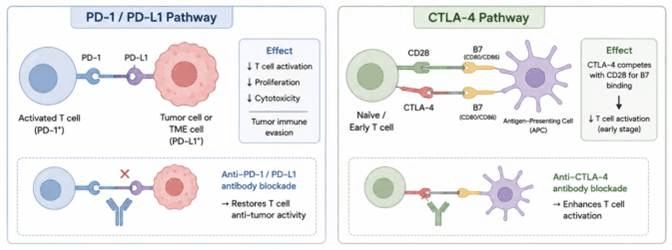
PD‑1/PD‑L1 and CTLA‑4 immune checkpoint axes and antibody‑mediated immune checkpoint blockade. PD‑1 on activated T cells binds PD‑L1 on tumor/TME cells to inhibit T‑cell function and drive tumor immune escape; anti‑PD‑1/PD‑L1 antibodies reverse this suppression. CTLA‑4 expressed by naïve/early T cells competes with CD28 for APC B7 (CD80/CD86) to limit early T‑cell activation. Anti‑CTLA‑4 blockade releases this inhibitory constraint and boosts T‑cell priming.
Common Research Strategies for Studying Immune Cells and Tumor Microenvironment
Research Area | Typical Research Model | Main Research Question | Common Experimental Methods | Key Readouts |
Immune Surveillance | Tumor cells + T cells / NK cells / DCs | Can immune cells recognize and eliminate tumor cells? | Flow cytometry, IF, ELISA/TR-FRET, cytotoxicity assays | CD8, CD69, CD25, Granzyme B, Perforin, IFN-γ |
T-cell Activation | Tumor cells + T cells | Do tumor antigens induce T-cell activation? | Flow cytometry, ELISA/TR-FRET, qPCR | CD69, CD25, IFN-γ, TNF-α, IL-2 |
Tumor Killing | Tumor cells + CD8⁺ T cells / NK cells | Can T/NK cells kill tumor cells? | Cell viability assay, LDH release assay, Annexin V/PI staining, Live-cell imaging | Tumor viability, cell death, apoptosis, cytotoxicity |
NK-cell Function | Tumor cells + NK cells | Can NK cells recognize and kill tumor cells? | Flow cytometry, degranulation assay, cytotoxicity assay | CD107a, Granzyme B, Perforin, IFN-γ |
T-cell Exhaustion | Tumor cells + T cells; chronic/persistent stimulation model | Does persistent antigen stimulation induce T-cell dysfunction? | Flow cytometry, ELISA/TR-FRET, functional cytotoxicity assays | PD-1, TIM-3, LAG-3, TIGIT, IFN-γ, TNF-α, Granzyme B |
PD-1/PD-L1 Signaling | Tumor cells + T cells | Does PD-1/PD-L1 signaling suppress anti-tumor T-cell function? | Flow cytometry, Western blot, IF, blocking antibody treatment | PD-1, PD-L1, T-cell activity, tumor killing |
CTLA-4 Signaling | T cells + APCs / tumor-associated immune cells | Does CTLA-4 regulate T-cell activation? | Flow cytometry, blocking antibody treatment, cytokine assays | CTLA-4, CD80, CD86, T-cell activation |
TAM Polarization | Tumor cells + macrophages | Do tumor cells alter macrophage phenotypes? | Flow cytometry, IF, qPCR, ELISA/TR-FRET | CD68, CD163, CD206, MHC-II, Arg1, iNOS |
TAM–Tumor Interaction | Tumor cells + TAMs | Do TAMs promote tumor growth, migration, and invasion? | Cell viability assay, EdU assay, wound healing assay, Transwell assay | Proliferation, migration, invasion, tumor viability |
MDSC-mediated Suppression | MDSCs + T cells | Do MDSCs suppress T-cell function? | T-cell proliferation assay, Flow cytometry, ELISA/TR-FRET | T-cell proliferation, IFN-γ, Granzyme B, activation markers |
Immune Cell Recruitment | Tumor-conditioned medium + immune cells | Do tumor-secreted factors recruit immune cells? | Transwell migration assay, chemotaxis assay, ELISA/TR-FRET | Migration, CCL2, CXCL8, CXCL12, etc. |
Immune Escape | Tumor + T cells / NK cells / MDSCs / TAMs | How do tumors evade immune recognition and immune-mediated killing? | Co-culture, Flow cytometry, IF, Western blot | PD-L1, MHC-I, immune checkpoints, cytotoxicity |
Immune Suppression | Tumor + T cells / NK cells / TAMs / MDSCs | How does the TME establish an immunosuppressive environment? | Co-culture, cytokine profiling, Flow cytometry | TGF-β, IL-10, PD-L1, immune-cell activity |
3D Immune Infiltration | Tumor spheroid + T cells / NK cells / macrophages | Can immune cells enter and infiltrate 3D tumor structures? | Confocal imaging, IF, Live-cell imaging | Immune-cell infiltration, spatial distribution, tumor killing |
Organoid–Immune Interaction | Tumor organoid + T cells / NK cells / macrophages | How do immune cells interact with more physiologically relevant tumor models? | 3D culture, IF, confocal microscopy, Flow cytometry, cytotoxicity assays | Infiltration, tumor cell death, immune activation |
Immunotherapy Evaluation | Tumor cells / spheroids / organoids + immune cells | Can drugs or cell-based therapies enhance anti-tumor immunity? | Drug treatment, blocking antibody treatment, cytotoxicity assays | Tumor viability, immune activation, cytokine production, immune infiltration |
Learn about our immune cell culture and detection reagents (cytokines, sera, media, etc.) and consumables
Learn about our monoclonal antibodies
Learn about our ELISA/TR-FRET kits
Learn about our fluorescent probes and TSA kits
Data display: