
info@ucallmlabs.com
T Cells
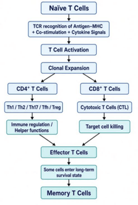
T cells are core lymphocytes of the adaptive immune system. They undergo development and selection primarily in the thymus and recognize antigens presented by the major histocompatibility complex (MHC) through the T cell receptor (TCR). After entering the peripheral immune system, mature T cells generally exist in a naïve state. When antigen-presenting cells, particularly dendritic cells, provide sufficient activation signals through antigen–MHC complexes, co-stimulatory signals, and cytokines, naïve T cells become activated and undergo clonal expansion and functional differentiation.
Based on CD4/CD8 expression and functional characteristics, T cells can further differentiate into distinct subsets. CD4⁺ T cells mainly include Th1, Th2, Th17, Tfh, and Treg subsets, which are involved in cellular immunity, humoral immunity, mucosal and inflammatory responses, and immune regulation, respectively. CD8⁺ T cells can differentiate into cytotoxic effector T cells, which eliminate virus-infected and tumor cells through perforin, granzymes, and other mechanisms.
Some activated T cells do not immediately disappear after completing their effector functions but instead develop into memory T cells. Upon re-exposure to the same antigen, memory T cells can rapidly expand and generate a stronger immune response, thereby providing long-term immune protection.
Basic Differentiation and Functional Pathways of T Cells:
Click to explore our animal-origin-free, carrier-protein-free, high-purity, low-endotoxin, highly bioactive recombinant cytokines with excellent lot-to-lot consistency.
We also provide customized services and professional technical support to meet a wide range of life science research needs. Contact our product specialists for more information.
Naïve T Cells
Naïve T cells are mature T cells that have completed their development in the thymus but have not yet encountered their specific antigens. They continuously monitor MHC–antigen complexes presented by antigen-presenting cells (APCs) through their T-cell receptors (TCRs). Upon receiving appropriate antigen-recognition, co-stimulatory, and cytokine signals, naïve T cells become activated and undergo rapid clonal expansion. Naïve T cells represent an important cell population that gives rise to subsequent effector T cells and memory T cells.
T Cell Activation
T-cell activation is a critical process that initiates the adaptive immune response and typically requires the coordinated action of three types of signals: antigen recognition, co-stimulatory signals, and cytokine signals.
First, the T-cell receptor (TCR) specifically recognizes antigenic peptides presented by major histocompatibility complex (MHC) molecules. CD4⁺ T cells primarily recognize antigens presented by MHC class II, whereas CD8⁺ T cells primarily recognize antigens presented by MHC class I.
Second, the co-stimulatory molecule CD28 binds to CD80/CD86 on the surface of antigen-presenting cells, providing the second signal required for full T-cell activation.
Third, the cytokine microenvironment provides additional signals that promote T-cell proliferation and influence their subsequent functional differentiation.
Together, these three signals drive resting naïve T cells into an activated state, followed by clonal expansion, effector differentiation, and memory T-cell formation. Ultimately, these processes enable T cells to participate in immune regulation, target-cell killing, and long-term immune protection.
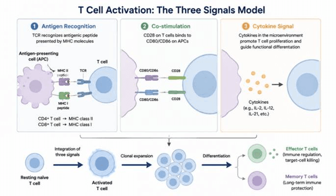
Schematic illustration of the three-signal model for T cell activation. Full T cell activation depends on three signals: antigen recognition by TCR-MHC-peptide interaction, CD28-CD80/CD86 co-stimulation, and cytokine signals from the microenvironment. Combined input of the three signals induces naïve T cell activation, clonal expansion and differentiation into effector T cells and memory T cells to execute immune responses and long-term immune protection.
CD4⁺ T Cells — Helper T Cells
CD4⁺ T cells are important regulatory and helper lymphocytes in the adaptive immune system. Naïve CD4⁺ T cells are activated through the coordinated action of TCR-mediated antigen recognition, co-stimulatory signals, and cytokine signals provided by antigen-presenting cells. Depending on the local cytokine environment, transcription factor regulatory networks, and tissue microenvironment, activated CD4⁺ T cells differentiate into distinct functional subsets. The major classical CD4⁺ T-cell subsets include Th1, Th2, Th17, Tfh, and Treg.
These subsets possess distinct transcriptional programs and effector functions. Through the secretion of specific cytokines and interactions with other immune cells, they collectively contribute to pathogen clearance, inflammatory responses, antibody production, immune tolerance, and maintenance of tissue homeostasis.
It is important to note that CD4⁺ T-cell subsets are not completely fixed or mutually exclusive cell types. Their functional states exhibit a degree of T-cell plasticity, allowing them to undergo functional changes or varying degrees of phenotypic remodeling in response to different antigenic stimuli, cytokine environments, and tissue microenvironments. Therefore, modern research increasingly views CD4⁺ T cells as a dynamic cell population with continuous functional states, rather than simply a collection of several independent subtypes.
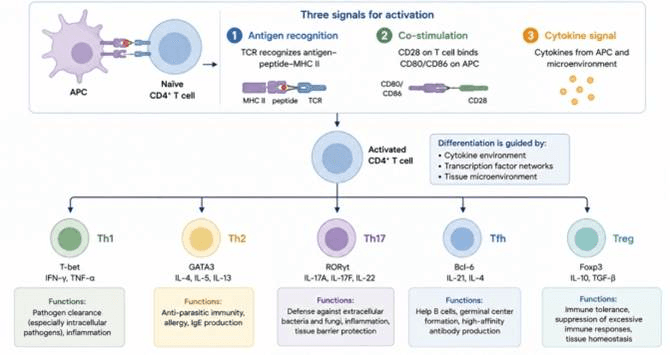
Schematic of CD4⁺ T cell activation and lineage differentiation. Full activation of naïve CD4⁺ T cells requires three signals: MHC II-peptide recognition by TCR, CD28-mediated co-stimulation, and cytokine signals from APCs and microenvironment. Activated CD4⁺ T cells differentiate into Th1, Th2, Th17, Tfh and Treg subsets under the control of cytokines, transcription factors and tissue context. Each subset is defined by specific transcription factors and cytokine profiles to mediate distinct immune responses.
Major Functional Subsets of CD4⁺ T Cells
| CD4⁺ T Cell Subset | Major Functions | Representative Transcription Factor | Representative Cytokines | Major Immune Processes |
| Th1 | Promotes cell-mediated immunity and enhances the killing capacity of macrophages and other immune cells | T-bet | IFN-γ, TNF | Defense against intracellular pathogens, anti-tumor immunity, inflammatory responses |
| Th2 | Promotes humoral and type 2 immune responses; regulates eosinophil- and mast cell-mediated reactions | GATA3 | IL-4, IL-5, IL-13 | Anti-parasitic immunity, allergic responses, tissue repair |
| Th17 | Promotes mucosal immunity and inflammatory responses; enhances neutrophil recruitment | RORγt | IL-17A, IL-17F, IL-22 | Defense against bacterial and fungal infections, mucosal protection, inflammation |
| Tfh (T follicular helper cells) | Assist B cells in germinal center reactions, promoting antibody production and affinity maturation | BCL6 | IL-21 | Antibody responses, B cell differentiation, immune memory formation |
| Treg (Regulatory T cells) | Suppress excessive immune responses and maintain immune tolerance and immune homeostasis | FOXP3 | IL-10, TGF-β | Peripheral immune tolerance, autoimmune control, inflammation regulation |
CD8⁺ T Cells——Cytotoxic T Cells
CD8⁺ T cells are important effector lymphocytes in the adaptive immune system. They mainly recognize antigenic peptides presented by MHC‑I molecules through the T‑cell receptor (TCR), so as to monitor host cells for viral infection, abnormal protein expression or tumour‑associated antigens.
Naïve CD8⁺T cells are activated upon receiving antigen‑recognition, co‑stimulation and cytokine signals. They undergo rapid clonal expansion and functional differentiation to generate potent cytotoxic effector CD8⁺T cells. The canonical effector cells are known as cytotoxic T lymphocytes (CTLs).
CTLs can form immunological synapses to make close contact with target cells, and release cytotoxic granules containing perforin and granzymes to induce programmed cell death of target cells. Meanwhile, CTLs can also facilitate target‑cell apoptosis via death‑receptor pathways such as Fas‑FasL.
After target‑cell killing, some activated CD8⁺ T cells differentiate into long‑lived memory CD8⁺ T cells, including central memory T cells (Tcm), effector memory T cells (Tem) and tissue‑resident memory T cells (Trm), which can elicit rapid immune responses upon re‑exposure to the same antigen.
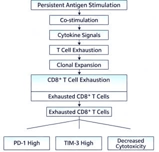
On the other hand, under persistent antigen‑stimulated conditions such as tumours or chronic infections, some CD8⁺ T cells may enter the state of T‑cell exhaustion, characterized by reduced cytotoxicity and cytokine production, together with up‑regulated expression of inhibitory receptors including PD‑1, TIM‑3 and LAG‑3.
Therefore, CD8⁺ T cells are not only key executors of host antiviral and anti‑tumour immunity, but also an important cell population for investigating immune memory, chronic infection and cancer immunotherapy.
Major Differentiation and Functional States of CD8⁺ T Cells
| English | Major Characteristics | Representative Markers | Major Functions |
| Naïve CD8⁺ T cells | Have not yet been exposed to cognate antigen stimulation | CD45RA, CCR7, CD62L | Antigen surveillance; undergo activation and differentiation upon antigen recognition |
| Effector CD8⁺ T cells | Rapidly expand following activation and acquire effector functions | CD69, CD25, Granzyme B, Perforin | Rapidly mediate cytotoxic immune responses |
| Cytotoxic T lymphocytes (CTLs) | Possess strong target-cell killing capacity | CD8, Granzyme B, Perforin | Kill virus-infected cells and tumor cells |
| Central memory T cells (Tcm) | Primarily circulate through lymphoid tissues and have strong proliferative capacity | CCR7, CD62L, CD45RO | Rapidly proliferate upon secondary antigen stimulation |
| Effector memory T cells (Tem) | Exhibit strong immediate effector functions | CCR7⁻, CD62L⁻, CD45RO⁺ | Rapidly migrate to peripheral tissues and exert effector functions |
| Tissue-resident memory T cells (Trm) | Persist long-term within specific tissues | CD69, CD103 | Provide rapid local immune protection within tissues |
| Exhausted CD8⁺ T cells | Progressive functional impairment caused by persistent antigen stimulation | PD-1, TIM-3, LAG-3, TIGIT | Represent an important T-cell state in chronic infections and tumors |
Differentiation Process of CD8⁺ T Cells
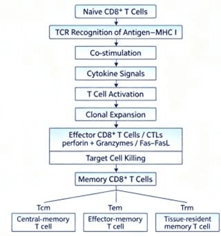
Under conditions of chronic infection and tumorigenesis
Treg — Regulatory T Cells
Regulatory T cells (Tregs) are an important subset of CD4⁺ T cells that play a central role in maintaining immune tolerance and immune homeostasis. Their classical characteristics include high expression of CD4 and CD25, together with expression of the transcription factor FOXP3.
Tregs can suppress excessive immune responses through multiple mechanisms, including inhibiting effector T cells, modulating antigen-presenting cells, and producing immunosuppressive cytokines, thereby reducing the risk of autoimmunity and tissue damage. On the other hand, within the tumor microenvironment, the immunosuppressive activity of Tregs can be exploited by tumors, thereby weakening anti-tumor immune responses.
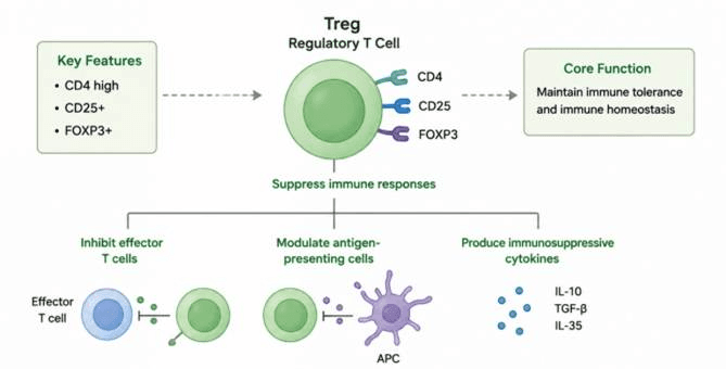
Schematic illustration of Treg markers and suppressive mechanisms. Tregs display signature markers CD4high, CD25⁺ and FOXP3⁺. They sustain immune tolerance and homeostasis by suppressing effector T cells, regulating antigen-presenting cells, and releasing inhibitory cytokines such as IL-10, TGF-β and IL-35.
Effector T Cells
Following T-cell activation, clonal expansion, and functional differentiation, T cells develop into effector T cells, which are capable of directly mediating immune responses. CD4⁺ effector T cells primarily exert their functions through cytokine secretion and regulation of other immune cells, whereas CD8⁺ effector T cells mainly eliminate infected cells and tumor cells through cytotoxic mechanisms. Effector T cells typically exhibit strong immediate immune effector functions; however, following antigen clearance, a subset of these cells can further differentiate into long-lived memory T cells.
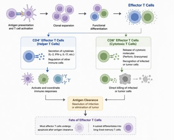
T cell activation, effector function and memory formation. Upon antigen presentation, T cells clonally expand and differentiate into CD4⁺ helper T cells and CD8⁺ cytotoxic T cells. CD4⁺ T cells coordinate immunity via cytokine secretion, while CD8⁺ T cells directly kill infected or tumor cells through perforin and granzymes. Their combined action clears antigen and resolves infection or tumors. Most effector T cells then undergo apoptosis, with a small subset persisting as long-lived memory T cells.
Memory T Cells
Following the resolution of an acute immune response, most effector T cells undergo contraction, while a subset survives long term and develops into memory T cells. Upon re-encountering the same antigen, memory T cells can rapidly expand and mount effector responses, providing an important basis for long-term protection by adaptive immunity. Based on their migratory properties and tissue distribution, memory T cells can be further classified into distinct subsets, including central memory T cells (Tcm), effector memory T cells (Tem), and tissue-resident memory T cells (Trm).
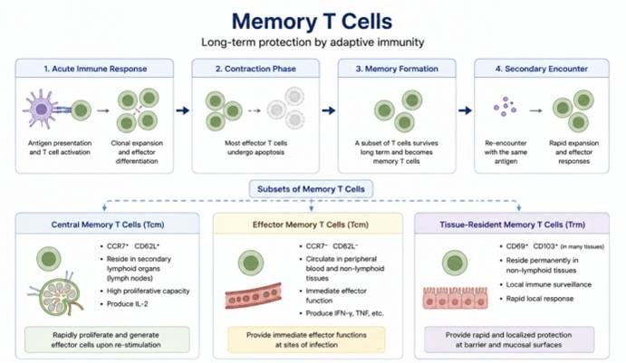
Memory T cell formation and subset classification. After acute immune activation and clonal expansion, most effector T cells die during contraction, and persistent memory T cells are formed. Re-stimulation by the same antigen triggers fast recall responses. Three memory T cell subsets (Tcm, Tem and Trm) differ in surface markers, tissue distribution and functions to sustain long-term adaptive immunity.
B Cells
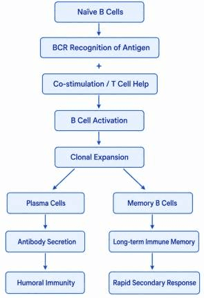
B cells are important lymphocytes of the adaptive immune system and are the central cells responsible for humoral immunity. B cells recognize specific antigens through their surface B-cell receptors (BCRs) and undergo activation, clonal expansion, and functional differentiation in response to antigen stimulation and signals from helper T cells and cytokines.
Activated B cells can differentiate into plasma cells, which produce and secrete large amounts of antigen-specific antibodies that eliminate pathogens through mechanisms such as neutralization, opsonization and phagocytosis, and complement activation. Some activated B cells develop into long-lived memory B cells, which can rapidly generate stronger antibody responses upon re-exposure to the same antigen. In addition, a subset of B cells can differentiate into regulatory B cells (Bregs) with immunoregulatory functions. Bregs help limit inflammation and maintain immune tolerance by secreting immunoregulatory factors such as IL-10, IL-35, and TGF-β. Therefore, B cells are not only responsible for antibody production, but also participate in antigen presentation, immune regulation, and the establishment of immunological memory.
Major B-Cell Subsets
| B-Cell Type | Major Characteristics | Major Functions |
| Naïve B Cells | Mature B cells that have not yet been exposed to specific antigen stimulation | Recognize antigens through B-cell receptors (BCRs) and initiate the activation process |
| Plasma Cells | Highly differentiated antibody-secreting cells | Produce and secrete large amounts of antibodies |
| Memory B Cells | Long-lived cells that maintain immune memory against specific antigens | Rapidly generate antibody responses upon re-exposure to the same antigen |
| Regulatory B Cells (Bregs) | B-cell subset with immunoregulatory functions | Suppress excessive inflammation and promote immune tolerance |
B cell activation and differentiation: an overview
Click to explore our animal-origin-free, carrier-protein-free, high-purity, low-endotoxin, highly bioactive recombinant cytokines with excellent lot-to-lot consistency.
We also provide customized services and professional technical support to meet a wide range of life science research needs. Contact our product specialists for more information.
NK Cells:
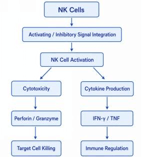
Natural Killer cells (NK cells) are important cytotoxic lymphocytes of the innate immune system that can recognize and eliminate virus-infected cells and certain tumor cells without prior antigen-specific sensitization. NK cells identify the “danger status” of target cells by integrating signals from multiple activating and inhibitory receptors.
When activating signals dominate, NK cells form an immunological synapse with target cells and release cytotoxic granules containing perforin and granzymes, inducing target-cell death. In addition, NK cells can mediate cytotoxicity through death receptor-dependent pathways, including Fas–FasL and TRAIL signaling.
Beyond direct cytotoxicity, NK cells produce large amounts of cytokines such as IFN-γ and TNF, which regulate the functions of other immune cells, including macrophages, dendritic cells, and T cells, thereby linking innate immunity and adaptive immune responses.
Based on the expression level of surface CD56 and functional characteristics, human NK cells are commonly classified into two major subsets: CD56^bright NK cells and CD56^dim NK cells. CD56^dim NK cells primarily exert strong cytotoxic functions, whereas CD56^bright NK cells generally exhibit higher cytokine-producing capacity and stronger immunoregulatory functions.
Major NK Cell Subsets:
| NK Cell Subset | Major Characteristics | Major Functions | Representative Features |
| CD56^bright NK cells | High CD56 expression and generally strong cytokine-producing capacity | Immune regulation and cytokine production | High production capacity of cytokines such as IFN-γ and TNF |
| CD56^dim NK cells | Relatively low CD56 expression and strong cytotoxic activity | Target-cell killing | High expression of Perforin and Granzyme B |
| NK cells (Natural Killer Cells) | Innate cytotoxic lymphocytes | Natural killing activity and cytokine production | Function independently of classical antigen-specific TCR recognition |
NK Cells:
Click to explore our animal-origin-free, carrier-protein-free, high-purity, low-endotoxin, highly bioactive recombinant cytokines with excellent lot-to-lot consistency.
We also provide customized services and professional technical support to meet a wide range of life science research needs. Contact our product specialists for more information.