
info@ucallmlabs.com
The mammary epithelium forms a bilayered structure, which can be classified into two principal lineages: luminal cells and basal cells, according to their spatial distribution. The majority of basal cells are terminally differentiated contractile myoepithelial cells, while a small subset of CD49⁺EpCAM⁻ basal cells possess properties of tissue-specific adult progenitor cells. The luminal cell population consists of two subtypes: mature luminal cells capable of sensing hormonal signals and mediating milk secretion, as well as luminal progenitor cells with restricted differentiation potential. Accumulating studies have verified that the hierarchical differentiation mechanism of mammary epithelial cells is highly intricate. Alterations in physiological conditions such as pregnancy and aging, together with inherited mutations in breast cancer susceptibility genes, can markedly reshape the proportion and distribution of mammary epithelial subpopulations.
In vitro culture techniques for mammary epithelial cells have provided crucial support for investigating the regular regulatory mechanisms and malignant transformation of mammary cells, facilitating fundamental research advances in mammary gland biology. Nevertheless, it remains challenging to establish an in vitro culture system that enables long-term stable cultivation and fully sustains all mammary epithelial lineages. Conventional culture approaches fail to efficiently preserve progenitor cells that serve as potential cells of origin for various breast cancer subtypes, which greatly hinders in-depth research on early oncogenic mechanisms of breast tumors. In contrast, mammary epithelial organoids derived from primary mammary tissues support sustained long-term passaging and completely retain all native mammary epithelial cell lineages. This model faithfully recapitulates the biomarker expression patterns of mammary tissues and stably reproduces the aberrant expansion phenotype of luminal progenitors triggered by BRCA1 heterozygous mutation, representing an excellent experimental system for exploring the mechanisms underlying mammary tumorigenesis and disease progression.
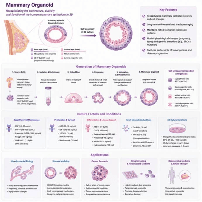
Application Scenarios of Mammary Organoids
Data display.
(A) Organoid culture status (bright-field morphology)
(B) Multicolor immunofluorescence staining
(C) Post-translational modification proteomics
(D) Epigenetic profiling (ChIP-seq / ATAC-seq)
(E) Gene editing validation (CRISPR)