
info@ucallmlabs.com
```
```
1.1 Mechanism Overview
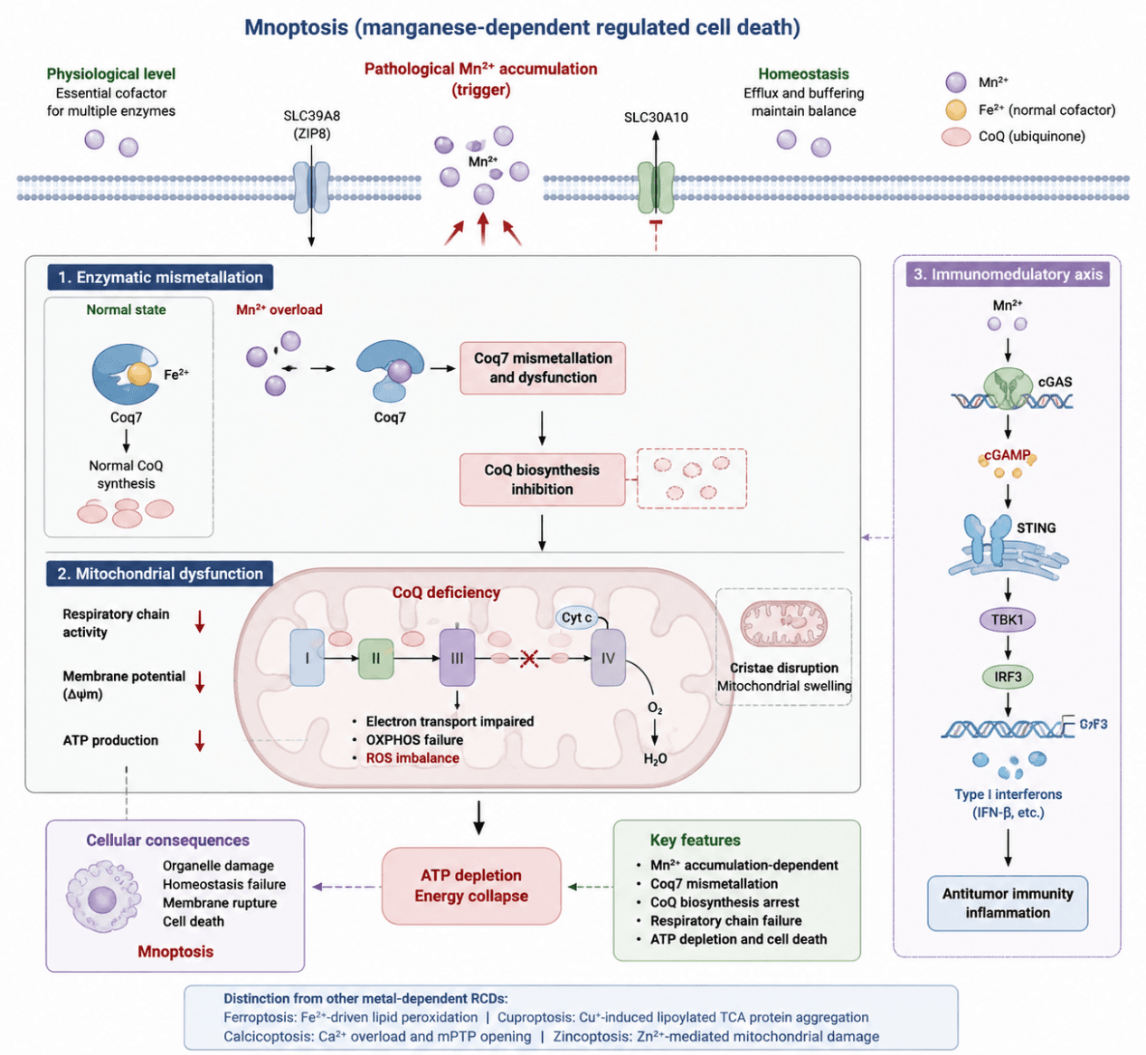
Mnoptosis is an emerging manganese‑dependent regulated cell death modality triggered by disrupted intracellular Mn²⁺ homeostasis and abnormal Mn²⁺ accumulation. Excessive Mn²⁺ replaces physiological metal cofactors in essential metabolic enzymes through enzymatic mismetallation. Coq7, the key enzyme for mitochondrial coenzyme Q (CoQ) synthesis, acts as the core target. Mn²⁺‑mediated Coq7 dysfunction blocks CoQ biosynthesis, disrupts the mitochondrial electron transport chain (ETC) and oxidative phosphorylation (OXPHOS), leading to impaired mitochondrial respiration, damaged membrane structure and inadequate ATP production, and ultimately induces cell death. Furthermore, Mn²⁺ promotes activation of the cGAS–STING pathway, elicits type I interferon responses and regulates anti‑tumor immunity。
Mnoptosis Mechanism Diagram. Aberrant Mn²⁺ accumulation induces mismetallation of mitochondrial Coq7, blocks CoQ biosynthesis, impairs the electron transport chain and oxidative phosphorylation, triggers mitochondrial structural damage and ATP depletion, and ultimately leads to Mnoptosis. Meanwhile, Mn²⁺ activates the cGAS‑STING pathway to regulate anti‑tumor immunity.
1.2 Target List
| Mechanism Module | Target (Gene/Protein) | Full Name | Biological Function | Mechanism in Mn²⁺‑dependent Cell Death | Research Value / Detection Index |
|---|---|---|---|---|---|
| Mn²⁺ Homeostasis Regulation | SLC39A8 (ZIP8) | Solute carrier family 39 member 8 | Metal ion transporter mediating Mn²⁺ uptake | Promotes intracellular Mn²⁺ accumulation and initiates Mn²⁺ toxicity | Indicator of Mn²⁺ influx |
| SLC30A10 | Solute carrier family 30 member 10 | Mn²⁺ efflux transporter maintaining manganese homeostasis | Reduced Mn²⁺ export increases intracellular Mn²⁺ accumulation | Indicator of Mn²⁺ efflux regulation | |
| COQ7‑dependent Core Execution Mechanism | COQ7 | Coenzyme Q7 hydroxylase | Enzyme involved in CoQ biosynthesis | Mn²⁺ induces COQ7 mismetallation, disrupting CoQ metabolism and mitochondrial function | Core execution marker |
| CoQ Metabolic Pathway Regulation | COQ3 | Ubiquinone biosynthesis O‑methyltransferase | Participates in CoQ biosynthetic modification | Dysfunction contributes to impaired CoQ synthesis | Marker of CoQ pathway alteration |
| COQ5 | Coenzyme Q biosynthesis protein COQ5 | Involved in CoQ maturation process | Regulates CoQ biosynthesis efficiency | Indicator of CoQ metabolism | |
| COQ8A | Coenzyme Q8A protein | Regulates CoQ biosynthesis and mitochondrial function | Alteration affects mitochondrial respiratory activity | CoQ pathway regulatory marker | |
| Mitochondrial Complex I Dysfunction | NDUFS1 | NADH dehydrogenase [ubiquinone] Fe‑S protein 1 | Component of respiratory Complex I involved in electron transfer | Mn²⁺ stress disrupts Complex I electron transport | Complex I damage marker |
| NDUFB8 | NADH dehydrogenase [ubiquinone] 1 beta subcomplex subunit 8 | Structural component of Complex I | Indicates mitochondrial respiratory impairment | Complex I functional marker | |
| Mitochondrial Complex II Dysfunction | SDHA | Succinate dehydrogenase flavoprotein subunit A | Catalyzes succinate oxidation and electron transfer | Mn²⁺ exposure decreases Complex II activity | Complex II function marker |
| Mitochondrial Complex III Dysfunction | UQCRC2 | Ubiquinol‑cytochrome c reductase core protein 2 | Component of mitochondrial Complex III | Electron transport inhibition increases mitochondrial oxidative stress | Complex III damage marker |
| Mitochondrial Complex IV Dysfunction | COX4 | Cytochrome c oxidase subunit 4 | Regulates cytochrome c oxidase activity and oxygen consumption | Mn²⁺ stress impairs Complex IV activity | Complex IV functional marker |
| MT‑CO1 | Mitochondrially encoded cytochrome c oxidase subunit 1 | Catalytic subunit of Complex IV | Reflects impaired oxidative phosphorylation | Respiratory chain damage marker | |
| ATP Synthesis Dysfunction | ATP5A | ATP synthase F1 subunit alpha | Catalyzes ATP production through oxidative phosphorylation | Mitochondrial dysfunction reduces ATP synthesis capacity | Mitochondrial energy metabolism marker |
| Mitochondrial Structural and Functional Damage | TOM20 | Translocase of outer mitochondrial membrane 20 | Mediates mitochondrial protein import and reflects mitochondrial mass | Indicates mitochondrial structural alteration | Mitochondrial integrity marker |
| VDAC1 | Voltage‑dependent anion‑selective channel protein 1 | Regulates mitochondrial metabolite exchange | Reflects mitochondrial membrane damage | Mitochondrial damage marker | |
| Oxidative Stress Response | 4‑HNE | 4‑Hydroxynonenal | Lipid peroxidation product | Indicates oxidative membrane damage caused by mitochondrial dysfunction | Lipid oxidative damage marker |
| 8‑OHdG | 8‑Hydroxy‑2'‑deoxyguanosine | Marker of oxidative DNA damage | Reflects oxidative stress‑induced DNA injury | DNA oxidative damage marker | |
| Terminal Stress Response | p‑AMPK | Phosphorylated AMP‑activated protein kinase | Regulates cellular energy stress response | Activated during ATP depletion and metabolic stress | Energy stress indicator |
| ATF4 | Activating transcription factor 4 | Regulates integrated stress response | Indicates mitochondrial and metabolic stress | Stress response marker | |
| CHOP | C/EBP homologous protein | Mediates stress‑induced cell death signaling | Reflects severe cellular stress and death response | Terminal stress marker |
1.3 Application Scheme
| Functional Module | Marker |
|---|---|
| Mn²⁺ accumulation | ICP‑MS Mn²⁺, SLC39A8, SLC30A10 |
| Core node of Mnoptosis | COQ7 |
| Impaired CoQ metabolism | CoQ10 |
| ETC injury | COX4, NDUFS1 |
| Defective ATP production | ATP5A, ATP level |
| Mitochondrial function | JC‑1/TMRE, OCR |
| Exclude ferroptosis | GPX4 |
| Exclude cuproptosis | FDX1/DLAT |
| Exclude apoptosis | Cleaved‑Caspase3 |
1.4 References
- Hu H, Chen Z, Li Y, Peng J, Cao J, Zhou H, Wang M, Du Y, Wu H, Zhao H, Huang S, Yu D, Liu M, Shevchenko OV, Matveeva NY, Yang Y, Huang K, Lv D, Min J, Chen L, Wang F. Metal‑dependent regulated cell death: Molecular architecture and translational frontiers. Imeta. 2026 Jul 6;5(3):e70141. doi: 10.1002/imt2.70141. PMID: 42491529; PMCID: PMC13377419.