
info@ucallmlabs.com
```
```
Multiplex Immunohistochemistry Solution for Human Colorectal Cancer Organoids
Comprehensive mIHC Workflow for Cancer Stemness, Epithelial Identity, and Tumor Microenvironment Profiling in FFPE Organoid Models
Unlock Spatial Insights into colorectal Cancer Organoid Biology with High‑Performance Tyramide Signal Amplification‑Based Multiplex Detection
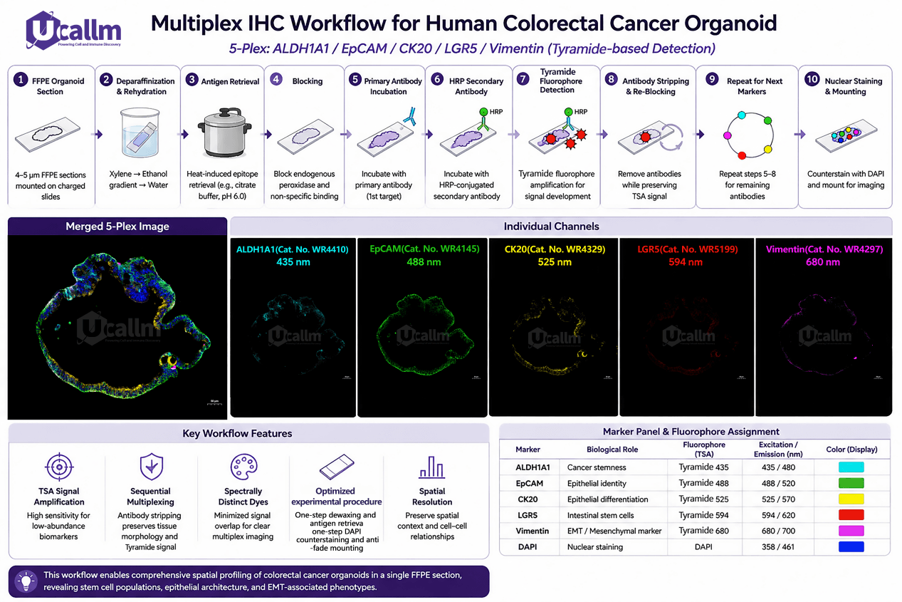
Multiplex IHC Workflow for Human Colorectal Cancer Organoid (5‑Plex: ALDH1A1 / EpCAM / CK20 / LGR5 / Vimentin (Tyramide‑based Detection))
Human cancer organoids preserve key architectural and molecular characteristics of primary tumors, providing a powerful model system for studying tumor heterogeneity, cancer stem cell populations, epithelial differentiation, and epithelial‑mesenchymal transition (EMT).
Our FFPE‑compatible multiplex immunohistochemistry (mIHC) solution enables simultaneous visualization of multiple biomarkers within human colorectal cancer organoid sections, combining high‑sensitivity Tyramide Signal Amplification signal amplification technology, validated antibodies, optimized detection reagents, and workflow‑compatible consumables.
The ALDH1A1/EpCAM/CK20/LGR5/Vimentin biomarker panel provides comprehensive characterization of:
- Cancer stem‑like cell populations
- Epithelial tumor identity
- Differentiation status
- EMT‑associated phenotypes
- Tumor heterogeneity and spatial organization
Application Overview
Spatial Profiling of colorectal Cancer Organoids Using a 5‑Plex Biomarker Panel
Recommended Marker Combination
| Biomarker | Biological Significance | Research Application |
|---|---|---|
| ALDH1A1 | Cancer stem cell‑associated marker | Identification of stem‑like tumor cell populations |
| EpCAM | Epithelial cell adhesion molecule | Identification of epithelial tumor compartments |
| CK20 | Colorectal epithelial differentiation marker | Assessment of intestinal lineage and tumor differentiation |
| LGR5 | Intestinal stem cell and colorectal cancer stem cell marker | Evaluation of tumor initiation and stemness |
| Vimentin | Mesenchymal marker | Detection of EMT and invasive phenotypes |
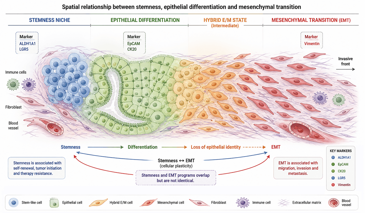
Spatial relationship between stemness, epithelial differentiation and mesenchymal transition
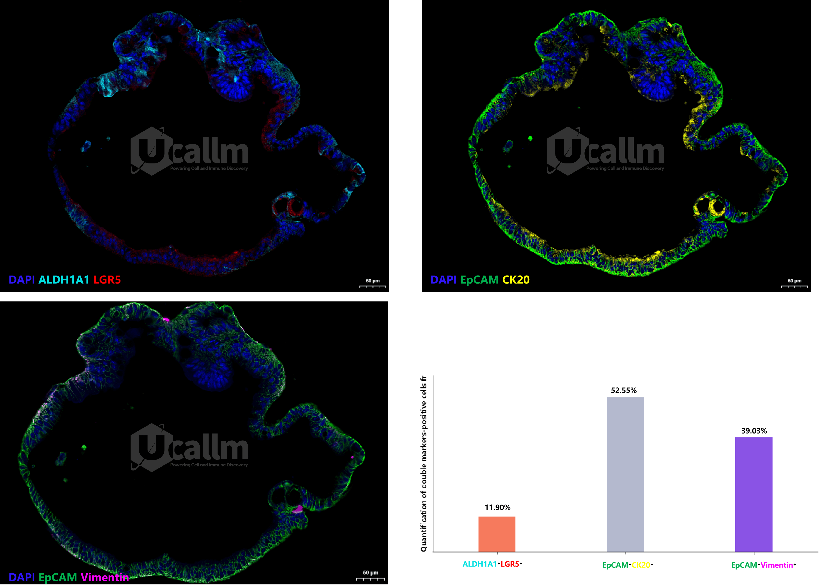
Multiplex immunohistochemistry (mIHC) staining of ALDH1A1, EpCAM, CK20, LGR5 and Vimentin in human colorectal cancer organoids, which enables researchers to investigate the relationship between stemness, epithelial differentiation, and mesenchymal transition within the same spatial context.
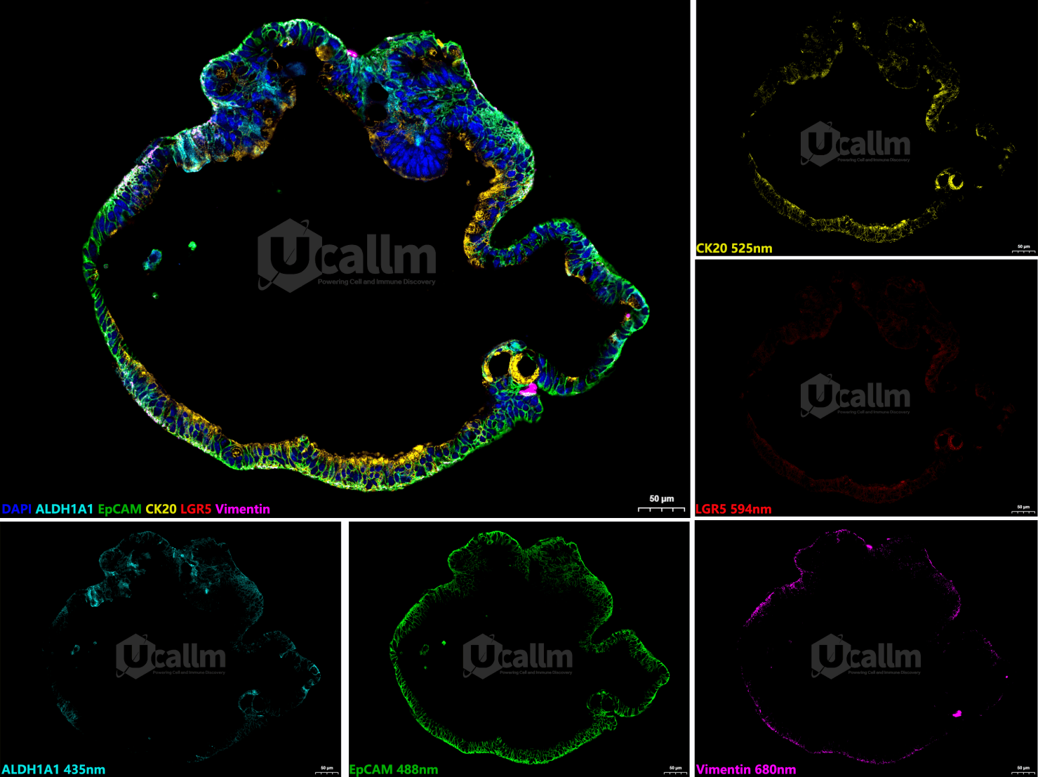
Figure 1. 5‑Plex 6 color mIHC staining images of human colorectal cancer organoids (Scale Bar: 50 μm)
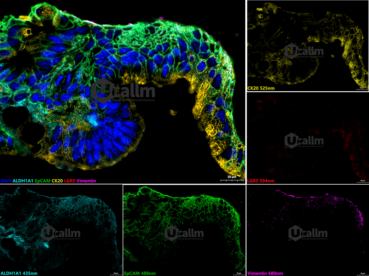
Representative 5‑Plex 6 color mIHC staining images of human colorectal cancer organoids, which was performed using the 5‑Plex mIHC Assay Kit (WT0018‑50) to detect the expression and spatial distribution of ALDH1A1 (WR4410, 1:500, 435 nm), EpCAM (WR4145, 1:500, 488 nm), CK20 (WR4329, 1:500, 525 nm), LGR5 (WR5199, 1:500, 594 nm), and Vimentin (WR4297, 1:500, 680 nm), and all above imaging data were acquired via the 3D‑Histech digital pathology scanning system. Scale bar = 50 μm.
Figure 2. High‑magnification 5‑Plex 6 color mIHC staining images of human colorectal cancer organoids (Scale Bar: 20 μm)
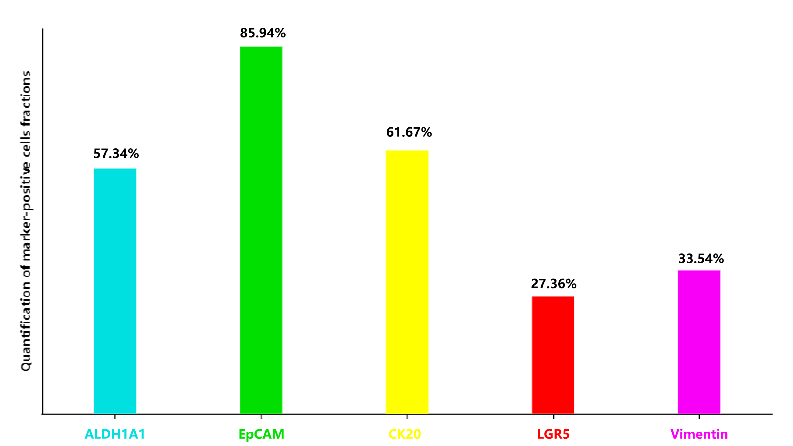
Figure 3. Quantification of marker‑positive cells fractions
EpCAM (85.94%) > CK20 (61.67%) ≈ ALDH1A1 (57.34%) > Vimentin (33.54%) > LGR5 (27.36%), which is biologically consistent consistent with a heterogeneous but predominantly epithelial CRC organoid model, rather than a purely differentiated epithelial culture or a fully mesenchymal tumor.
The dominant EpCAM⁺ epithelial population (85.94%), together with substantial CK20⁺ differentiation (61.67%), demonstrates preservation of colorectal epithelial identity. At the same time, the high ALDH1A1⁺ fraction (57.34%) and distinct LGR5⁺ population (27.36%) indicate coexistence of multiple stemness‑associated cellular states. The presence of 33.54% Vimentin⁺ cells further suggests substantial epithelial–mesenchymal plasticity or partial EMT.
Overall, these organoids are best characterized as a heterogeneous CRC model containing epithelial, differentiated, stem‑like, and EMT‑associated cellular subpopulations. This is consistent with the established literature showing that colorectal cancer organoids can retain both tumor characteristics and cellular heterogeneity of the parental cancer.
Figure 4. Quantification of double markers‑positive cells fractions
A substantial EpCAM⁺CK20⁺ population (52.55%) demonstrated preservation of epithelial identity and colorectal differentiation. In contrast, the limited ALDH1A1⁺LGR5⁺ overlap (11.90%) suggested that stemness‑associated features were distributed across partially distinct cellular subpopulations rather than a single homogeneous cancer stem‑cell compartment. Furthermore, a substantial EpCAM⁺Vimentin⁺ population (39.03%) indicated the presence of hybrid epithelial‑mesenchymal cellular states, consistent with epithelial‑mesenchymal plasticity. Collectively, these findings suggest that the colorectal cancer organoids preserve differentiation, heterogeneous stemness, and phenotypic plasticity, supporting their value as a representative model of colorectal cancer heterogeneity.
Complete Multiplex Immunohistochemistry Solution
1. mIHC Fluorescence Amplification System
High‑Sensitivity Signal Enhancement for Multiplex Biomarker Detection
Our Tyramide Signal Amplification‑based fluorescence amplification solution provides:
- Strong signal amplification for low‑abundance biomarkers
- High fluorescence intensity with minimal background
- Excellent compatibility with FFPE tissue sections
- Sequential multiplex staining capability
- Reliable performance across automated and manual workflows
Recommended Tyramide Signal Amplification Workflow Components
| ProductCategory | CatalogNo. | ProductName |
|---|---|---|
| mIHC Kits | WT0018 | 5‑Plex mIHC Assay Kit (6‑color, Anti‑Rabbit Secondary Antibody) |
| Fluorescence Amplification Reagents | WT1001 | Antibody Diluent (IHC) |
| Antigen Retrieval | WT1003 | Citrate Antigen Retrieval Buffer (pH 6.0)(20X) |
| WT1004 | TRIS‑EDTA Antigen Retrieval Buffer (pH 9.0)(20X) | |
| WT1005 | Citrate Antigen Retrieval Buffer (for automated IHC stainer) (pH 6.0)(20X) | |
| WT1006 | TRIS‑EDTA Antigen Retrieval Buffer (for automated IHC stainer) (pH 9.0)(20X) | |
| WT1007 | One‑step Deparaffinization Antigen Retrieval Solution (pH 6.0)(20X) | |
| WT1008 | One‑step Deparaffinization Antigen Retrieval Solution (pH 9.0)(20X) | |
| Antibody Stripping | WT1010 | High‑Temperature Antibody Stripping Buffer(20X) |
| WT1011 | Mild Antibody Stripping Buffer | |
| Mounting | WT1012 | Anti‑Fade DAPI Mounting Buffer (One‑Step) |
| Inactivation & Blocking | WT1013 | One‑Step Inactivation & Blocking Solution (For Auto IHC Stainer) |
2. Primary Antibody Portfolio
Validated Antibodies for colorectal Cancer Organoid Phenotyping
A carefully optimized antibody panel is essential for reliable mIHC performance.
Recommended Primary Antibody Panel
| Biomarker | Catalog No. | Product Name | Application | Species reactivity | Recommended mIHC dilution |
|---|---|---|---|---|---|
| ALDH1A1 | WR4410 | U‑Blot® ALDH1A1 Rabbit mAb | WB,mIHC,IHC‑P,IF/ICC,IP,ELISA | Human,Mouse,Rat | 1:500 |
| EpCAM | WR4145 | U‑Blot® EpCAM Rabbit mAb | WB,mIHC,IHC‑P,IF/ICC,IP,ELISA | Human,Mouse,Rat | 1:500 |
| CK20 | WR4329 | U‑Blot® CK20 Rabbit mAb | WB,mIHC,IHC‑P,IF/ICC,IP,ELISA | Human,Mouse,Rat | 1:500 |
| LGR5 | WR5199 | U‑Blot® LGR5 Rabbit mAb | WB,mIHC,IHC‑P,IF/ICC,IP,ELISA | Human,Mouse,Rat | 1:500 |
| Vimentin | WR4297 | U‑Blot® Vimentin Rabbit mAb | WB,mIHC,IHC‑P,IF/ICC,IP,ELISA | Human,Mouse,Rat | 1:500 |
3. Secondary Antibody Solutions
Optimized Detection Reagents for Multiplex Fluorescence Imaging
Our secondary antibody portfolio supports high‑quality fluorescence‑based detection:
Features
- High affinity and low background
- Multiple host species compatibility
- Broad fluorophore selection
- Excellent Tyramide Signal Amplification workflow compatibility
Recommended options:
- HRP‑conjugated secondary antibodies
- Species‑specific secondary antibodies
- Polymer‑based detection systems
4. Auxiliary Reagents
Complete Workflow Support from Sample Preparation to Imaging
Reliable mIHC performance requires optimized supporting reagents.
Sample Preparation
Products include:
- FFPE tissue deparaffinization reagents
- Antigen retrieval buffers
- Tissue permeabilization solutions
- Endogenous enzyme blocking reagents
- Matrigel
Staining Optimization
Products include:
- Protein blocking buffers
- Antibody dilution buffers
- Washing solutions
- Mounting media
Fluorescence Preservation
Products include:
- Antifade mounting medium
- Nuclear counterstaining reagents
- Fluorescence preservation solutions
5. Consumables and Workflow Accessories
Reliable Tools for Consistent mIHC Performance
Recommended consumables:
- Pipette tips and reservoirs
- Microcentrifuge Tubes
Key Advantages
Why Choose Our colorectal Cancer Organoid mIHC Solution?
- Complete Workflow Integration
From antibodies to Tyramide Signal Amplification reagents and consumables, every component is designed for compatibility. - High Sensitivity Detection
Tyramide Signal Amplification amplification enables visualization of low‑expression biomarkers in complex tumor models. - Multiparameter Spatial Profiling
Analyze multiple biological pathways simultaneously within a single FFPE section. - Reproducible Research Results
Optimized reagents support consistent staining quality across experiments. - Flexible Biomarker Expansion
The platform can be customized for additional oncology, immunology, and stem cell markers. - Optimized experimental procedure
One‑step dewaxing and antigen retrieval, as well as integrated one‑step DAPI counterstaining and anti‑fade mounting, enhanced your research efficiency
Applications
Suitable for:
- colorectal cancer organoid characterization
- Cancer stem cell research
- Tumor heterogeneity studies
- EMT mechanism investigation
- Drug response evaluation
- Translational oncology research
- Spatial biology applications
Advance Your colorectal Cancer Organoid Research with Precision Multiplex Imaging
- Our integrated mIHC solution provides researchers with the tools needed to transform complex tumor organoid models into actionable spatial insights.
- From biomarker selection to fluorescence imaging, we deliver a complete workflow designed for reliable, high‑quality multiplex analysis.