
info@ucallmlabs.com
1.1 Monocytes
Monocytes are bone marrow-derived myeloid innate immune cells that are primarily present in the peripheral blood. In response to signals associated with infection, inflammation, or tissue injury, monocytes can migrate into tissues and further differentiate into macrophages or subsets of dendritic cells.
Monocytes detect pathogen- and damage-associated signals through pattern recognition receptors (PRRs) and participate in multiple immune processes, including phagocytosis, antigen presentation, inflammatory cytokine production, tissue repair, and immune regulation.
Based on the expression of surface markers such as CD14 and CD16 and their functional characteristics, human peripheral blood monocytes are commonly classified into three major subsets: Classical Monocytes (CD14⁺⁺CD16⁻), Intermediate Monocytes (CD14⁺⁺CD16⁺), and Non-classical Monocytes (CD14⁺CD16⁺⁺).
These three monocyte subsets are not completely independent cell types but represent a continuum of functional states. They can undergo phenotypic and functional changes under different physiological and pathological conditions.
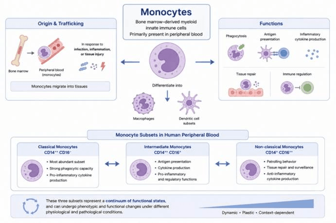
Schematic overview of monocyte development, subsets and functions. Monocytes are bone marrow-originated myeloid innate immune cells. They circulate in blood, migrate into inflamed tissues and differentiate into macrophages or dendritic cells. Monocytes mediate phagocytosis, antigen presentation, cytokine release, tissue repair and immune regulation. Human monocytes are divided into classical, intermediate and non-classical subsets. These subsets exhibit dynamic plasticity and shift their phenotypes and functions in response to different microenvironmental cues.
| Monocyte Subset | Typical Phenotype | Major Characteristics | Major Functions |
| Classical Monocytes | CD14⁺⁺CD16⁻ | The most abundant monocyte population in peripheral blood, with strong inflammatory responses and phagocytic capacity | Rapid recruitment, pathogen phagocytosis, and inflammatory cytokine production |
| Intermediate Monocytes | CD14⁺⁺CD16⁺ | Exhibit enhanced antigen presentation capacity and inflammatory regulatory functions | Antigen presentation, cytokine production, and immune regulation |
| Non-classical Monocytes | CD14⁺CD16⁺⁺ | Possess strong vascular patrolling and tissue surveillance capabilities | Vascular endothelial monitoring, clearance of damaged cells, and inflammatory regulation |
Click to explore our animal-origin-free, carrier-protein-free, high-purity, low-endotoxin, highly bioactive recombinant cytokines with excellent lot-to-lot consistency.
We also provide customized services and professional technical support to meet a wide range of life science research needs. Contact our product specialists for more information.
1.2 Macrophages:
Macrophages are important myeloid innate immune cells that are widely distributed throughout different tissues and play essential roles in pathogen clearance, inflammatory responses, antigen presentation, tissue repair, and maintenance of immune homeostasis.
Macrophages recognize pathogen- and damage-associated signals through pattern recognition receptors (PRRs) and participate in immune defense through various mechanisms, including phagocytosis, degranulation, production of reactive oxygen species (ROS) and reactive nitrogen species (RNS), and secretion of cytokines.
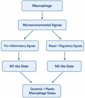
Macrophages exhibit highly dynamic functional plasticity, and their phenotypes and functions are influenced by multiple factors, including local cytokine signals, metabolic states, tissue microenvironments, and disease conditions.
In traditional studies, M1-like macrophages and M2-like macrophages are commonly used to describe two representative functional states of macrophages. The M1-like state is generally associated with pro-inflammatory responses, antimicrobial activity, and anti-tumor immunity, whereas the M2-like state is typically linked to resolution of inflammation, tissue repair, and immune regulation.
However, it is important to emphasize that M1/M2 classification represents a functional state description used in experimental studies rather than absolute and mutually exclusive cell categories. In physiological tissue environments, macrophages usually exhibit continuous, dynamic, and highly heterogeneous functional states, rather than belonging strictly to either the M1 or M2 category.
| Functional State | Typical Inducing Signals / Microenvironment | Representative Markers | Major Functions |
| M1-like | Inflammatory stimuli such as IFN-γ and LPS | iNOS/NOS2, CD80, CD86, MHC II | Pro-inflammatory responses, antimicrobial activity, anti-tumor immunity, and enhancement of immune activation |
| M2-like | Type 2 immune signals such as IL-4 and IL-13 | CD206, CD163, Arg1 | Resolution of inflammation, tissue repair, immune regulation, and tissue remodeling |
Click to explore our animal-origin-free, carrier-protein-free, high-purity, low-endotoxin, highly bioactive recombinant cytokines with excellent lot-to-lot consistency.
We also provide customized services and professional technical support to meet a wide range of life science research needs. Contact our product specialists for more information.
1.3 Dendritic Cells:
Dendritic cells (DCs) are essential antigen-presenting cells (APCs) that serve as a critical link between innate immunity and adaptive immunity. They are also key cells responsible for initiating primary T-cell immune responses. DCs capture pathogens, cellular debris, and other exogenous or endogenous antigens in peripheral tissues through mechanisms such as phagocytosis, macropinocytosis, and receptor-mediated endocytosis. These antigens are then processed through proteolytic degradation into antigenic peptides that can be recognized by T cells. The processed peptides are subsequently loaded onto major histocompatibility complex (MHC) molecules and transported to the cell surface for antigen presentation. Upon stimulation by pathogen-associated molecular patterns (PAMPs) or damage-associated molecular patterns (DAMPs), DCs undergo maturation, characterized by increased expression of MHC molecules and co-stimulatory molecules such as CD80 and CD86, followed by migration to draining lymph nodes. Within lymph nodes, mature DCs activate naïve T cells through the coordinated interaction of antigen–MHC complexes, co-stimulatory signals, and cytokines, thereby initiating and regulating adaptive immune responses. Based on their origin, phenotype, and functional characteristics, conventional dendritic cells (cDCs) are mainly classified into cDC1 and cDC2 subsets. In addition, plasmacytoid dendritic cells (pDCs) represent a specialized DC population with unique antiviral functions and a strong capacity for type I interferon production.
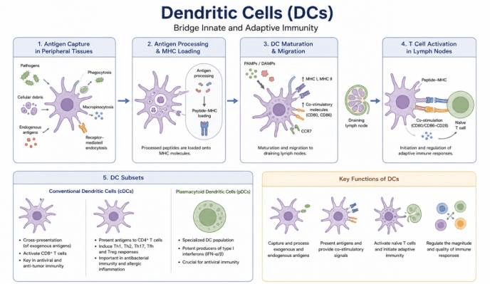
Schematic workflow and subsets of dendritic cells linking innate and adaptive immunity. DCs capture antigens in peripheral tissues, process antigens and load peptides onto MHC molecules. Stimulation by PAMPs/DAMPs induces DC maturation and CCR7-guided migration to lymph nodes, where they activate naïve T cells to launch adaptive immunity. Major DC subsets include conventional DCs (cDCs) and plasmacytoid DCs (pDCs). cDCs mediate antigen cross-presentation and prime CD4⁺ and CD8⁺ T cells, while pDCs secrete large amounts of type I interferons for antiviral immunity. DCs orchestrate immune responses by antigen processing, co-stimulation and tuning immune response strength.
Click to explore our animal-origin-free, carrier-protein-free, high-purity, low-endotoxin, highly bioactive recombinant cytokines with excellent lot-to-lot consistency.
We also provide customized services and professional technical support to meet a wide range of life science research needs. Contact our product specialists for more information.
1.4 Neutrophils
Neutrophils are among the most abundant innate immune cells in peripheral blood and are one of the first immune cell populations to arrive at sites of infection and tissue injury. Neutrophils can sense chemokines, pathogen-associated molecular patterns (PAMPs), and damage-associated signals, and rapidly migrate to infected or damaged tissues through chemotaxis.
Upon reaching local tissues, neutrophils eliminate pathogens such as bacteria and fungi through phagocytosis. They also exert antimicrobial and tissue-remodeling functions through degranulation, releasing granule components including myeloperoxidase (MPO), elastase, and cathepsins. In addition, neutrophils generate large amounts of reactive oxygen species (ROS) through respiratory burst, further enhancing their ability to kill pathogens.
Under specific stimulation conditions, neutrophils can undergo NETosis and release neutrophil extracellular traps (NETs), which are composed of DNA, histones, and granule proteins. NETs contribute to innate immune defense by capturing and restricting the spread of pathogens.
It is important to note that although these mechanisms are essential for rapid pathogen clearance, excessive or prolonged production of ROS, proteases, and NETs can also cause surrounding tissue damage and contribute to pathological processes, including chronic inflammation, autoimmune diseases, and tumor microenvironments.
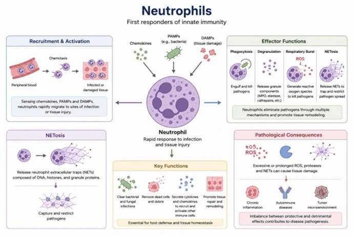
Recruitment, effector functions and pathological roles of neutrophils. Neutrophils are primary innate immune first responders. Guided by chemokines, PAMPs and DAMPs, they migrate to injured or infected tissues. Neutrophils kill pathogens through phagocytosis, degranulation, ROS production and NETosis. They also clear dead cells, secrete inflammatory mediators and support tissue repair. Uncontrolled neutrophil activation and release of ROS, proteases and NETs can induce tissue injury, driving chronic inflammation, autoimmunity and tumor microenvironment remodeling.
Major Functions of Neutrophils
| Function | Major Mechanism | Major Role |
| Chemotaxis | Sensing chemotactic signals such as CXCL8/IL-8, C5a, and LTB4 | Migration toward sites of infection and inflammation |
| Phagocytosis | Recognition and engulfment of pathogens and cellular debris | Clearance of bacteria, fungi, and other pathogens |
| Degranulation | Release of granule components such as myeloperoxidase (MPO) and elastase | Antimicrobial activity, pathogen degradation, and regulation of inflammatory responses |
| ROS Production | NADPH oxidase-mediated respiratory burst | Oxidative killing of pathogens |
| NET Formation / NETosis | Release of DNA, histones, and granule proteins to form neutrophil extracellular traps | Capture and restriction of pathogen dissemination |
Click to explore our animal-origin-free, carrier-protein-free, high-purity, low-endotoxin, highly bioactive recombinant cytokines with excellent lot-to-lot consistency.
We also provide customized services and professional technical support to meet a wide range of life science research needs. Contact our product specialists for more information.
1.5 Eosinophils:
Eosinophils are important innate immune cells that primarily participate in parasite immunity, type 2 immune responses, allergic inflammation, and tissue immune regulation. After developing and maturing in the bone marrow, eosinophils enter the peripheral blood and can be recruited to tissues in response to signals such as IL-5 and CCL11 (Eotaxin-1).
Upon stimulation by antigens, cytokines, or immune complexes, eosinophils exert their effector functions through multiple mechanisms, including chemotaxis, degranulation, lipid mediator production, and cytokine secretion.
Their cytoplasmic granules contain abundant cytotoxic molecules, including Major Basic Protein (MBP), Eosinophil Peroxidase (EPO), Eosinophil Cationic Protein (ECP), and Eosinophil-Derived Neurotoxin (EDN), which can directly damage large parasites and other target cells.
Meanwhile, eosinophils produce leukotrienes, prostaglandins, and type 2 immune-related cytokines such as IL-4, IL-5, and IL-13, contributing to allergic responses, mucosal immunity, and tissue inflammation.
Therefore, eosinophils function not only as important effector cells responsible for defense against helminths and other large parasites but also as key immune cells involved in asthma, allergic rhinitis, and other eosinophil-associated inflammatory diseases.
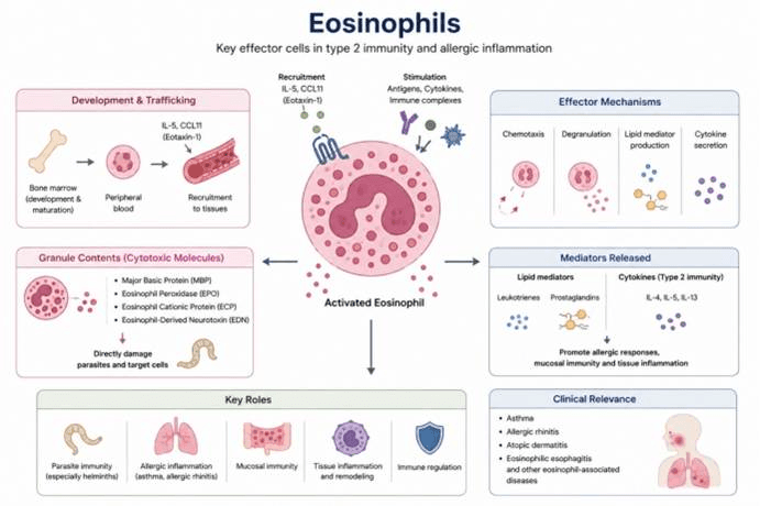
Development, effector mechanisms and clinical relevance of eosinophils. Eosinophils mature in bone marrow and traffic to tissues driven by IL‑5 and CCL11. Upon activation, they release granule cytotoxic proteins, lipid mediators and type‑2 cytokines. These mediators mediate anti-parasite immunity and drive allergic inflammation and tissue remodeling. Eosinophils are central players in type 2 immune responses and are implicated in asthma, allergic rhinitis, atopic dermatitis, eosinophilic esophagitis and other eosinophil-associated diseases.
Major Functions of Eosinophils
| Function | Major Mechanism | Major Role |
| Chemotaxis & Recruitment | IL-5, CCL11/Eotaxin, and other chemotactic signals | Migration to sites of inflammation and allergic responses |
| Degranulation | Release of MBP, EPO, ECP, EDN, and other granule proteins | Parasite killing and promotion of local inflammation |
| Parasite Defense | Cytotoxic granule proteins and reactive oxygen species (ROS) | Clearance of large parasites, especially helminths |
| Type 2 Immunity | Production of IL-4, IL-5, IL-13, and other type 2 immune mediators | Regulation of Th2-associated immune responses |
| Allergic Inflammation | Release of cytokines, lipid mediators, and granule proteins | Involvement in inflammatory diseases such as asthma and allergic rhinitis |
| Tissue Immunoregulation | Growth factors, cytokines, and lipid mediators | Regulation of tissue homeostasis and inflammatory responses |
Click to explore our animal-origin-free, carrier-protein-free, high-purity, low-endotoxin, highly bioactive recombinant cytokines with excellent lot-to-lot consistency.
We also provide customized services and professional technical support to meet a wide range of life science research needs. Contact our product specialists for more information.
2.2.6 Basophils
Basophils are rare but important innate immune cells in peripheral blood that play key roles in type I hypersensitivity reactions, type 2 immunity, and anti-parasite immune responses.
Basophils highly express the high-affinity IgE receptor (FcεRI) on their surface. When antigen-specific IgE antibodies cross-link and activate FcεRI, basophils rapidly undergo degranulation, releasing histamine, leukotrienes, and other inflammatory mediators. These mediators promote increased vascular permeability, smooth muscle contraction, and local inflammatory responses.
In addition, basophils can produce and secrete type 2 immune-related cytokines such as IL-4 and IL-13, thereby promoting Th2-type immune responses and IgE-mediated immunity.
Therefore, basophils not only contribute to the development and progression of allergic diseases, but also participate in parasite defense and immune regulation through interactions with other immune cells, including B cells, T cells, and eosinophils.
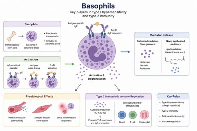
Development, activation and immune functions of basophils. Basophils are uncommon innate immune cells circulating in blood. Antigen-mediated cross-linking of FcεRI on IgE-primed basophils induces activation and degranulation. Basophils release histamine, heparin, proteases and lipid mediators, causing vascular leakage, smooth muscle contraction and local inflammation. By secreting IL‑4 and IL‑13, basophils promote Th2 responses and IgE production. They participate in type I hypersensitivity, anti-parasite defense and immune regulation via crosstalk with B cells, T cells and eosinophils.
Major Functions of Basophils
| Function | Major Mechanism | Major Role |
| IgE-mediated Activation | IgE–FcεRI cross-linking | Recognition of allergens and rapid cellular activation |
| Degranulation | Release of preformed mediators such as histamine | Rapid induction of allergic inflammation |
| Type 2 Cytokine Production | Production of IL-4, IL-13, and other type 2 cytokines | Promotion of Th2 and type 2 immune responses |
| Lipid Mediator Production | Release of leukotrienes, prostaglandins, and other lipid mediators | Amplification of inflammation and vascular responses |
| Anti-parasite Immunity | IgE-mediated responses and type 2 cytokine production | Contribution to defense against helminths and other parasites |
| Immune Regulation | Interactions with T cells, B cells, and other myeloid immune cells | Regulation of the local immune microenvironment |
Click to explore our animal-origin-free, carrier-protein-free, high-purity, low-endotoxin, highly bioactive recombinant cytokines with excellent lot-to-lot consistency.
We also provide customized services and professional technical support to meet a wide range of life science research needs. Contact our product specialists for more information.
2.2.7 Mast Cells:
Mast cells are innate immune cells derived from hematopoietic stem cells (HSCs). After maturation, they are primarily distributed in skin, respiratory tract, gastrointestinal tract, perivascular regions, and other tissue barrier sites, where they serve as important tissue-resident immune sentinels linking external stimuli to local immune responses.
Mast cells highly express the high-affinity IgE receptor (FcεRI) on their surface and can rapidly recognize allergens and become activated through IgE-dependent mechanisms. They can also sense pathogen- and tissue damage-associated signals through pattern recognition receptors (PRRs), complement receptors, and other activation pathways.
Upon activation, mast cells rapidly undergo degranulation, releasing preformed mediators such as histamine and proteases. They also synthesize and release lipid mediators, including leukotrienes and prostaglandins, as well as cytokines such as TNF, IL-4, IL-5, IL-6, and IL-13. These mediators rapidly alter local vascular permeability, recruit additional immune cells, and regulate inflammatory responses.
Therefore, mast cells play essential roles in type I hypersensitivity reactions, allergic inflammation, anti-parasite immunity, innate immune defense, tissue repair, and maintenance of tissue homeostasis. Unlike basophils, which are mainly found in peripheral blood, mast cells are typically long-lived tissue-resident cells and function as rapid-response sentinels at tissue barrier sites.
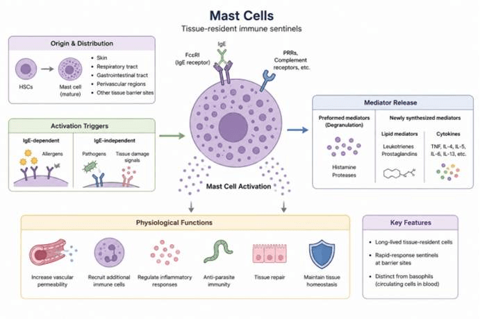
Origin, activation and biological functions of mast cells. Mast cells originate from hematopoietic stem cells and reside long-term in barrier tissues such as skin, airway and gut. They can be activated through IgE-dependent allergen stimulation or IgE-independent signals from pathogens, tissue injury and complement. Activated mast cells release pre-stored granule mediators, lipid mediators and multiple cytokines. These products enhance vascular permeability, recruit immune cells, regulate inflammation, defend against parasites and support tissue repair. Mast cells function as rapid tissue sentinels and are distinct from circulating basophils.
Major Functions of Mast Cells
| Function | English Term | Major Mechanism | Major Role |
| IgE-mediated Activation | IgE-mediated Activation | IgE–FcεRI cross-linking | Rapid recognition of allergens |
| Degranulation | Degranulation | Release of histamine, tryptase, and other preformed mediators | Rapid initiation of local inflammation and allergic responses |
| Lipid Mediator Production | Lipid Mediator Production | Production of leukotrienes, prostaglandins, and other lipid mediators | Regulation of vascular responses, smooth muscle responses, and amplification of inflammation |
| Cytokine Production | Cytokine Production | Secretion of TNF, IL-4, IL-5, IL-6, IL-13, and other cytokines | Regulation of inflammation and type 2 immune responses |
| Anti-parasite Immunity | Anti-parasite Immunity | IgE-dependent responses and release of inflammatory mediators | Contribution to defense against helminths and other parasites |
| Tissue Homeostasis & Repair | Tissue Homeostasis & Repair | Release of growth factors, proteases, and cytokines | Contribution to angiogenesis, tissue remodeling, and repair |
Click to explore our animal-origin-free, carrier-protein-free, high-purity, low-endotoxin, highly bioactive recombinant cytokines with excellent lot-to-lot consistency.
We also provide customized services and professional technical support to meet a wide range of life science research needs. Contact our product specialists for more information.