
info@ucallmlabs.com
```
```
Necrosis by Sodium Overload (NECSO)
1.1 Mechanism Overview
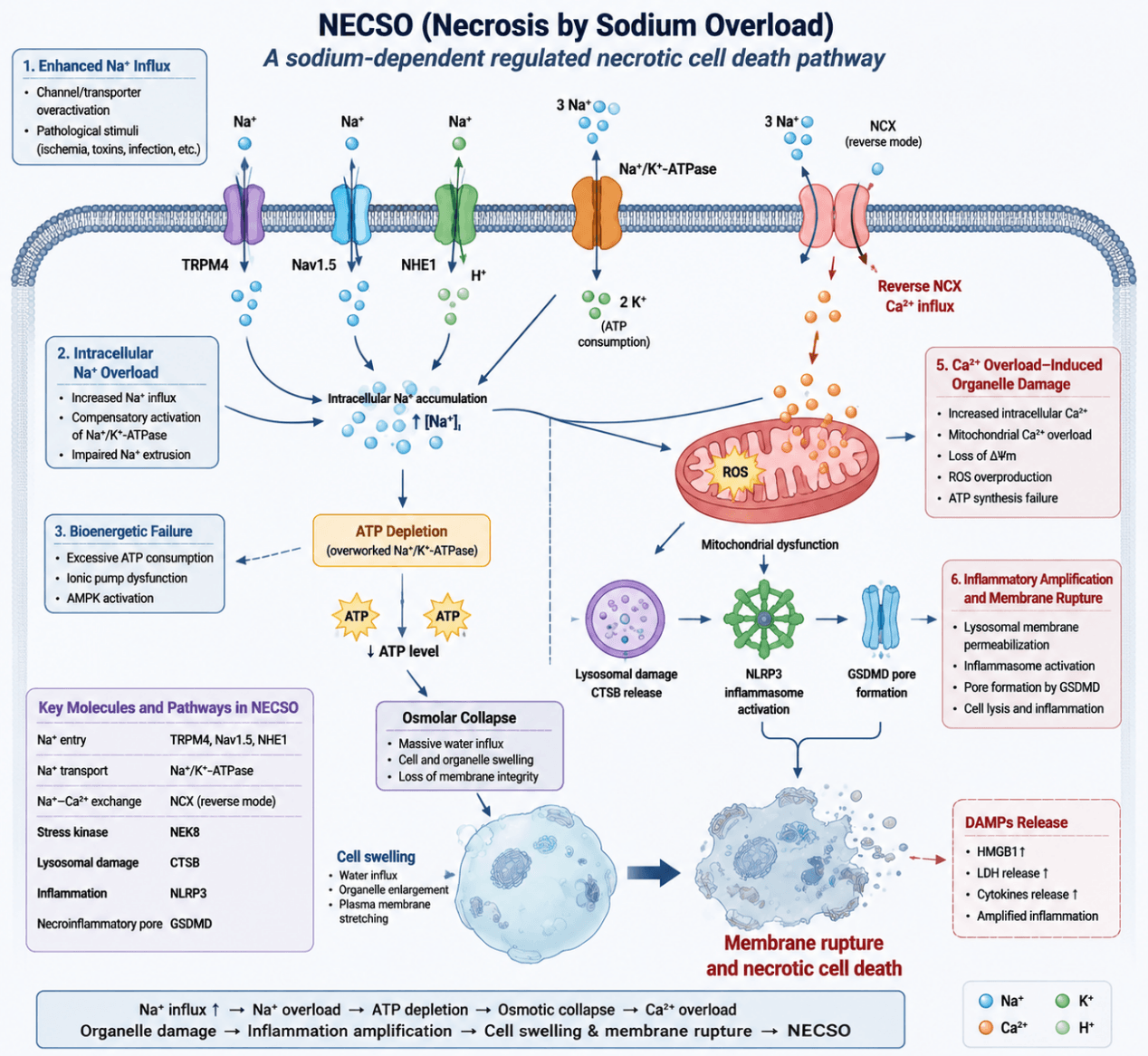
Necrosis by sodium overload (NECSO) represents a novel metal/ion‑dependent regulated cell death driven by disrupted intracellular Na⁺ homeostasis, which is characterized by osmotic collapse, energy exhaustion and secondary ionic toxicity triggered by aberrant sodium accumulation. Under physiological conditions, cells maintain intracellular Na⁺ homeostasis via Na⁺/K⁺‑ATPase, Na⁺/H⁺ exchanger 1 (NHE1), voltage‑gated sodium channels (Nav), transient receptor potential channel TRPM4 and other transporters. Abnormal activation or impairment of these ion regulatory systems triggers sustained Na⁺ influx and elevates cytoplasmic Na⁺ concentrations. This forces Na⁺/K⁺‑ATPase to continuously consume ATP to sustain ionic gradients, eventually resulting in severe energy depletion. Once sodium accumulation exceeds cellular buffering capacity, osmotic equilibrium collapses, massive water influx occurs, leading to cellular swelling, organelle distension and disrupted plasma membrane integrity, presenting typical necrotic morphological features.
Meanwhile, sodium overload provokes secondary Ca²⁺ influx via reverse‑mode operation of the Na⁺/Ca²⁺ exchanger (NCX), aggravating mitochondrial Ca²⁺ burden, loss of mitochondrial membrane potential, reactive oxygen species (ROS) accumulation and mitochondrial functional failure. Furthermore, Na⁺‑mediated cellular stress initiates lysosomal injury and inflammatory signaling cascades, including CTSB release, NLRP3 inflammasome activation and GSDMD‑dependent pore formation, which further amplify cell lysis and inflammatory responses. Accordingly, the execution cascade of NECSO can be summarized as: disrupted Na⁺ homeostasis → ATP depletion → osmotic collapse → secondary Ca²⁺ overload → mitochondrial damage → inflammatory necrosis.
NECSO Mechanism Diagram. Necrosis by sodium overload (NECSO) is initiated by massive Na⁺ influx and disrupted sodium homeostasis, triggering ATP depletion and osmotic collapse. Subsequently, Na⁺/Ca²⁺ exchanger (NCX)‑mediated calcium overload, mitochondrial dysfunction and amplified inflammatory signaling cascades occur, ultimately leading to necrotic plasma membrane rupture.
1.2 Target List
| Mechanism Module | Target (Gene/Protein) | Full Name | Biological Function | Mechanism in Na⁺‑dependent Cell Death | Research Value / Detection Index |
|---|---|---|---|---|---|
| Na⁺ Overload (Core Event) | TRPM4 | Transient receptor potential melastatin 4 | Calcium‑activated non‑selective cation channel mediating Na⁺ influx | Excessive activation promotes Na⁺ entry and intracellular Na⁺ accumulation | Marker of Na⁺ influx activation |
| Nav1.5 (SCN5A) | Sodium voltage‑gated channel alpha subunit 5 | Voltage‑gated sodium channel responsible for Na⁺ transport | Increased sodium channel activity contributes to Na⁺ overload | Indicator of sodium channel activation | |
| NHE1 (SLC9A1) | Sodium/hydrogen exchanger 1 | Exchanges intracellular H⁺ for extracellular Na⁺ to regulate intracellular pH | Enhanced Na⁺/H⁺ exchange increases intracellular Na⁺ accumulation | Marker of Na⁺ influx regulation | |
| Na⁺/K⁺‑ATPase (ATP1A1) | Sodium/potassium‑transporting ATPase alpha‑1 subunit | Maintains Na⁺ and K⁺ gradients through ATP‑dependent ion transport | Initially compensates for Na⁺ overload by promoting Na⁺ extrusion, but fails during late‑stage injury | Indicator of Na⁺ homeostasis disruption | |
| Energy Metabolic Collapse | ATP5A | ATP synthase F1 subunit alpha | Catalyzes ATP synthesis through oxidative phosphorylation | Mitochondrial dysfunction reduces ATP production capacity | Marker of mitochondrial energy failure |
| p‑AMPK | Phosphorylated AMP‑activated protein kinase | Energy stress sensor regulating metabolic adaptation | Activated during ATP depletion and cellular energy crisis | Energy stress indicator | |
| COX4 | Cytochrome c oxidase subunit 4 | Component of mitochondrial respiratory Complex IV involved in oxygen consumption | Impaired respiratory chain decreases oxidative phosphorylation efficiency | Respiratory chain damage marker | |
| NDUFS1 | NADH dehydrogenase [ubiquinone] Fe‑S protein 1 | Core component of mitochondrial Complex I involved in electron transfer | Dysfunction disrupts electron transport and mitochondrial respiration | Complex I functional marker | |
| Na⁺‑Ca²⁺ Coupled Injury | NCX1 (SLC8A1) | Sodium/calcium exchanger 1 | Exchanges intracellular Ca²⁺ and extracellular Na⁺ to regulate calcium homeostasis | Reverse‑mode activation promotes Ca²⁺ influx and secondary calcium overload | Marker of Na⁺/Ca²⁺ exchange dysfunction |
| SERCA2 | Sarco/endoplasmic reticulum Ca²⁺‑ATPase 2 | Pumps Ca²⁺ into the endoplasmic reticulum and maintains Ca²⁺ homeostasis | Reduced activity contributes to intracellular Ca²⁺ accumulation | Calcium homeostasis marker | |
| Mitochondrial Injury | JC‑1 / TMRE | Mitochondrial membrane potential fluorescent probes | Detect mitochondrial membrane potential (ΔΨm) | Na⁺ overload induces mitochondrial depolarization and dysfunction | Detection of mitochondrial membrane potential loss |
| ROS / MitoSOX | Reactive oxygen species / mitochondrial superoxide indicator | Detect mitochondrial oxidative stress | Mitochondrial dysfunction increases ROS production | Oxidative stress indicator | |
| HMOX1 | Heme oxygenase 1 | Antioxidant enzyme involved in cellular stress response | Activated as a protective response to oxidative stress | Oxidative stress response marker | |
| NQO1 | NAD(P)H quinone dehydrogenase 1 | Antioxidant and detoxification enzyme | Reflects activation of cellular antioxidant defense | Antioxidant response marker | |
| TOM20 | Translocase of outer mitochondrial membrane 20 | Mediates mitochondrial protein import and reflects mitochondrial integrity | Altered distribution indicates mitochondrial structural damage | Mitochondrial integrity marker | |
| COX4 | Cytochrome c oxidase subunit 4 | Maintains respiratory Complex IV function | Reduced expression reflects mitochondrial respiratory injury | Mitochondrial damage marker | |
| Osmotic Collapse (NECSO Hallmark Event) | Cell swelling | Cellular volume expansion | Morphological consequence of ionic imbalance | Na⁺ accumulation drives osmotic water influx and cellular swelling | Hallmark morphological feature |
| PI uptake | Propidium iodide uptake | Detects loss of membrane integrity | Increased membrane permeability occurs during terminal necrotic damage | Cell death validation marker | |
| LDH release | Lactate dehydrogenase release | Reflects plasma membrane rupture | Indicates terminal necrotic membrane damage | Necrotic cell death marker | |
| Lysosomal Damage | CTSB | Cathepsin B | Lysosomal cysteine protease involved in protein degradation | Lysosomal injury causes CTSB release and amplifies cell death signaling | Marker of lysosomal membrane damage |
| Inflammatory Amplification | NLRP3 | NLR family pyrin domain containing 3 | Forms inflammasome complexes in response to cellular stress | Activation promotes inflammatory cell death signaling | Inflammasome activation marker |
| Caspase‑1 | Cysteine‑aspartic acid protease 1 | Inflammatory caspase responsible for IL‑1β/IL‑18 maturation | Activation promotes inflammatory amplification | Marker of inflammasome activation | |
| GSDMD‑N | Gasdermin D N‑terminal domain | Forms membrane pores after GSDMD cleavage | Mediates membrane rupture and inflammatory cell death execution | Pyroptosis‑like execution marker | |
| DAMP Release / Terminal Necrosis | HMGB1 | High mobility group box 1 protein | Nuclear DNA‑binding protein released during necrotic cell death | Release indicates inflammatory necrosis and DAMP signaling | DAMP release marker |
| IL‑1β | Interleukin‑1 beta | Pro‑inflammatory cytokine | Increased secretion reflects inflammatory activation | Inflammatory output marker | |
| IL‑18 | Interleukin‑18 | Cytokine involved in immune activation | Released during inflammasome‑associated cell death | Inflammatory cytokine marker |
1.3 Application Scheme
| Judgement Purpose | Recommended Marker Panel |
|---|---|
| Na⁺ overload | Na⁺ probe + TRPM4 + NHE1 |
| Disrupted ion homeostasis | Na⁺/K⁺‑ATPase + NCX1 |
| Secondary Ca²⁺ injury | Ca²⁺ probe + NCX1 |
| Mitochondrial damage | ROS + ΔΨm + COX4/ATP5A |
| Necrosis occurrence | LDH + PI + HMGB1 |
| Inflammatory amplification | NLRP3 + GSDMD‑N |
1.4 References
- Hu H, Chen Z, Li Y, Peng J, Cao J, Zhou H, Wang M, Du Y, Wu H, Zhao H, Huang S, Yu D, Liu M, Shevchenko OV, Matveeva NY, Yang Y, Huang K, Lv D, Min J, Chen L, Wang F. Metal‑dependent regulated cell death: Molecular architecture and translational frontiers. Imeta. 2026 Jul 6;5(3):e70141. doi: 10.1002/imt2.70141. PMID: 42491529; PMCID: PMC13377419.