
info@ucallmlabs.com
1. Overview
Nipah virus (NiV) is the prototype species of the genus Henipavirus within the family Paramyxoviridae and is the causative agent of Nipah virus disease. It is an enveloped, negative‑sense, single‑stranded, non‑segmented RNA virus with pleiomorphic virions, mostly irregularly spherical, approximately 150–500 nm in diameter.
NiV was first identified in Malaysia in 1999, named after the village of Kampung Sungai Nipah where the outbreak occurred. The virus is transmitted primarily through fruit bats (Pteropus species, also known as flying foxes) to humans, as well as through contaminated food or direct human‑to‑human transmission. NiV can cause severe respiratory illness and fatal encephalitis in humans, with case fatality rates of 40%–75%. The World Health Organization (WHO) has classified Nipah as a priority pathogen due to its alarming potential to cause widespread outbreaks and even trigger the next pandemic. Currently, no specific drugs or vaccines are approved for NiV infection.
2. Background Information
2.1 What is Nipah Virus?
Nipah virus is an enveloped, negative‑sense, single‑stranded, non‑segmented RNA virus belonging to the family Paramyxoviridae and genus Henipavirus. The virion is pleiomorphic, mostly irregularly spherical, approximately 150–500 nm in diameter. The viral envelope is derived from the host cell membrane and contains two surface glycoproteins——attachment glycoprotein (G) and fusion protein (F) . The inner side of the envelope is lined by the matrix protein M.
The NiV genome is a negative‑sense, single‑stranded RNA of approximately 18.2 kb, non‑segmented. The genome contains six genes in the order 3′ to 5′: nucleocapsid (N), phosphoprotein (P), matrix (M), fusion (F), attachment (G), and long polymerase (L) . The P gene also encodes three non‑structural proteins—C, V, and W—through RNA editing.
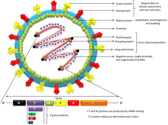
Structure of Nipah virus. (PMID: 31006350)
2.2 Nipah Virus Genome & Classification
CNiV belongs to the genus Henipavirus in the family Paramyxoviridae. The genus Henipavirus also includes Hendra virus (HeV) , with which NiV shares high structural, sequence, and antigenic similarity. The G proteins of NiV and HeV can provide heterologous cross‑protective immunity.
NiV is divided into two major genetic lineages:
l Malaysian lineage (NiV-M) : from the 1999 Malaysian outbreak
l Bangladesh lineage (NiV-B) : circulating in Bangladesh and India
2.3 What Are Nipah Virus Structural Proteins?
NiV encodes six major structural proteins and three non‑structural proteins:
Protein | Gene | Main Function |
N (Nucleoprotein) | N gene | Binds tightly to viral RNA, forms the nucleocapsid core |
P (Phosphoprotein) | P gene | Polymerase cofactor, involved in transcription and replication |
M (Matrix protein) | M gene | Inner envelope protein, mediates morphogenesis and budding |
F (Fusion protein) | F gene | Class I fusion protein, mediates membrane fusion |
G (Attachment protein) | G gene | Surface attachment glycoprotein (602 aa), recognizes host receptors |
L (Large polymerase) | L gene | RNA‑dependent RNA polymerase (RdRp), catalyzes transcription and replication |
C, V, W (Non‑structural) | P gene (RNA editing) | Host immune evasion; V protein interferes with JAK–STAT signaling |
2.4 Mechanism of Cell Entry and Replication
2.4.1 Viral Attachment and Receptor Recognition
NiV infection begins with the binding of the viral G protein to host cell surface receptors. G protein recognizes and binds Ephrin-B2 or Ephrin-B3 receptors. Ephrin-B2 and Ephrin-B3 are the functional receptors for henipaviruses. G protein–Ephrin receptor binding triggers conformational changes in G that activate F protein.
2.4.2 Viral Entry
Following G protein–Ephrin receptor binding, F protein mediates fusion of the viral envelope with the host cell membrane. NiV membrane fusion is pH‑independent, occurring either at the endosome or at the cell surface. F protein rearranges from prefusion to postfusion conformation, inserting the fusion peptide into the host membrane and releasing the viral nucleocapsid into the cytoplasm.
2.4.3 Viral RNA Replication
After RNP release into the cytoplasm, the L protein (RdRp) initiates transcription and replication:
l Transcription: negative‑sense RNA genome is transcribed into mRNAs, which are translated into viral proteins
l Replication: positive‑sense RNA intermediates are synthesized using negative‑sense RNA as template
l Progeny negative‑sense genomic RNAs are synthesized using positive‑sense RNA as template
2.4.4 Viral Assembly and Release
Newly synthesized N protein associates with progeny genomic RNA and L protein to form new RNPs. M protein recruits RNPs to the cell membrane. The virus acquires its envelope by budding from the host cell membrane, releasing new virions.
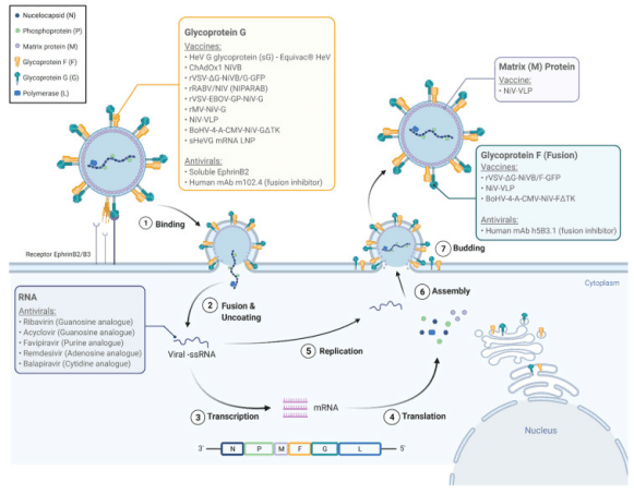
The Nipah virus life cycle and molecular targets for pharmacologic agents. (PMID: 33672796)
2.5 Symptoms and Treatment
2.5.1 Clinical Symptoms
The incubation period for NiV infection is typically 3–14 days, with rare cases up to 45 days. Approximately 20% of infections are asymptomatic. Common symptoms in symptomatic patients include:
l Fever
l Headache
l Muscle pain (myalgia)
l Vomiting
l Sore throat
l Dizziness, drowsiness
The disease can rapidly progress to acute encephalitis, pneumonia, and severe respiratory problems. Severe cases may develop brain swelling (encephalitis) and frequently death. Approximately 1 in 5 survivors experience long‑term neurologic conditions.
2.5.2 Treatment
Currently, no specific antiviral therapy is approved for NiV infection. Treatment is primarily early intensive supportive care:
l Intensive care support
l Respiratory support
l Management of neurologic complications
2.6 Targets for Intervention
2.6.1 Viral Targets
Target | Protein | Biological Role |
G | Attachment protein | Ephrin receptor binding |
F | Fusion protein | Membrane fusion |
L (RdRp) | RNA polymerase | RNA transcription & replication |
N | Nucleoprotein | RNP formation |
M | Matrix protein | Assembly & budding |
2.6.2 Host Receptors & Entry Factors
Host Target | Type | Role in Infection |
Ephrin-B2 | Cell surface receptor | G protein‑mediated viral attachment |
Ephrin-B3 | Cell surface receptor | G protein‑mediated viral attachment |
2.6.3 Host Signaling Pathways
Pathway | Key Targets | Biological Role |
JAK–STAT signaling | JAK1, STAT1/2 | Interferon signaling, suppressed by V protein |
Interferon pathway | IFN‑α/β | Antiviral response |
2.7 Vaccine Types and Development Progress
Currently, no Nipah virus vaccine is licensed worldwide. However, several candidates have entered clinical trials, with major breakthroughs in 2025.
2.7.1 HeV‑sG‑V (Hendra Virus Soluble G Glycoprotein Vaccine)
HeV‑sG‑V is a vaccine candidate based on the Hendra virus soluble G glycoprotein. Due to the structural and sequence similarities between NiV and HeV G glycoproteins, HeV‑sG provides heterologous cross‑protective immunity.
2.7.2 ChAdOx1 NipahB (Adenovirus‑Vectored Vaccine)
ChAdOx1 NipahB was developed by scientists at the University of Oxford's Pandemic Sciences Institute (PSI), using the same ChAdOx1 adenovirus vector platform as the Oxford/AstraZeneca COVID‑19 vaccine.
2.7.3 Other Vaccine Candidates
l Nanoparticle‑based G head (GH) vaccines: induced neutralizing antibodies against NiV in animal models
l VEEV replicon‑based vaccines: in preclinical development
l mRNA vaccines: in preclinical development
2.8 Drugs
Currently, no specific small‑molecule antiviral is approved for NiV infection. Treatment relies primarily on supportive care. Several novel antiviral candidates have shown promise:
2.8.1 Virus-Targeted Drugs
Target | Drug/candidate | Mechanism | Stage |
L protein (RdRp) | VV116 (Deuremidevir hydrobromide) | Oral nucleoside prodrug targeting RdRp | In vitro |
Functionalized phytochemical‑conjugated fullerene quantum dots | Inhibits viral chaperone activity and RdRp | Computational design | |
F protein (fusion) | VQ‑P1‑EK3‑C3 (dePEGylated lipopeptide) | High affinity for NiV‑HR1, inhibits 6‑HB formation | In vitro |
G/F proteins | Bispecific nanobody (DS90 + m102.4) | Dual‑targeting (anti‑F nanobody + anti‑G mAb), resistant to viral escape | Preclinical |
F protein | Rhesus‑derived monoclonal antibodies | Target prefusion F conformation | Preclinical |
2.9 Epidemiology
l First identified: 1999, Malaysia
l Geographic distribution: Bangladesh, India, Malaysia, Philippines, Singapore
l Bangladesh: since the first outbreak in 2001, cases have been detected almost every year. As of 2025, Bangladesh has documented 347 cases with a case fatality rate of 71.7%
l India: multiple outbreaks since 2001—West Bengal (2001, 2007), Kerala (regularly since 2018). In 2025, Kerala reported 4 confirmed cases (including 2 deaths). In the last week of December 2025, West Bengal reported 2 confirmed cases
l 2025 outbreaks: Bangladesh—4 confirmed fatal cases; India (Kerala)—4 confirmed cases (2 deaths)
l Transmission routes:
Ø Animal‑to‑human: contact with infected fruit bat saliva, urine, and excreta, or consumption of contaminated raw date palm sap
Ø Human‑to‑human: through close contact with infected individuals
Ø Approximately half of primary cases are linked to raw date palm sap consumption, while about one‑third result from human‑to‑human transmission
l Natural reservoir: fruit bats (Pteropus species, flying foxes)
2.10 Diagnosis
Diagnosis of NiV infection relies primarily on laboratory testing:
l RT‑PCR (Reverse Transcription Polymerase Chain Reaction) : gold standard for acute‑phase diagnosis; detects viral RNA in blood and cerebrospinal fluid
l ELISA (Enzyme‑Linked Immunosorbent Assay) : detects anti‑NiV IgM and IgG antibodies in serum and cerebrospinal fluid
l LAMP (Loop‑mediated Isothermal Amplification) : molecular detection method
2.11 Prevention
l Avoid contact: avoid contact with fruit bats, pigs, and other animals
l Food safety: do not drink raw date palm sap; avoid eating fruit that may have been contaminated by animals; wash and peel fruit before eating
l Personal hygiene: practice good handwashing, especially after contact with animals, visiting sick people, or after being in public places
l Healthcare settings: healthcare providers should wear gowns, gloves, eye protection, and N95 respirators or higher
3. Related Products & Services
l Recombinant Antigens & Receptors
l Antibodies
l Detection Kits
l Recombinant Protein Expression Services
l Antibody Development Services
4. Resources
5. References
1. Spengler, J. R., Lo, M. K., Welch, S. R., et al. (2024). Henipaviruses: epidemiology, ecology, disease, and the development of vaccines and therapeutics. Clinical Microbiology Reviews, 37(4), e00128-23.
2. Eaton, B. T., Broder, C. C., Middleton, D., et al (2006). Hendra and Nipah viruses: different and dangerous. Nature Reviews Microbiology, 4, 23–35.
3. Diederich, S., Dietzel, E. (2021). Evolution of Nipah Virus Infection: Past, Present, and Future Considerations. Viruses, 13.
4. Kumar, S., et al. (2019). Nipah virus: epidemiology, pathology, immunobiology and advances in diagnosis, vaccine designing and control strategies—a comprehensive review.
5. Negrete, O. A., Levroney, E. L., Aguilar, H. C., et al. (2005). EphrinB2 is the entry receptor for Nipah virus, an emergent deadly paramyxovirus. Nature, 436, 401–405.
6. Xu, K., Chan, Y. P., Bradel-Tretheway, B., et al. (2012). Crystal structure of the pre-fusion Nipah virus fusion glycoprotein reveals a novel hexamer-of-trimers assembly. PLoS Pathogens, 8, e1003026.
7. Goh, K. J., Tan, C. T., Chew, N. K., et al. (2000). Clinical features of Nipah virus encephalitis among pig farmers in Malaysia. New England Journal of Medicine, 342, 1229–1235.
8. Chadha, M. S., Comer, J. A., Lowe, L., et al. (2006). Nipah virus-associated encephalitis outbreak, Siliguri, India. Emerging Infectious Diseases, 12, 235–240.
9. Luby, S. P., Gurley, E. S., & Hossain, M. J. (2009). Transmission of human infection with Nipah virus. Clinical Infectious Diseases, 49, 1743–1748.
10. Luby, S. P., et al. (2006). Foodborne transmission of Nipah virus, Bangladesh. Emerging Infectious Diseases, 12, 1888–1894.
11. Sejvar, J. J., Hossain, J., Saha, S. K., et al. (2007). Long-term neurological and functional outcome of Nipah virus infection. Annals of Neurology, 62, 235–242.
12. Pallister, J., Middleton, D., Wang, L. F., et al. (2013). A recombinant Hendra virus G glycoprotein-based vaccine protects against both Hendra and Nipah viruses. PLoS ONE, 8, e70023.
13. McEachern, J. A., Bingham, J., Crameri, G., et al. (2008). A recombinant subunit vaccine candidate for Nipah virus and Hendra virus. Vaccine, 26, 3842–3852.
14. Aponte, J. J., Hamm, S., Rathi, N., et al. (2025). Safety and immunogenicity of a Nipah virus vaccine (HeV-sG-V) in adults: a single-centre, randomised, observer-blind, placebo-controlled, phase 1 study. The Lancet, 406(10521), 2792–2803.
15. Isaacs, A., Valenzuela Nieto, G., Zhang, X., et al. (2025). A nanobody-based therapeutic targeting Nipah virus limits viral escape. Nature Structural & Molecular Biology, 32, 1920–1931.
16. Nipah Virus: An Overview of the Current Status of Diagnostics and Their Role in Preparedness in Endemic Countries. (2023). Viruses.
17. Current status of diagnostic assays for emerging zoonotic viruses: Nipah and Hendra. (2024). Expert Review of Molecular Diagnostics.