
info@ucallmlabs.com
Osteoarthritis and articular cartilage defects caused by sports injuries are highly prevalent chronic orthopedic diseases worldwide. Due to the avascular nature and extremely limited self-repair capacity of articular cartilage, clinical cartilage repair has long been challenged by the shortage of reliable cellular resources. Traditional research and development commonly relies on primary human articular chondrocytes; however, primary cartilage harvesting is invasive, and chondrocytes rapidly lose their functional phenotype during in vitro culture, severely limiting standardized drug screening and tissue engineering development. In 2010, a research team from the University of Manchester published a landmark standardized differentiation technology in Nature Biotechnology. Based on the authentic signaling pathways involved in human limb cartilage embryonic development, the team established a three-step, time-sequential, chemically defined, serum-free, feeder-free, and scalable 2D induction process, enabling efficient directed differentiation of human embryonic stem cells (hESCs) into hyaline chondrocytes. This approach strictly recapitulates the in vivo developmental sequence from primitive streak formation to mesoderm specification and cartilage development: during days 1–3, graded Wnt3a and Activin-A signaling induces mesendodermal progenitors; during days 4–8, follistatin is added to block the generation of endodermal contaminating cells, while BMP4 and FGF signaling promotes enrichment of pure lateral plate mesoderm; during days 9–14, GDF5 signaling is introduced to activate the cartilage master transcription factor SOX9, ultimately generating highly purified chondrocytes. Validation using multiple hESC lines demonstrated that SOX9-positive chondrocytes could reach up to 97%. These cells extensively secreted cartilage-specific extracellular matrix components, including type II collagen, aggrecan, and glycosaminoglycans. Throughout the process, no hypertrophic marker COL10A1 expression was detected, with no contamination from endodermal or neural ectodermal cells. Undifferentiated hESCs were also completely eliminated, reducing the potential risk of teratoma formation. Compared with traditional cellular resources, this system offers multiple advantages for industrial applications: the starting hESCs can be expanded indefinitely with high homogeneity, completely overcoming donor scarcity and passage-related functional decline; fully defined culture media eliminate interference from animal-derived components and greatly improve experimental reproducibility; the differentiated products are non-hypertrophic hyaline chondrocytes, which more closely resemble the physiological characteristics of human articular cartilage. Based on this classical induction technology, the standardized chondrocyte platform has been widely applied in in vitro efficacy and toxicity screening of osteoarthritis drugs, cartilage tissue engineering scaffold development, fundamental research on early human cartilage development, and preclinical development of cell therapies for cartilage injuries, providing a stable, high-purity, and scalable standardized human chondrocyte solution for orthopedic regenerative medicine.
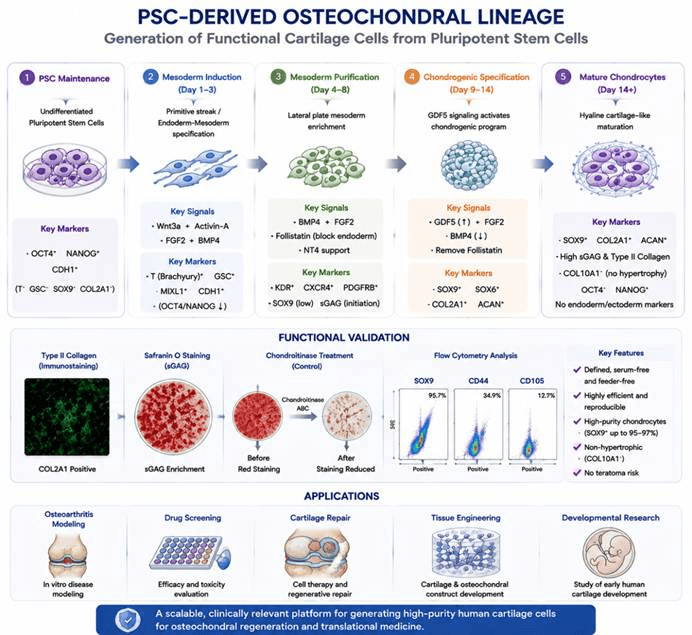
The directed differentiation process of pluripotent stem cells into chondrocytes. Human embryonic stem cells (hESCs) or induced pluripotent stem cells (iPSCs) first undergo mesoderm induction, followed by the formation of chondroprogenitors under the regulation of signaling factors such as BMP, FGF, and GDF5, and further mature into functional chondrocytes. The expression of cartilage markers, including SOX9, COL2A1, and ACAN, can be detected to verify cell identity and maturation status. This technology enables the generation of high-purity, scalable human chondrocytes, providing a stable cellular resource for osteoarthritis models, drug screening, cartilage tissue engineering, and regenerative medicine research.
Differentiation Stage | Added Factors / Culture Conditions | Core Biological Functions | Corresponding Cell Type / Developmental Stage | Stage-specific Identification Markers |
Undifferentiated hESC Maintenance Stage | 1. Matrix: Fibronectin (FN) coating2. Culture medium: Feeder-free, serum-free chemically defined medium3. Maintenance of pluripotency without differentiation-inducing factors | Provides homogeneous undifferentiated starting cells and ensures consistency of the differentiation starting population | Undifferentiated human embryonic stem cells (pluripotent state) | Positive: OCT4, NANOG, CDH1Negative: T, GSC, SOX9, COL2A1 |
Stage 1: Primitive Streak / Mesendoderm Induction (D1–D3) | 1. Matrix: Fibronectin (FN)2. D1: Wnt3a 25 ng/mL + Activin-A 50 ng/mL3. D2: Activin-A reduced to 25 ng/mL + FGF2 20 ng/mL added4. D3: Activin-A reduced to 10 ng/mL + BMP4 40 ng/mL added5. Serum-free and chemically defined conditions throughout | Activates Wnt and Nodal signaling, mimics primitive streak formation, induces mesendoderm fate specification, and initiates early germ layer differentiation | Primitive streak-like cells / Mesendoderm progenitor cells | Positive: T (Brachyury), GSC, MIXL1, CDH1Pluripotency genes: OCT4/NANOG slightly decreasedEndoderm gene: transient GATA4 expression |
Stage 2: Early Mesoderm Purification and Specification (D4–D8) | 1. Remove Wnt3a and Activin-A2. Add Follistatin 100 ng/mL (inhibits endoderm differentiation)3. Maintain BMP4 40 ng/mL + FGF2 20 ng/mL4. Add NT4 neurotrophic factor to maintain cell survival5. Matrix transition: FN initially, followed by FN + gelatin mixture; two passages for expansion | Blocks endodermal differentiation pathways, enriches and purifies mesodermal cells; BMP/FGF signaling drives lateral plate mesoderm fate and initiates early cartilage progenitor programs | Early lateral plate mesoderm cells / Chondroprogenitor cells | Mesoderm markers: KDR, CXCR4, PDGFRBEndoderm markers significantly reduced: GATA4, FOXA2Early cartilage initiation: low-level SOX9 expression, initial sGAG secretionCell morphology: transparent 3D cell clusters |
Stage 3: Mature Chondrocyte Differentiation (D9–D14) | 1. Gradually reduce BMP4 and increase GDF5 to 40 ng/mL2. Maintain FGF2 and remove Follistatin3. Replace matrix with pure gelatin coating4. Serum-free chemically defined medium | Activates the SOX9 cartilage master regulatory pathway, induces mesenchymal condensation, promotes deposition of cartilage extracellular matrix components including type II collagen and aggrecan; suppresses hypertrophic differentiation and generates stable hyaline chondrocytes | Mature hyaline chondrocytes / Chondrocyte aggregates | Cartilage core genes strongly upregulated: SOX9, SOX6, COL2A1, ACANMatrix staining: Safranin O positive (sGAG enrichment), type II collagen immunofluorescence positiveNegative: COL10A1 (hypertrophic marker), AFP/PAX6 (endoderm/ectoderm contamination), complete silencing of OCT4/NANOGFlow cytometry: SOX9⁺ cells up to 95–97% purity |
Endpoint Functional Validation (Chondrocyte Phenotype) | 1. Chondroitinase treatment control2. Flow cytometric quantification of SOX9, CD44, CD1053. sGAG quantitative analysis | Confirms cartilage matrix specificity, cell purity, and functional maturation status | Functional mature chondrocytes (non-hypertrophic phenotype) | Functional indicators: extensive sGAG deposition; Safranin O staining abolished after chondroitinase treatmentSurface markers: CD44⁺ approximately 34.9%, CD105⁺ approximately 12.7%Key advantage: no COL10A1 expression throughout the process, avoiding hypertrophic calcification tendency |