
info@ucallmlabs.com
Pluripotent Stem Cell‑Derived Mesenchymal Stem Cell (MSC) Lineage
Bone‑related diseases, including bone defects, osteoporosis, and joint injuries, represent a major clinical challenge. Bone regeneration and repair, orthopedic drug development, and biomaterial evaluation all highly depend on standardized mesenchymal stem cell resources. Currently, adult human bone marrow‑derived mesenchymal stem cells (hBMSCs) are widely used in related research and preclinical studies. However, hBMSCs have several significant limitations: bone marrow collection requires invasive procedures, donor‑derived samples are limited, in vitro expansion capacity is restricted, osteogenic differentiation potential declines significantly after repeated passages, and substantial batch‑to‑batch variation exists among donors. These challenges have greatly limited the development of standardized and scalable cell manufacturing platforms for skeletal regeneration applications.
Human embryonic stem cells (hESCs) possess the key advantages of unlimited in vitro expansion capacity and the ability to differentiate into derivatives of all three germ layers, providing a continuous and renewable source of mesenchymal progenitor cells. In 2009, a research team at the University of Michigan reported a classical standardized protocol using BG01 human embryonic stem cells as the starting material. Through a 7‑day embryoid body suspension induction process combined with adherent culture expansion and purification, the researchers successfully generated human embryonic stem cell‑derived mesenchymal stem cells (hES‑MSCs).
These hES‑MSCs exhibited immunophenotypic characteristics highly similar to bone marrow‑derived mesenchymal stem cells, with stable expression of mesenchymal markers including CD73 and STRO‑1, while lacking the hematopoietic marker CD45. After long‑term expansion, the cells maintained stable chromosomal karyotypes without detectable genetic abnormalities. Functionally, hES‑MSCs could be efficiently directed toward mature osteogenic and adipogenic differentiation. Under osteogenic induction conditions, cells formed mineralized nodules and showed high expression of osteogenic‑specific genes such as Runx2 and osteocalcin. Following adipogenic induction, cells accumulated characteristic intracellular lipid droplets and upregulated the master adipogenic regulator PPAR‑γ.
The study also established a Col2.3‑GFP osteogenic fluorescent tracing system, in which a bone‑specific promoter drives green fluorescent protein expression, enabling non‑invasive real‑time monitoring of osteogenic differentiation in living cells. This system allows rapid evaluation of osteogenic potential and facilitates the isolation of highly pure mature osteoblast populations.
Based on this hES‑MSC differentiation technology, the limitations associated with traditional bone marrow‑derived mesenchymal stem cell sources and batch variability can be overcome, enabling large‑scale production of genetically uniform human mesenchymal stem cells with stable differentiation capacity. This cell platform is now widely applied in orthopedic tissue engineering scaffold development, preclinical cell therapy studies for bone defects, in vitro screening of osteogenic and bone‑toxic drugs, and investigation of early human skeletal development mechanisms. It provides a standardized, reproducible human cell platform for skeletal regenerative medicine and innovative orthopedic drug development.
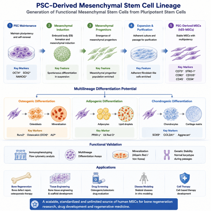
Using hESCs/iPSCs, PSC‑derived MSCs can be generated through mesenchymal lineage induction and further differentiated into osteoblasts, adipocytes, and chondrocytes. PSC‑derived MSCs overcome the limitations of traditional bone marrow MSCs, including donor availability constraints and batch‑to‑batch variability, providing a standardized and expandable human mesenchymal cell platform for bone regeneration, tissue engineering, disease modeling, and drug screening.
| Differentiation Stage | Added Factors / Culture Conditions | Core Biological Function | Corresponding Cell Type / Developmental Stage | Stage‑Specific Identification Markers |
|---|---|---|---|---|
| BG01 hESC Maintenance and Expansion Stage | 1. Feeder layer: irradiated mouse embryonic fibroblasts (MEFs), 0.1% gelatin‑coated plates 2. Basal medium: 80% DMEM/F12 + 20% KnockOut Serum Replacement (KSR) 3. Supplements: L‑glutamine, non‑essential amino acids, β‑mercaptoethanol, 4 ng/mL bFGF 4. Culture at 37°C, 5% CO₂, daily medium change, weekly manual passaging | Maintains long‑term human embryonic stem cell pluripotency, suppresses spontaneous differentiation, and enables continuous homogeneous expansion | Undifferentiated human embryonic stem cells (derived from blastocyst inner cell mass) | No mesenchymal marker expression detected; negative for CD73 and STRO‑1; negative for hematopoietic marker CD45 |
| Embryoid Body (EB) Suspension Induction Stage (EB Differentiation D0–7) | 1. hESCs dissociated and transferred to low‑attachment culture plates for suspension culture 2. hESC maintenance medium used without differentiation medium replacement or additional induction factors 3. Entire process maintained under suspension conditions without attachment | Releases monolayer pluripotency constraints, initiates spontaneous multilineage differentiation, and promotes generation of mesodermal/mesenchymal progenitor cells | Mixed‑lineage embryoid bodies, early mesenchymal progenitor cells (heterogeneous cell population) | No stable specific mesenchymal phenotype; pluripotency markers gradually decrease; weak expression of mesenchymal markers |
| EB Adherent Expansion and Purification Stage (0–14 Days After Attachment) | 1. Approximately 70 EBs seeded per well on 0.1% gelatin‑coated 6‑well plates 2. Medium switched to hBMSC expansion medium: α‑MEM + 10% FBS + L‑glutamine, non‑essential amino acids + 4 ng/mL bFGF 3. Cells passaged at 1:3 ratio using trypsin after reaching confluence; repeated passaging removes unwanted cell populations | Selects adherent fibroblast‑like cells, gradually eliminates epithelial and hematopoietic contaminants, and enriches purified hES‑MSCs | Hybrid mesenchymal progenitors → homogeneous hES‑MSCs (final purified passage) | Stable positive markers: CD73, STRO‑1 Stable negative marker: CD45 (hematopoietic marker) Fibroblast‑like spindle‑shaped morphology; normal karyotype without abnormalities |
| hES‑MSC Osteogenic Differentiation (28‑Day Induction) | Osteogenic differentiation medium (OM); conventional expansion medium (CM) used as control | Drives hES‑MSCs toward mature osteoblast differentiation and induces extracellular matrix mineralization | Osteogenic progenitor cells → mature mineralized osteoblasts | Gene expression: high Runx2 and osteocalcin (OCN) expression Staining: positive Alizarin Red staining and Von Kossa mineralization nodules Col2.3‑GFP fluorescence increases progressively during differentiation |
| hES‑MSC Adipogenic Differentiation (28‑Day Induction) | Adipogenic differentiation medium (AM); conventional expansion medium (CM) used as control | Promotes differentiation of hES‑MSCs into mature adipocytes and induces intracellular lipid droplet formation | Adipogenic progenitor cells → mature adipocytes | Gene expression: specific upregulation of PPAR‑γ Staining: positive Oil Red O lipid droplet staining |
| Osteogenic Tracking Gene Modification Stage | 1. Cell seeding density: 10,000 cells/cm² 2. Col2.3‑GFP lentiviral transduction with 5 μg/mL polybrene for 4 h 3. After transduction, cells cultured in either normal medium or osteogenic differentiation medium for 28 days | Establishes osteoblast‑specific fluorescent reporter cells and enables dynamic visualization of the entire osteogenic maturation process | Genetically modified hES‑MSCs and staged mature osteoblast populations | GFP fluorescence gradually increases only in the osteogenic induction group over time; no GFP expression detected in the normal culture group |