
info@ucallmlabs.com
Type 1 diabetes is caused by autoimmune destruction of pancreatic β cells, leaving patients dependent on exogenous insulin for life and prone to severe complications such as diabetic nephropathy, blindness, and cardiovascular diseases. The current curative strategy is allogeneic islet transplantation; however, the extreme shortage of healthy donor pancreases prevents this approach from meeting the needs of the large patient population. This limitation severely restricts the clinical translation of diabetes cell replacement therapy, glucose-lowering drug discovery, and studies of human pancreatic development. In 2008, Nature Biotechnology published a milestone standardized differentiation protocol in the field of pancreatic islet regeneration, which fully recapitulated the developmental sequence of human embryonic pancreatic formation. A four-step chemically defined, serum-free, two-dimensional directed induction system was established using human embryonic stem cells (hESCs) as the starting material. Through precise temporal regulation of key developmental signals, including Activin A, Wnt3a, KGF, retinoic acid, and Noggin, cells sequentially underwent fate specification into definitive endoderm, primitive gut tube, posterior foregut, and pancreatic endoderm. Within only 12 days, high-purity PDX1⁺NKX6-1⁺ pancreatic endoderm progenitors could be consistently generated. At this in vitro stage, the cells contain only a small proportion of immature polyhormonal endocrine cells and require the in vivo microenvironment for terminal organ maturation. Gold-standard transplantation studies in immunodeficient mice demonstrated that implanted pancreatic endoderm cells spontaneously differentiated into functional human islets with complete structural organization. Endocrine cell populations, including α, β, δ, and PP cells, were independently distributed and specifically expressed mature β-cell markers such as MAFA and PCSK1. The cells possessed complete proinsulin processing pathways, and glucose-stimulated human insulin and C-peptide secretion levels were comparable to those of adult primary human islets. In streptozotocin-induced severe diabetic mouse models, transplanted cells provided long-term and stable regulation of blood glucose levels. Removal of the grafts immediately resulted in recurrence of hyperglycemia, further demonstrating the independent glucose-lowering therapeutic potential of the transplanted cells. Long-term pathological evaluation showed an extremely low incidence of teratoma formation, indicating that the safety profile of the cells meets preclinical development standards. This differentiation platform features clearly defined components, standardized procedures, and compatibility with large-scale cell production. Based on this pancreatic endoderm platform, it can be widely applied in patient-specific in vitro disease modeling for type 1 diabetes, high-throughput efficacy screening of glucose-lowering drugs and islet-protective therapeutics, investigation of human islet developmental mechanisms, preclinical development of regenerative diabetes cell therapies, and evaluation of biomaterials for islet transplantation. It provides a stable, traceable, and functionally complete standardized pancreatic progenitor cell solution for islet regenerative medicine.
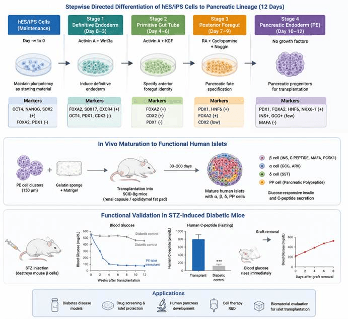
This process recapitulates human embryonic pancreatic development. Through sequential regulation of key signaling factors, including Activin A, Wnt3a, KGF, retinoic acid (RA), and Noggin, human embryonic stem cells/induced pluripotent stem cells (hESCs/iPSCs) are sequentially induced through the stages of definitive endoderm, primitive gut tube, posterior foregut, and pancreatic endoderm, ultimately generating PDX1⁺/NKX6-1⁺ pancreatic progenitor cells. Following transplantation into immunodeficient mice, pancreatic endoderm cells further mature into functional human islets containing β, α, δ, and PP cells. In STZ-induced diabetic models, these cells exhibit glucose-responsive insulin secretion and blood glucose regulation capacity. This technology provides a standardized cell source for diabetes cell replacement therapy, pancreatic developmental biology research, and drug screening applications.
Differentiation Stage | Added Factors / Culture Conditions | Core Biological Function | Corresponding Cell Type / Embryonic Development Stage | Stage-specific Identification Markers |
Human ES Cell Maintenance | Feeder-free standard human ES cell culture medium; maintain pluripotent state as the starting material for differentiation | Maintains unlimited self-renewal capacity and full three-germ-layer differentiation potential | Undifferentiated human embryonic stem cells (hESCs) | Positive: OCT4, NANOG, SOX2 Negative: FOXA2, PDX1, NKX6-1 and other endoderm/pancreatic markers |
Stage 1: Definitive Endoderm Induction (Day 0–3) | Serum-free RPMI basal medium; 100 ng/mL Activin A + 25 ng/mL Wnt3a; daily medium replacement | Mimics embryonic gastrulation, activates mesendoderm signaling pathways, efficiently induces definitive endoderm while excluding ectodermal and mesodermal contaminants | Embryonic definitive endoderm (the earliest precursor for pancreatic development) | Positive: FOXA2, SOX17, BRA (Brachyury), CXCR4 Negative: OCT4, PDX1, CDX2 |
Stage 2: Primitive Gut Tube Induction (Day 4–6) | RPMI + 0.2% FBS; continuous Activin A and KGF supplementation | Drives definitive endoderm conversion into foregut epithelium and establishes early intestinal/foregut characteristics | Primitive foregut epithelial cells | Maintained: FOXA2 Upregulated: CDX2 No PDX1 expression |
Stage 3: Posterior Foregut Specification (Day 7–9) | DMEM + 1% B27; 2 μM retinoic acid (RA) + 0.25 μM cyclopamine + 50 ng/mL Noggin | Suppresses hepatic and intestinal differentiation pathways, directs cells toward pancreatic fate, and activates pancreatic master transcription factors | Embryonic posterior foregut pancreatic progenitor cells | Maintained: FOXA2⁺ Upregulated: PDX1, HNF6 Downregulated: CDX2 |
Stage 4: Pancreatic Endoderm (PE) (Day 10–12, transplantation-ready cells) | DMEM + 1% B27 without additional exogenous growth factors; cell clusters mechanically cut into ~150 μm aggregates after differentiation | Stably enriches pancreatic epithelial progenitors; only a small proportion of immature polyhormonal cells are generated in vitro, requiring in vivo maturation for β-cell development | Embryonic pancreatic endoderm progenitors (PE, fetal pancreatic epithelial-like cells) | Core co-expression: PDX1, FOXA2, HNF6, NKX6-1 Small population expressing INS⁺GCG (immature fetal endocrine characteristics) Negative: MAFA (absence of mature β-cell marker) |
In Vivo Maturation Stage (PE transplantation into immunodeficient mice, Day 30–200) | PE cell clusters combined with gelatin sponge + Matrigel, implanted into SCID-Bg mouse epididymal fat pad or kidney capsule | Utilizes the in vivo microenvironment to complete terminal pancreatic islet differentiation, generating mature α/β/δ/PP endocrine cells and establishing glucose-responsive secretion pathways | Mature human pancreatic islet endocrine cells (functionally equivalent to adult islets) | 1. β cells: INS, C-PEPTIDE, MAFA, PCSK1 2. α cells: GCG, ARX 3. δ cells: SST 4. PP cells: Pancreatic polypeptide Structural features: Independent endocrine cell distribution, mature insulin secretory granules; glucose stimulation induces human insulin/C-peptide secretion |
STZ Diabetes Functional Validation | Mice continuously injected with streptozotocin (STZ) to destroy endogenous mouse β cells after transplantation | Evaluates the ability of human-derived islets to independently regulate blood glucose and validates therapeutic potential | Functional human pancreatic islets (diabetes-repair units) | High levels of human C-peptide detected under fasting and glucose stimulation conditions; normal glucose tolerance; removal of grafts immediately results in recurrent hyperglycemia |