
info@ucallmlabs.com
Duchenne muscular dystrophy (DMD), sarcopenia, and large-scale traumatic skeletal muscle defects represent major unmet clinical needs and significant challenges for drug development. However, conventional skeletal muscle cell generation approaches have long faced multiple technical limitations. Previous strategies mainly relied on spontaneous embryoid body differentiation or exogenous gene overexpression to induce myogenic differentiation. The former exhibits low differentiation efficiency and heterogeneous lineage composition, often resulting in contamination with non-muscle cell types such as cardiomyocytes and endothelial cells. The latter requires genetic modification of cells, introducing risks associated with genomic integration mutations and limited clinical translation potential. Moreover, many existing methods only generate short, immature myotubes lacking organized sarcomere structures, failing to fully recapitulate the complete process of human embryonic primary and secondary myogenesis. They also fail to simultaneously generate regenerative muscle satellite stem cells, resulting in poor transplantation outcomes, incomplete disease phenotypes in vitro models, and significant limitations for muscular disease drug screening, muscle tissue engineering, and cell therapy development.
In 2015, Nature Biotechnology published a landmark serum-free directed differentiation technology that established a standardized skeletal muscle induction system based on the natural developmental regulatory pathways of paraxial mesoderm (PSM) during vertebrate embryogenesis. This approach enables skeletal muscle differentiation from mouse and human pluripotent stem cells without transgene introduction or fluorescence-activated cell sorting. Through precise temporal regulation of Wnt activation using the small molecule CHIR and inhibition of BMP signaling using LDN, pluripotent stem cells are sequentially induced into posterior primitive streak mesoderm and paraxial mesoderm, followed by stepwise myogenic differentiation driven by HGF, IGF-1, and FGF2. This system faithfully recapitulates the two major stages of in vivo myogenesis, including primary and fetal secondary muscle development. The resulting cultures generate millimeter-scale, multinucleated mature muscle fibers with organized sarcomeres and spontaneous contractile activity. In addition, quiescent Pax7-positive muscle satellite stem cells are spontaneously established beneath the basement membrane of muscle fibers. Transplantation of purified Pax7-positive satellite cells into Duchenne muscular dystrophy mdx mouse models enables robust generation of dystrophin-expressing functional muscle fibers, restoration of muscle stem cell reservoirs, and long-term maintenance without teratoma formation.
Application of this differentiation process to DMD patient-derived pluripotent stem cells enables stable in vitro recapitulation of disease-associated skeletal muscle phenotypes, including abnormal myofiber branching and disrupted myofibrillar organization, providing highly human-relevant disease models. This differentiation platform is compatible with mouse embryonic stem cells, human embryonic stem cells (H9), and human induced pluripotent stem cells (hiPSCs), and is performed under fully defined, serum-free conditions with high batch-to-batch reproducibility and scalability. Based on this established technology, standardized skeletal muscle cell platforms have been widely applied in genetic muscle disease modeling, high-throughput screening of anti-muscle-wasting therapeutics, preparation of skeletal muscle tissue engineering seed cells, preclinical evaluation of satellite cell-based regenerative therapies, and fundamental studies of human skeletal muscle development, providing a safe, efficient, and functionally complete human cell resource for muscle regeneration research and translational applications.
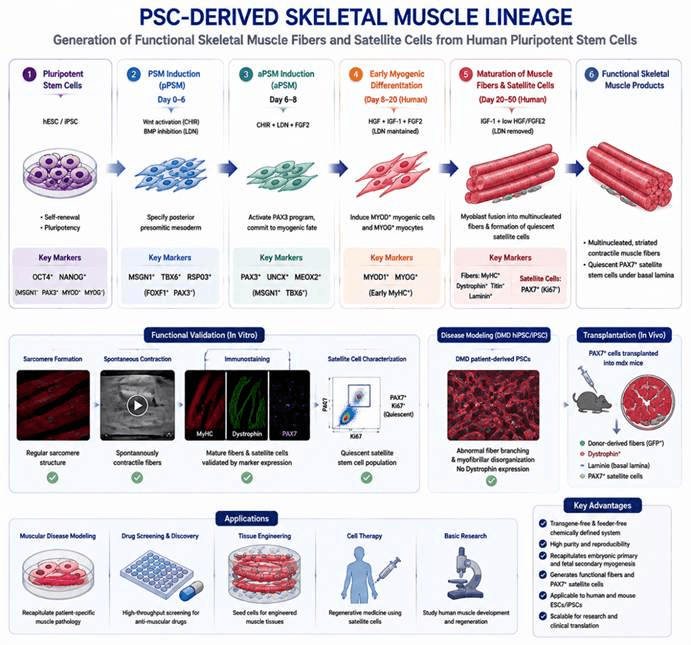
The directed differentiation process of pluripotent stem cell-derived skeletal muscle lineage. Human hESC/iPSC are first induced to form presomitic mesoderm (PSM) through the regulation of signaling pathways such as Wnt and BMP. Subsequently, under the stimulation of factors including FGF, HGF, and IGF-1, cells gradually develop into myogenic progenitor cells and further differentiate into mature skeletal muscle fibers with contractile function and PAX7⁺ satellite cells with regenerative potential. This system recapitulates human skeletal muscle development and provides a stable, scalable human cell platform for genetic muscle disease modeling, drug screening, skeletal muscle tissue engineering, and regenerative medicine research.
Differentiation Stage | Added Factors / Culture Conditions | Core Biological Function | Corresponding Cell Type / Developmental Stage | Stage-Specific Identification Markers |
|---|---|---|---|---|
Pluripotent Stem Cell Maintenance (Mouse ES / Human hiPS/H9 hES) | Mouse ES: 2i + LIF feeder-based culture mediumHuman iPS/hES: Matrigel-coated plates, mTeSR1 complete medium | Maintains pluripotent state and provides homogeneous starting materials for differentiation | Undifferentiated pluripotent stem cells (inner cell mass-derived / induced pluripotent state) | Positive: Oct4, NanogNegative: Msgn1, Pax3, Myod, Myog, MyHC |
Stage 1: Posterior Presomitic Mesoderm (pPSM) Induction (Mouse D0–6 / Human D0–6, CL medium) | Serum-free DMEM + N2B27/KSR; supplemented with 3 μM CHIR (Wnt activator) + 0.5 μM LDN193189 (BMP inhibitor); ROCK inhibitor added during early human differentiation | Activates Wnt signaling and blocks BMP-mediated lateral plate mesoderm (cardiac/endothelial lineage) formation, efficiently generating posterior presomitic mesoderm and restricting skeletal muscle lineage specification | Posterior presomitic mesoderm (pPSM), earliest embryonic muscle progenitors | High expression: Msgn1, Tbx6, Rspo3Low/Negative: Foxf1 (lateral plate mesoderm marker), Pax3 |
Stage 2: Anterior Presomitic Mesoderm (aPSM) Induction (Mouse D6–8 / Human D6–8, CFL medium) | Maintain CHIR + LDN; add 20 ng/mL FGF2; serum-free basal system | Suppresses Msgn1 expression, activates Pax3 signaling, promotes transition toward anterior presomitic mesoderm and establishes myogenic fate | Anterior presomitic mesoderm (aPSM), myogenic progenitor precursor cells | Upregulated: Pax3, Uncx, Meox2, Ripply2Downregulated: Msgn1, Tbx6 |
Stage 3: Early Myogenic Cell Induction (Mouse D8–12 / Human D8–20, K-HIF medium) | Remove CHIR; maintain LDN; add 10 ng/mL HGF, 2 ng/mL IGF-1, and 20 ng/mL FGF2 | Activates myogenic differentiation cascade and induces Myod-positive myoblasts and Myog-positive mononuclear muscle cells (primary myogenesis) | Embryonic primary myoblasts and mononuclear myocytes | Positive: Myod1, Myogenin (Myog)Early expression: Fast MyHC |
Stage 4: Mature Myofiber and Satellite Cell Formation (Mouse D12–50 / Human D20–50, K-I/K-HI medium) | Maintain IGF-1; low concentrations of HGF/FGF2; remove LDN; long-term serum-free maturation culture | Completes fetal secondary myogenesis; promotes fusion of mononuclear myocytes into multinucleated striated contractile muscle fibers; spontaneously generates Pax7 quiescent satellite cells and reconstructs the muscle extracellular matrix niche | Mature functional multinucleated skeletal muscle fibers and muscle satellite stem cells | Muscle fibers: Fast MyHC, Titin, Laminin, Dystrophin, NfixSatellite cells: Pax7 (Ki67⁻ quiescent state) |
In Vitro Disease Modeling Culture (mdx Deficient ES Specific) | Same complete differentiation protocol without additional factors; long-term maturation culture | Recapitulates Duchenne muscular dystrophy pathological features and generates abnormal branched muscle fibers | DMD pathological muscle fibers | Absence of Dystrophin; high proportion of branched myofibers; disrupted myofibrillar organization |
In Vivo Transplantation Stage (Pax7⁺ Cell Transplantation into mdx Mice) | Sort Pax7-GFP positive cells, resuspend in PBS, and inject into muscle/systemically in immunodeficient mdx mice | Evaluates in vivo regenerative capacity of satellite cells and reconstruction of dystrophin-positive muscle fibers and satellite cell pools | Newly generated mature skeletal muscle fibers and host muscle satellite cells | Donor GFP labeling, Fast MyHC, Dystrophin, fiber basement membrane Laminin, sublaminar Pax7⁺ satellite cells |