
info@ucallmlabs.com
The retina is a delicate neural tissue lining the inner wall of the eyeball and serves as the core structure sustaining human visual function. Retinal diseases have become a public health issue attracting global attention. As an important component of the central nervous system, retinal tissue lacks inherent regenerative capacity, and injuries generally lead to permanent and irreversible visual impairment. Epidemiological data indicate a huge population suffering from various retinal diseases worldwide, including approximately 8.4 million patients with age-related macular degeneration, 4 million with glaucoma, 2.6 million with diabetic retinopathy, and more than 1 million with retinitis pigmentosa. Accordingly, developing innovative therapeutic strategies bears great practical significance for alleviating patients’ suffering, easing social medical burdens and reducing economic costs.
At present, preclinical research on retinal diseases mainly relies on conventional cell culture and animal models, yet both systems have obvious shortcomings in new drug development and efficacy screening. Monolayer cell culture models feature a simple structure and fail to recapitulate the complex in vivo physiological microenvironment. They lack diverse cell subtypes and intact tissue architecture, making it difficult to simulate dynamic mechanical and biochemical signal regulation in organisms. Animal models are inherently limited by interspecies differences; they cannot accurately reproduce the genuine pathogenesis of human retinal disorders and tend to produce false-positive or false-negative experimental results. In addition, marked disparities exist among species in responses to drug efficacy and toxicity, which greatly restricts the reference value and translational potential of experimental findings.
Retinal organoids can faithfully recapitulate the in vivo structural characteristics and physiological functions of the human retina. They contain all retinal cell types and are capable of reconstructing the complete developmental process of the retina in vitro. When integrated with microfluidic technology, this model precisely mimics the biochemical and mechanical properties of the retinal microenvironment and supports high-throughput screening for drug efficacy and toxicity. Meanwhile, retinal organoids can reflect individual variations among different donors and accurately predict human drug responses, effectively overcoming the drawbacks of animal models and substantially shortening the timeline for translating basic research into clinical applications.
Compared with traditional research systems, retinal organoids can effectively cut down drug research and development cycles, reduce experimental costs and improve the success rate and overall efficiency of new drug development, acting as an innovative platform with great application value in translational medicine and drug screening. Combined with cutting-edge technologies such as stem cell engineering, tissue engineering, single-cell sequencing, gene editing and viral transduction, this model enables high-precision biomimetic reconstruction of human retinal structure and function. Equipped with comprehensive multicellular components, three-dimensional architecture, intact microenvironmental regulatory signals and compatibility with high-throughput assays, retinal organoids can remarkably accelerate preclinical drug research for retinal diseases and provide an advanced solution to address the low efficiency in mechanistic exploration and drug development targeting retinal disorders.
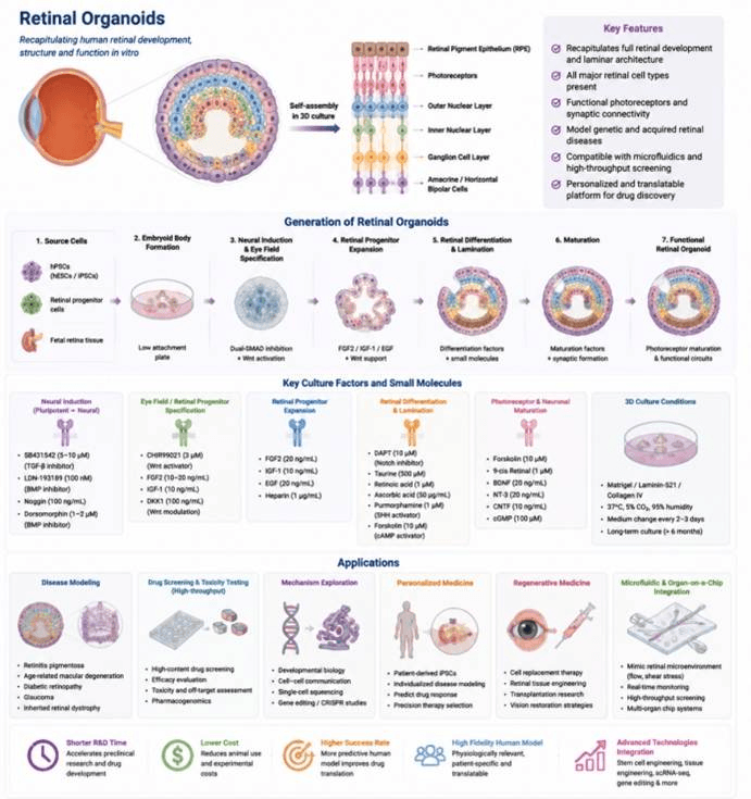
Applications of Human Retinal Organoids. Human retinal organoids (ROs) serve as versatile research tools across four core fields. First, they support fundamental biological investigations, covering the exploration of disease-related pathological pathways, the developmental trajectory of retinal organoids, and crosstalk between retinal tissue and surrounding vascular, immune and stromal cell populations. Second, patient-derived retinal organoids can be preserved as biobank resources. After dissociation into single-cell suspensions or assembly into intact retinal sheets, these samples are transplantable for regenerative therapies; additionally, they act as ideal research materials to uncover rules governing organ formation. Third, these organoid platforms enable the construction of eye disease models. Researchers can leverage them to analyze genomic and epigenetic mutations, as well as dissect the pathogenic pathways of congenital ocular developmental disorders. Fourth, retinal organoids lay a solid foundation for developing novel therapeutic strategies. They support the creation of gene editing and gene therapy regimens tailored to precision medical needs, and also act as high-throughput screening tools for candidate drugs, alongside advancing the development of cell-based therapeutic approaches .
Data display.
(A) Organoid culture status (bright-field morphology)
(B) Multicolor immunofluorescence staining
(C) Post-translational modification proteomics
(D) Epigenetic profiling (ChIP-seq / ATAC-seq)
(E) Gene editing validation (CRISPR)