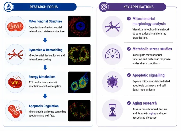
Mitochondria——Powerhouse of Cellular Adaptation
Key Markers
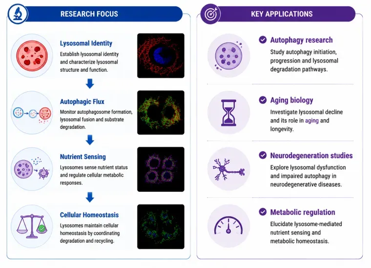
- TOM20
- COX IV
- VDAC1
- ATP5A
- HSP60

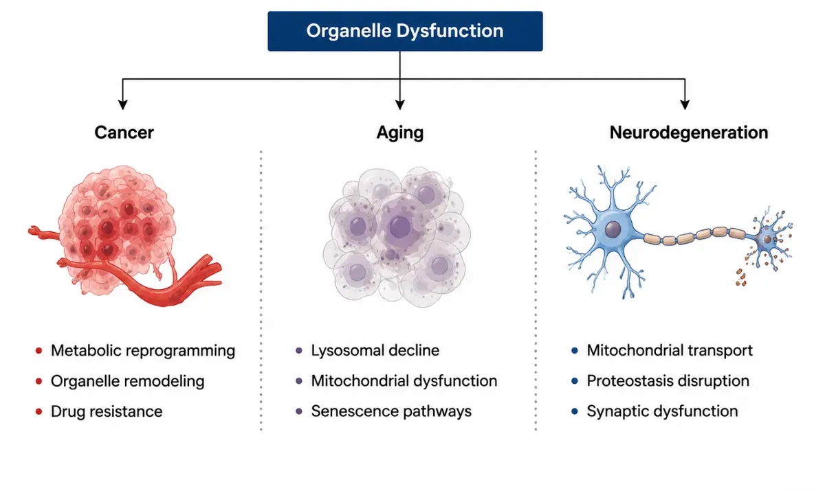
From Cellular Architecture to Biological Insight — Empowering Precision Research with Validated Organelle Markers
Cellular function depends on the precise coordination of specialised organelles. Understanding their structure, localisation, and dynamic changes is essential for uncovering fundamental biological mechanisms and disease-associated pathways.
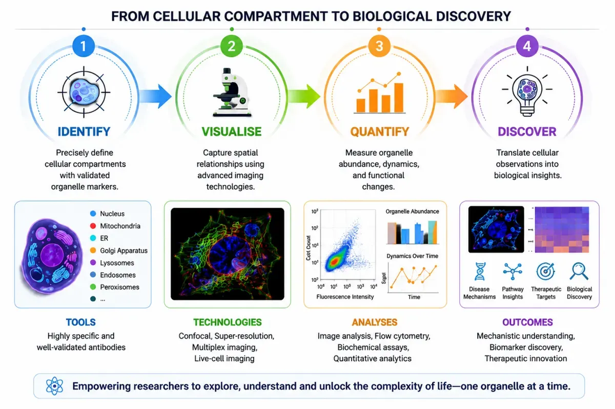
Our organelle marker solutions provide researchers with a reliable toolkit to visualise, validate, and investigate key cellular compartments — enabling deeper insights into metabolism, stress responses, protein homeostasis, and cellular recycling pathways.
——Precision tools for visualizing the cellular landscape.
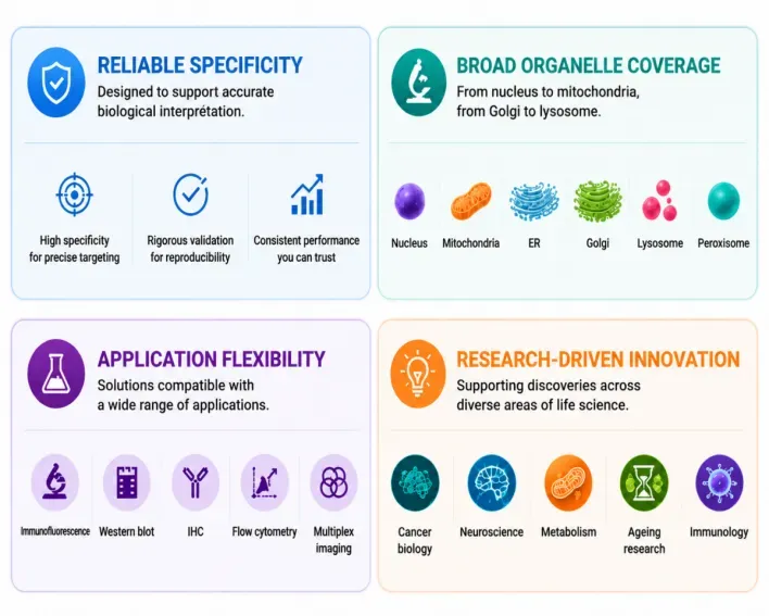
Understanding cellular organization requires reagents that combine uncompromised specificity with exceptional imaging clarity. We offer a rigorously validated collection of antibodies optimized for high-resolution organelle visualization.
Our comprehensive portfolio supports researchers throughout the discovery journey:
| Cellular Compartment | Representative Markers | Research Applications | Biological Role |
|---|---|---|---|
| Mitochondria | TOM20, COXIV, HSP60, VDAC1, AIF, ATP5A1, CPS1, Mitofilin, Prohibitin | Mitochondrial morphology, bioenergetics, metabolism, ageing, apoptosis | Energy production, apoptosis regulation, metabolic signalling |
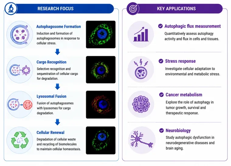
| Autophagosome & Lysosome | LC3B, p62/SQSTM1, LAMP1, LAMP2, ATG12, Cathepsin D | Autophagy flux, lysosomal function, neurodegeneration, stress response | Cellular recycling, degradation, organelle turnover |
| Proteasome | PSMA1, PSMA2, PSMB5, PSMB6, PSMB7, PSMD2 | Protein quality control, ubiquitin–proteasome pathway, ageing | Protein degradation and proteostasis maintenance |
| Exosome | CD9, CD63, CD81, TSG101, ALIX, Flotillin-1 | Biomarker discovery, liquid biopsy, cell communication | Intercellular communication and cargo transport |
| Endoplasmic Reticulum (ER) | CANX (Calnexin), CALR, Erp72, HSP90B1, PDIA3/4 | ER stress, unfolded protein response, proteostasis | Protein folding, calcium regulation, stress response |
| Cytoskeleton | β-Actin, α-Tubulin, β-Tubulin, Vimentin, Lamin A/C, Desmin | Cell migration, morphology, intracellular trafficking | Cellular architecture, transport, mechanical integrity |
| Plasma Membrane | ATP1A1, β-catenin, Caveolin-1, Flotillin-1, EGFR, CD44 | Membrane dynamics, signalling, cell interaction | Cell signalling, transport, receptor organisation |
| Migrasome | TSPAN4, Integrin α5β1, PIGK, NDST1, CD9 | Cell migration, developmental biology, tissue repair | Migration-associated extracellular vesicular structures |
| Nucleolus | FBL, NIFK, NCL, NOP2 | rRNA synthesis, ribosome assembly, cancer biology | Ribosome biogenesis and RNA processing |
| Nucleus | Histone H3, Histone H2A.X, Ki67, PCNA,POLR2A, LMNA, LMNB1 | Chromatin biology, transcription, DNA replication | Genome organisation and transcription regulation |
| Nuclear Membrane | Lamin A/C, Lamin B1, Emerin, SUN1, Nesprin-1 | Nuclear architecture, mechanotransduction | Nuclear–cytoplasmic organisation and transport |
| Ribosome | RPLP0, RPL10, RPS6, RPS3, RPL7A | Translation regulation, protein synthesis | Protein translation machinery |
| Cilia | ADCY3, ARL13B,IFT20,IFT88,Acetyl-α-Tubulin (Lys40) | Ciliogenesis, Hedgehog signalling, developmental biology | Cell sensing, signalling and motility |
| Centrosome | CENPJ, CP110, CEP250, CETN1, PCM1, PCNT, γ-Tubulin | Cell cycle, mitosis, centrosome dysfunction | Microtubule organisation and cell division |
| Intermediate filament | GFAP, Cytokeratin, Nestin, Vimentin | Cell type identification, differentiation, tumour subtyping, cytoskeleton integrity | Mechanical support, cell shape maintenance, organelle anchoring |
| Caveolae | Caveolin-1, Caveolin-2, Cavin-1/PTRF, Cavin-2, EHD2 | Mechanotransduction, metabolism, membrane trafficking | Membrane signalling and lipid regulation |
| Tight Junction & Adherens Junction | ZO-1, Claudin-1, Occludin, E-cadherin, β-Catenin | Epithelial integrity, cancer invasion, barrier biology | Cell–cell adhesion and barrier formation |
| Focal Adhesion Fibres | Vinculin, Paxillin, FAK, Talin-1, Integrin β1 | Migration, invasion, mechanotransduction | Cell–ECM attachment and mechanosensing |
| Golgi Apparatus | GOLG2, STX 6, GORASP2, GP73, TGOLN2 | Vesicle trafficking, secretion pathways | Protein modification and trafficking |
| Endosome | EEA1, Rab5, Rab7, Rab11, VPS35 | Endocytosis, receptor recycling | Intracellular sorting and trafficking |
| Peroxisome | Catalase, ABCD3, PEX1, PEX3, PEX5, DAO | Lipid metabolism, ROS regulation | Lipid metabolism and oxidative regulation |
| Desmosome | Desmoplakin, Plakoglobin, DSG2 , PKP2 | Tissue integrity, skin biology | Strong cell–cell adhesion |
| Gap Junction | Connexin 43, Connexin 26, Connexin 32 | Intercellular communication, cardiac biology | Direct cell communication |
| Cell Junction (Pan-Junctional Markers) | E-cadherin,VE-cadherin, Connexin 43, ZO-1, Vinculin, Claudin-1 | Tissue architecture, tumour biology | Tissue organisation and cell connectivity |
| Coated Pit | Clathrin Heavy Chain, AP2, Dynamin-2, Eps15, Amphiphysin | Endocytosis, receptor internalisation | Endocytic cargo uptake |
| Microsome | Calnexin, PDI, Ribophorin I, Sec61α, GRP78/BiP | ER protein processing, biochemical fractionation | ER-derived vesicular fractions |
| Melanosome | TYRP1, TYR, PMEL, MART-1/Melan-A, Rab27A | Pigmentation biology, melanoma research | Melanin synthesis and storage |
| Lipid Droplet | PLIN2, PLIN3, LSS, LDAH, PLIN1 | Lipid droplet dynamics, obesity/NAFLD, lipotoxicity, adipogenesis | Neutral lipid storage, energy homeostasis, lipid mediator synthesis |
| Cytoplasm / Cytosol | GAPDH, β-Actin, Tubulin, LDHA, HSP90 | Metabolism, signalling, protein expression control | Metabolic reactions and signalling environment |
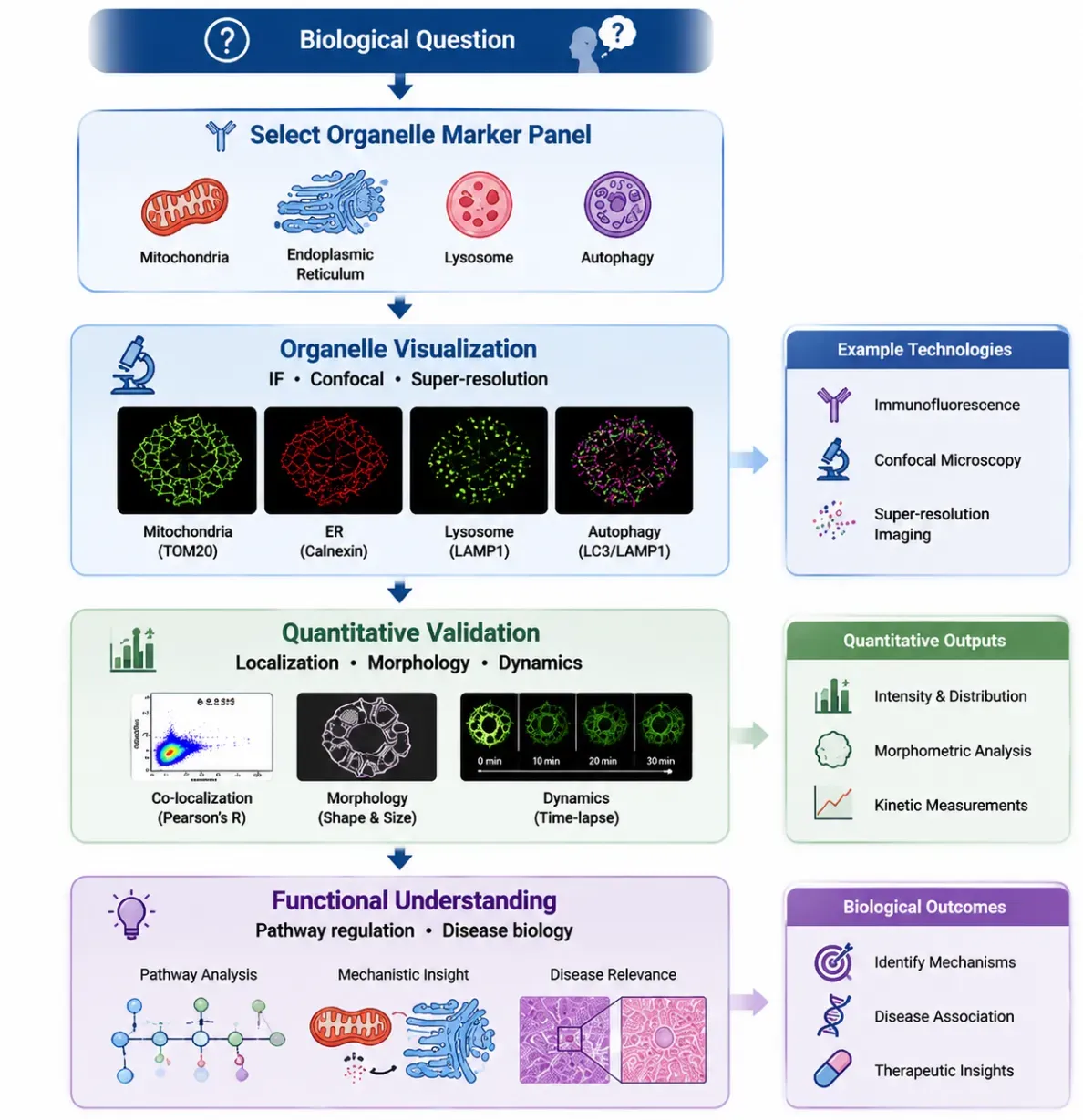
——Mapping Organelle Identity and Function
Our organelle markers provide researchers with reliable tools to visualize, validate, and investigate key intracellular compartments — enabling a seamless transition from structural observation to mechanistic insight.

Membrane contact sites regulate: Calcium exchange, Lipid transfer, Metabolic adaptation and Stress responses
Visualize the connections that define cellular homeostasis.
AI-assisted imaging pipelines now enable automated segmentation, tracking, and analysis of organelle morphology and interactions.
Reliable markers are the foundation of reproducible spatial biology.
New imaging technologies are enabling researchers to map metabolic states at individual organelle resolution, revealing previously hidden cellular heterogeneity.
Markers transform invisible biology into measurable insights.
Precision Tools. Comprehensive Coverage. Accelerated Discovery.
Our organelle marker portfolio is designed to provide researchers with the confidence, flexibility, and performance needed to explore cellular complexity — from fundamental organelle biology to advanced disease research.
| Research Challenge | Our Solutions |
|---|---|
| “How do I confirm organelle identity?” | Highly specific recombinant antibodies to ensure unambiguous organelle detection |
| “How can I prove protein localisation?” | Co-localisation-tested antibodies for multi-color IF/ICC, with ready-to-use protocols |
| “How do I improve reproducibility?” | Recombinant monoclonal antibodies delivering lot-to-lot consistency |
| “How can I study multiple organelles?” | Pre-designed organelle marker panels with compatible species and fluorophore combinations |
| “How do I connect structure with function?” | Functional antibodies against phospho-/cleaved forms, activity probes, and matched assay kits |
Reach our technical and product support team through your preferred channel.
info@ucallmlabs.com
+(1)-866-986-9598
Online Quote Submission
+(1)-866-986-9598