
info@ucallmlabs.com
```
```
ChIP‑seq
ChIP‑seq (Chromatin Immunoprecipitation followed by Sequencing): Principles, Workflow, and Applications
Chromatin immunoprecipitation followed by sequencing (ChIP‑seq) is a foundational technology for genome‑wide mapping of protein‑DNA interactions. Since its introduction in 2007, ChIP‑seq has become the gold standard for profiling transcription factor binding sites, histone post‑translational modifications, and chromatin‑associated proteins across diverse biological systems.
1. What Is ChIP‑seq?
ChIP‑seq combines chromatin immunoprecipitation (ChIP) with next‑generation sequencing (NGS) to identify the genomic loci where proteins of interest—such as transcription factors, RNA polymerases, or modified histones—associate with DNA. The technique was first demonstrated in 2007 by three landmark studies that applied NGS to map transcription factor binding sites and histone methylation marks genome‑wide, establishing ChIP‑seq as a superior alternative to microarray‑based ChIP‑chip. By directly sequencing the enriched DNA fragments, ChIP‑seq provides base‑pair‑level resolution, whole‑genome coverage, and quantitative measurements of binding enrichment, enabling researchers to construct comprehensive maps of regulatory elements, distinguish active versus repressed chromatin states, and identify transcriptional networks that govern development and disease.
2. Core Principles: A Multistep Workflow
The ChIP‑seq workflow is conceptually straightforward but technically demanding. It comprises two principal variants:
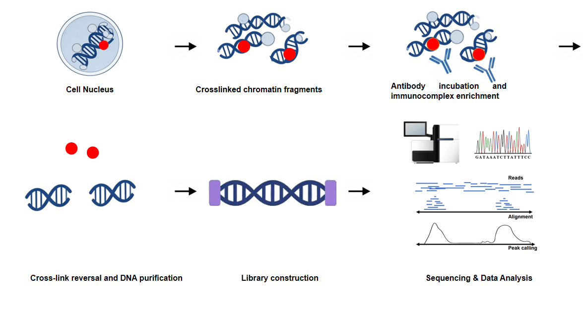
X‑ChIP‑seq (Cross‑linking ChIP‑seq) is the standard approach for transcription factors and other transiently or indirectly bound proteins. Cells are treated with formaldehyde to covalently cross‑link proteins to their associated DNA. Chromatin is then sheared by sonication or enzymatic digestion into fragments of 200–600 bp. A specific antibody against the target protein is used to immunoprecipitate the protein‑DNA complexes. After washing to remove non‑specifically bound material, cross‑links are reversed by heating, and the released DNA is purified. The DNA fragments then undergo end‑repair, A‑tailing, adapter ligation, and PCR amplification to generate sequencing‑ready libraries.
Native ChIP‑seq (N‑ChIP‑seq) is performed without cross‑linking and relies on micrococcal nuclease (MNase) digestion to fragment chromatin into mononucleosomes. It is primarily used for histone modifications, as histones remain tightly associated with DNA under native conditions. This variant avoids cross‑linking artefacts and epitope masking, but cannot capture weak or transient protein‑DNA interactions, limiting its application to histones and stably, tightly bound transcription factors such as CTCF.
3. Experimental Workflow
A typical X‑ChIP‑seq experiment follows these stages:
(1) Cross‑linking: Formaldehyde fixation of cells or tissues to covalently preserve protein‑DNA interactions in their native state.
(2) Chromatin fragmentation: Sonication or enzymatic digestion to generate DNA fragments of 200–600 bp.
(3) Immunoprecipitation: Incubation with a target‑specific antibody coupled to magnetic or agarose beads to enrich protein‑DNA complexes. A parallel total Input DNA sample (chromatin that does not undergo immunoprecipitation) and/or a mock IgG control should be processed alongside to assess non‑specific enrichment and background, as recommended by ENCODE guidelines.
(4) Washing and elution: Removal of non‑specifically bound material through stringent washing steps, followed by elution of the enriched complexes.
(5) Cross‑link reversal and DNA purification: Heating to reverse formaldehyde cross‑links, followed by proteinase K digestion and DNA purification.
(6) Library construction: End‑repair, A‑tailing, adapter ligation, and PCR amplification to generate sequencing‑ready libraries.
(7) Sequencing: High‑throughput sequencing (typically Illumina platforms) to read the enriched fragments.
Fig. 1. Workflow of ChIP‑seq
4. Key Advantages
Genome‑wide coverage provides unbiased mapping of binding sites across the entire genome without requiring prior knowledge of target loci.
Standard ChIP‑seq utilizes 100–600 bp sheared fragments to resolve binding peaks within a ~50–100 bp genomic window; optimized narrow‑fragment protocols improve resolution within this window, while ChIP‑exo derivatives achieve near single‑nucleotide resolution by utilizing exonucleases to digest DNA up to the protein‑DNA boundary.
Quantitative detection enables comparison of binding enrichment across different conditions and cell types.
Extensive validation is supported by a large body of published data and community standards (e.g., ENCODE guidelines) for antibody validation and data processing.
Broad applicability is demonstrated across diverse sample types, including cultured cells, primary tissues, and FFPE samples.
5. Limitations
High input requirements demand 105–107 cells per experiment, limiting application to rare or precious samples such as early embryos, FACS‑sorted populations, or clinical biopsies.
Variable signal‑to‑noise ratio arises from multiple sources. Formaldehyde cross‑linking not only masks antigenic epitopes to produce false negatives but also creates random non‑specific protein‑DNA cross‑links that generate false‑positive binding peaks. Non‑specific antibody binding and sonication bias further contribute to background noise.
Antibody dependency remains a critical bottleneck, as many targets lack high‑quality ChIP‑grade antibodies, and antibody specificity testing is required for both histones and transcription factors.
Multistep workflow is time‑consuming (3–7 days) and prone to batch effects, requiring extensive optimization of sonication conditions, antibody concentrations, and washing stringency.
GC bias arises primarily from uneven formaldehyde cross‑linking efficiency at protein‑dense regulatory regions (e.g., GC‑rich promoters and CpG islands), enzymatic digestion preferences (if MNase or other nucleases are used, which favor AT‑rich regions), and PCR/Illumina sequencing amplification preferences for moderate‑GC fragments.
6. Applications of ChIP‑seq
ChIP‑seq is a foundational technique for genome‑wide mapping of protein‑DNA interactions, enabling the identification of transcription factor binding sites, histone post‑translational modifications, and chromatin‑associated proteins across diverse biological systems. Compared to CUT&RUN and CUT&Tag, ChIP‑seq remains the preferred approach for samples with abundant starting material, FFPE tissues, and targets requiring crosslinking for epitope stabilization. It has been extensively applied in oncology, immunology, developmental biology, metabolism, and neuroscience research.
Representative case studies:
| Study | Target | Key Finding |
|---|---|---|
| Metformin reduces competitive advantage of Dnmt3aR878H HSPCs (Nature, 2025) | H3K27me3 | Metformin targets mutant HSPCs through epigenetic mechanisms |
| Targeting β‑catenin degradation induces cell death in ALL (Nat Cancer, 2026) | β‑catenin | GSK3β inhibitors induce ALL cell death via β‑catenin degradation |
| Nrf2 Drives Epigenetic Reprogramming as Master Regulator of KLF4 (Adv Sci, 2025) | H3K4me3, H3K27ac, KLF4 | Nrf2 orchestrates epigenetic reprogramming in transformation |
| Proteogenomic reprogramming via PARP‑DUX4 axis (Cell Rep, 2025) | BRD4, H3K27ac, SOX2 | PARP‑DUX4 reprograms cells to blastomere‑like state |
| EZH2 loss restores MHC class I in melanoma (iScience, 2025) | H3K27me3 | Metabolic stress drives EZH2 loss and immune restoration |
| Estrogen receptor alpha and beta govern ovarian folliculogenesis (Endocrinology, 2026) | ERα, ERβ | ER subtypes differentially regulate follicle development |
| Circadian reprogramming of salt handling in the colon (Sci Adv, 2026) | BMAL1, MR | Circadian clock regulates sodium absorption |
| Constitutive AMPK activation prevents hepatocellular carcinoma development through inhibition of HNF4α activity (Sci Adv, 2026) | HNF4A | Constitutive AMPK activation suppresses hepatocellular carcinoma development via inhibiting HNF4α activity |
| Aberrant Hippo‑YAP/TEAD signaling drives malignant transcriptional reprogramming in external auditory canal squamous cell carcinoma (Cancer Res Commun, 2026) | H3K27ac | Aberrant Hippo‑YAP/TEAD signaling mediates malignant transcriptional reprogramming in external auditory canal squamous cell carcinoma |
7. Considerations for Method Selection
ChIP‑seq remains the gold standard for applications where sample material is abundant (≥105 cells), where well‑validated ChIP‑grade antibodies are available, or where experiments involve FFPE or heavily cross‑linked tissues.
However, its high input requirements, variable signal‑to‑noise ratio, and complex workflow have led to the development of more streamlined enzyme‑tethering alternatives such as CUT&RUN and CUT&Tag. CUT&RUN applies pAG‑MNase to cleave and release target DNA fragments in situ, significantly increasing signal‑to‑noise ratio and reducing input requirements to 100–1,000 cells. CUT&Tag applies pA/G‑Tn5 transposase to simultaneously cleave DNA and insert sequencing adapters in a single step, eliminating end‑repair and adapter ligation, and enabling single‑cell profiling from as few as 60 cells.
For experiments with scarce starting material, low‑abundance targets or high‑throughput screening needs, CUT&Tag or CUT&RUN represent superior alternatives. For applications where large cell numbers are available, where established ChIP‑grade antibodies exist, or where FFPE or cross‑linked tissues are being analysed, ChIP‑seq remains the most robust and widely accepted approach.
Summary
ChIP‑seq has been instrumental in mapping transcription factor networks, histone modifications, and chromatin states across diverse biological systems. The ENCODE and Roadmap Epigenomics projects have generated comprehensive reference epigenomes using ChIP‑seq, providing foundational resources for understanding gene regulation in development and disease. While ChIP‑seq is increasingly complemented by newer technologies in low‑input and high‑throughput applications, it remains a robust and widely accepted method for chromatin profiling where sample availability and established antibodies are not limiting factors.
CUT&RUN
CUT&RUN (Cleavage Under Targets and Release Using Nuclease): Principles, Workflow, and Applications
The exploration of chromatin states has advanced significantly with the development of enzyme‑tethering strategies that overcome the limitations of conventional ChIP‑seq. Among these, Cleavage Under Targets and Release Using Nuclease (CUT&RUN) has emerged as a robust and widely adopted method for high‑resolution genomic mapping of chromatin‑associated proteins and histone post‑translational modifications. Originally developed in Dr. Steven Henikoff's laboratory and building upon the chromatin immunocleavage (ChIC) strategy pioneered by Dr. Ulrich Laemmli, CUT&RUN offers a streamlined, sensitive, and cost‑effective alternative to traditional chromatin profiling approaches.
1. What Is CUT&RUN?
CUT&RUN is an in situ chromatin profiling technique that employs a chimeric Protein A/G‑Micrococcal Nuclease (pAG‑MNase) fusion protein to selectively cleave antibody‑targeted chromatin within intact, permeabilized cells. Following antibody binding to the target chromatin protein, the pAG‑MNase fusion protein is tethered via its immunoglobulin‑binding domains to the antibody Fc region. Upon activation by calcium (Ca²⁺), MNase cleaves DNA in the immediate vicinity of the antibody‑bound site. The resulting chromatin fragments are released into the supernatant, where they can be separated from bulk chromatin, purified, and analyzed by next‑generation sequencing (NGS).
Unlike ChIP‑seq, which relies on cross‑linking, sonication, and immunoprecipitation, CUT&RUN is performed under native (unfixed) conditions, preserving chromatin structure and minimizing experimental artifacts.
2. Core Principles: The pAG‑MNase Tethering Strategy
The mechanistic basis of CUT&RUN can be summarized in five key steps:
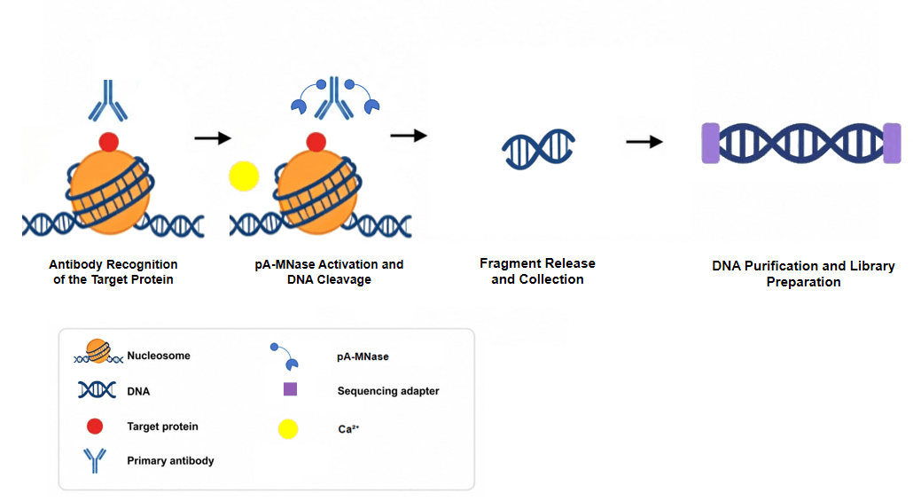
(1) Antibody Recognition of the Target Protein
Cells are permeabilized with digitonin, a mild nonionic detergent that creates small pores in the cell membrane without compromising nuclear integrity. A target‑specific antibody is introduced and binds to its cognate chromatin‑associated protein within the intact nucleus.
(2) pAG‑MNase Fusion Protein Tethering
The pAG‑MNase fusion protein is added to the reaction. The Protein A/G domains bind to the Fc region of the antibody, effectively tethering the MNase enzyme to antibody‑bound chromatin sites. Multiple washes are performed to remove unbound pAG‑MNase, minimizing off‑target cleavage.
(3) MNase Activation and DNA Cleavage
Calcium (Ca²⁺) is added to activate the tethered MNase. The enzyme cleaves DNA on both sides of the antibody‑bound site, generating fragments that correspond to the chromatin region occupied by the target protein. Because MNase is a processive enzyme, precise timing and quenching are essential to prevent over‑digestion.
(4) Fragment Release and Collection
Cleaved chromatin fragments diffuse out of the permeabilized cells into the supernatant. The reaction is quenched with a stop buffer containing EDTA and EGTA, which chelate free calcium ions and halt enzymatic activity. Cells and bulk chromatin remain immobilized on Concanavalin A (ConA)‑coated magnetic beads and are magnetically separated from the released target fragments.
(5) DNA Purification and Library Preparation
Released DNA fragments are purified using columns optimized for low DNA concentrations and small fragment sizes. Purified DNA then undergoes end‑repair, adapter ligation, and PCR amplification to generate sequencing‑ready libraries. The resulting libraries typically show a predominant mononucleosome‑sized peak of ~300 bp (~170 bp DNA fragments + adapters).
Fig. 2. Workflow of CUT&RUN
3. Experimental Workflow: A Streamlined Protocol
The CUT&RUN workflow is notably simpler than ChIP‑seq. A standard manual protocol finishes within 1–2 days; extended handling for large batches or multi‑step quality control may take up to four days. The key steps are as follows:
(1) Cell Immobilization
Cells are harvested, counted, and bound to Concanavalin A (ConA)‑coated magnetic beads, which bind to cell surface glycoproteins. This immobilization facilitates magnetic handling throughout the procedure.
(2) Permeabilization
Immobilized cells are treated with a buffer containing digitonin, a nonionic detergent that permeabilizes the plasma and nuclear membranes at low concentrations. Optimizing digitonin concentration is critical: under‑permeabilization prevents antibody and pAG‑MNase entry, while over‑permeabilization compromises cell integrity.
(3) Primary Antibody Incubation
The target‑specific antibody is added and incubated overnight at 4 °C to ensure sufficient binding. Positive controls (e.g., H3K4me3 or H3K27me3) and negative controls (e.g., IgG) should be included in every experiment.
(4) pAG‑MNase Binding
pAG‑MNase is added and incubated to allow tethering to antibody‑bound chromatin. Cells are washed several times to remove unbound enzyme and reduce nonspecific background.
(5) MNase Activation and Fragment Release
Ca²⁺‑containing buffer is added to activate MNase. After a precisely timed incubation, the reaction is quenched with a stop buffer containing EDTA and EGTA to chelate free calcium ions. Cleaved chromatin fragments diffuse into the supernatant, while bulk chromatin remains within the bead‑immobilized cells.
(6) DNA Purification
The supernatant containing released fragments is collected. DNA is purified using columns specifically designed to retain small fragments (down to ~50 bp) and eluted in low volume to maximize concentration.
Critical quality control note: Unamplified raw DNA from CUT&RUN typically yields concentrations below the detection limit of fragment analysis instruments (e.g., TapeStation, Bioanalyzer). Fluorometric quantification (e.g., Qubit) serves as the primary pre‑library quality control metric, while fragment size distribution can only be reliably assessed after library amplification. Exogenous E. coli spike‑in DNA is routinely added during reactions to normalize sequencing read counts across samples, eliminating technical batch‑to‑batch variability.
(7) Library Preparation
Purified DNA undergoes end‑repair, adapter ligation, and PCR amplification using conditions optimized for low yields and small fragment sizes. Barcoded primers enable multiplexed sequencing.
(8) Sequencing
Libraries are pooled and loaded onto an NGS platform. Only 3–8 million reads per sample are typically required for robust peak calling (vs. >20 million for ChIP‑seq), enabling high‑multiplexing of dozens to hundreds of samples per sequencing run.
4. Key Advantages: What Makes CUT&RUN Stand Out
CUT&RUN offers several distinct advantages over ChIP‑seq:
Low Input Requirements
CUT&RUN generates high‑resolution profiles from standard inputs of 100–1,000 native cells; commercial kits recommend 5,000 cells for routine validation, and optimized uliCUT&RUN protocols support single‑cell epigenomic profiling. This makes it feasible to analyze rare or precious samples, such as primary tissues, FACS‑sorted populations, or embryonic cells.
Exceptional Signal‑to‑Noise Ratio
Because target‑bound fragments are selectively released into the supernatant while bulk chromatin remains with the cells, background noise is dramatically reduced. This allows confident peak calling with far fewer sequencing reads and less primary antibody compared to ChIP‑seq.
Reduced Cost and Faster Turnaround
The high signal‑to‑noise ratio enables robust data generation with ~10‑fold fewer sequencing reads than ChIP‑seq. The streamlined protocol—free from sonication, cross‑linking, and immunoprecipitation—can be completed with minimal hands‑on time, significantly reducing reagent and sequencing costs.
Elimination of Technically Challenging Steps
CUT&RUN bypasses the most technically demanding steps of ChIP‑seq: cell lysis, chromatin fragmentation (sonication or enzymatic), and immunoprecipitation. This reduces optimization burden, minimizes batch effects, and improves reproducibility.
Automation Compatibility
CUT&RUN can be adapted to 96‑well plate formats and automated liquid handling systems, enabling high‑throughput epigenomic profiling for clinical research and drug discovery applications.
Compatibility with Native Chromatin
As a native technique performed on unfixed cells, CUT&RUN avoids cross‑linking artifacts that can mask epitopes or introduce spurious signals. For labile epitopes such as histone acetylation or weakly associated bromodomain proteins, mild cross‑linking can be applied as an optional modification; however, native unfixed conditions remain the preferred baseline for most chromatin targets.
5. Applications of CUT&RUN
CUT&RUN is an in situ chromatin profiling technique that employs a pAG‑MNase fusion protein to selectively cleave and release antibody‑targeted chromatin fragments. Compared to ChIP‑seq, CUT&RUN offers significantly lower background, reduced cell input requirements, and compatibility with direct qPCR on unamplified DNA. It has been widely applied to map histone modifications and transcription factor binding in primary tissues, FACS‑sorted cells, and disease models.
Representative case studies:
| Study | Target | Key Finding |
|---|---|---|
| A cell‑state axis underlying colonization in carcinomas with implications for metastasis risk prediction and interception (Cell Rep, 2025) | c‑Fos | A carcinoma cell‑state axis governed by c‑Fos underpins tumour colonization, supporting metastasis risk prediction and therapeutic interception |
| Resident memory T cell development is gradual and shows AP‑1 gene expression in mature cells (JCI Insight, 2025) | JunB | Resident‑memory T‑cell differentiation proceeds gradually, with JunB‑driven AP‑1 transcriptional programs activated in mature cells |
| Ganglioside GM3 Protects Against Abdominal Aortic Aneurysm by Suppressing Ferroptosis in VSMCs | KLF9, H3K27ac, Pol II | CUT&RUN reveals KLF9/H3K27ac/Pol II co‑occupancy at ferroptosis‑related genes |
| E2F7 drives oncogenic transcriptional programs in small‑cell lung neuroendocrine tumours (Cancer Res, 2025) | E2F7 | E2F7 genome‑wide binding remodels oncogenic transcriptome and sustains malignant phenotypes in neuroendocrine small‑cell lung cancer |
| TBX3 engages with the Wnt/β‑catenin transcriptional complex in colorectal cancer to regulate metastasis genes (Oncogene, 2024) | TBX3 | TBX3 collaborates with β‑catenin chromatin complex to activate metastatic‑related transcriptional programs in colorectal cancer cells |
| The transcription factor ZNF469 regulates collagen production in liver fibrosis (JCI Insight, 2025) | ZNF469 | ZNF469 occupies collagen‑related gene loci to drive collagen biosynthesis and promotes progression of liver fibrosis |
| ASB7 is a Negative Regulator of H3K9me3 Homeostasis (Science, 2025) | H3K9me3, SUV39H1, HP1α, ASB7 | ASB7 negatively regulates H3K9me3 homeostasis through SUV39H1 interaction |
6. Method Selection Guidance
The choice between CUT&Tag, CUT&RUN, and ChIP‑seq should be driven by experimental priorities:
Choose CUT&Tag when:
- Sample material is extremely limited (as few as 60 cells)
- Single‑cell epigenomic profiling is required
- Sequencing cost is a priority (far fewer reads yield high‑quality peaks)
- High‑throughput automation (96‑well format) is desired
- Simultaneous capture of target binding and chromatin accessibility is of interest
Choose CUT&RUN when:
- Sufficient cell input is available (≥100–1,000 cells)
- Single‑cell resolution is not required
- Weakly bound transcription factors benefit from milder wash conditions (e.g., 150 mM NaCl)
- Preliminary antibody validation via direct qPCR on unamplified DNA is desired
Choose ChIP‑seq when:
- Experiments involve FFPE tissues or heavily cross‑linked samples
- Target proteins rely on well‑validated ChIP‑grade antibodies that perform poorly under native, unfixed conditions
- No single‑cell or low‑input demands are present
- Mature, lab‑established ChIP‑seq workflows are already in place
- Complex protein targets that are poorly characterized under native conditions need to be studied
Summary
CUT&RUN represents a significant advance in chromatin profiling, offering high sensitivity, low background, reduced cost, and a streamlined workflow compared to conventional ChIP‑seq. By eliminating cross‑linking, sonication, and immunoprecipitation, it simplifies optimization and improves reproducibility while opening new avenues for analyzing rare and precious samples. With its adaptability to automation and scalable epigenomic applications, CUT&RUN is poised to complement or replace ChIP‑seq in many research and clinical settings, driving forward our understanding of gene regulation and epigenetic mechanisms.
CUT&Tag
CUT&Tag (Cleavage Under Targets and Tagmentation): Principles, Workflow, and Applications
The exploration of chromatin states and gene regulatory mechanisms has advanced rapidly, moving from traditional ChIP‑seq to more sensitive and streamlined approaches. Among these, Cleavage Under Targets and Tagmentation (CUT&Tag) has emerged as a powerful tool, distinguished by its unique enzyme‑tethering design. By combining antibody‑directed targeting with in situ transposase activity, CUT&Tag enables robust, high‑resolution epigenomic profiling from very small samples, including single cells.
1. What Is CUT&Tag?
CUT&Tag is a highly sensitive epigenomic profiling technique based on an enzyme‑tethering strategy. A specific antibody binds to a target chromatin‑associated protein (e.g., histone modifications, transcription factors) in unfixed, intact permeabilized cells, and this antibody subsequently tethers a Protein A‑Tn5 transposase fusion protein pre‑loaded with sequencing adapters. Upon activation, the transposase cleaves DNA and integrates adapters in situ at the target sites, generating fragment libraries ready for PCR enrichment and sequencing.
Compared to the long‑standing "gold standard" ChIP‑seq, CUT&Tag offers superior sensitivity, drastically lower background, and minimal input requirements. Relative to CUT&RUN—another enzyme‑tethering method—CUT&Tag eliminates the need for end‑polishing and adapter ligation, significantly simplifying library preparation and making it more amenable to single‑cell platforms. The entire procedure, from live cells to sequencing‑ready libraries, can be completed within a single day and in a single tube.
2. Core Principles: A Stepwise Mechanism of In Situ Tethering and Tagmentation
The mechanistic basis of CUT&Tag can be broken down into four key steps:
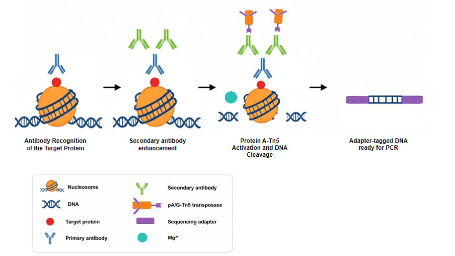
(1) Primary antibody recognition
A specific antibody against the target protein (e.g., H3K27me3, H3K4me2, or a transcription factor) is introduced to permeabilized unfixed cells and binds to its cognate chromatin epitope.
(2) Secondary antibody enhancement
A secondary antibody directed against the primary antibody is added to increase the local concentration of antibody at chromatin sites, thereby amplifying subsequent pA‑Tn5 recruitment. This step is a standard component of the CUT&Tag protocol.
(3) Protein A‑Tn5 fusion protein tethering
A pre‑assembled transposome, consisting of the pA‑Tn5 fusion protein loaded with sequencing adapters, is added. The Protein A moiety binds to the Fc region of the antibodies, anchoring the Tn5 transposase to the vicinity of the target protein. Because Tn5 has an inherent affinity for exposed DNA, stringent washing with Dig‑med buffer (300 mM NaCl) is performed to remove unbound transposome, minimising background. (Note: earlier washes with primary and secondary antibodies use Dig‑wash buffer at 150 mM NaCl.)
(4) Mg²⁺‑triggered cleavage and adapter integration
Addition of Mg²⁺ activates the tethered Tn5 transposase, which simultaneously fragments the DNA and inserts the pre‑loaded sequencing adapters at the cleavage sites. This combined fragmentation‑tagmentation step eliminates separate end‑repair and ligation steps, streamlining library construction.
Fig. 3. Workflow of CUT&Tag
3. Experimental Workflow: A Streamlined "One‑Day" Protocol
All steps of CUT&Tag are performed in a single reaction tube, using Concanavalin A‑coated paramagnetic beads for convenient magnetic handling:
(1) Sample preparation and bead immobilisation
Unfixed cells are harvested, counted, and washed. They are permeabilised with digitonin and then bound to Concanavalin A‑coated magnetic beads.
(2) Primary antibody incubation
The bead‑bound cells are incubated with the primary antibody for 2 hours at room temperature or overnight at 4 °C.
(3) Secondary antibody incubation
A secondary antibody is added and incubated for 30 minutes at room temperature to boost tethering efficiency.
(4) pA‑Tn5 transposome binding
The pre‑loaded pA‑Tn5 transposome is added and incubated for 1 hour at room temperature in Dig‑med buffer (300 mM NaCl). After binding, multiple washes with Dig‑med buffer are performed to remove unbound transposome.
(5) Tagmentation and adapter integration
Mg²⁺‑containing tagmentation buffer (10 mM MgCl₂ in Dig‑med buffer) is added, and the reaction is incubated at 37 °C for 1 hour to activate the transposase.
(6) DNA recovery and library amplification
The reaction is stopped by adding EDTA, SDS, and Proteinase K, followed by incubation at 55 °C for 30 minutes. After digestion, centrifuge at 16,000 × g for 5 minutes to pellet debris, and transfer the supernatant to a fresh tube. DNA is purified using AMPure XP beads, which removes Proteinase K along with the supernatant. For library amplification, a gap‑filling step at 72 °C for 5 minutes is performed first, followed by limited‑cycle PCR (typically 14 cycles: 98 °C for 10 s, 63 °C for 30 s) with indexed primers, yielding sequencing‑ready libraries.
The small amount of E. coli tracer DNA that remains associated with the pA‑Tn5 transposase preparation can serve as an internal calibration standard in certain formulations. However, many modern 'low‑carry‑over' pA‑Tn5 enzymes are highly purified and lack sufficient E. coli DNA for reliable normalization; for such preparations, exogenous spike‑in normalization is recommended.
4. Key Advantages: What Makes CUT&Tag Stand Out
CUT&Tag overcomes several limitations of traditional methods, offering distinct benefits:
Ultra‑low input requirement
CUT&Tag performs reliably across a wide range of cell numbers, from 100,000 down to as few as 60 cells, with high‑quality data maintained even at the lowest inputs. This makes it ideal for rare or limited samples.
Exceptional signal‑to‑noise and read efficiency
Because tagmentation is confined to antibody‑tethered sites, background is extremely low. Quantitative comparisons show that CUT&Tag populates peaks much more rapidly at low sequencing depths—~2 million reads are equivalent to ~8 million for CUT&RUN or ~20 million for ChIP‑seq. In the H3K4me2 benchmarking experiment, CUT&Tag was the only method that reached a fraction of 0.6 reads within called peaks. The dynamic range of ChIP‑seq signals is approximately 1/20 that of CUT&Tag.
High resolution
CUT&Tag produces a footprint of ~80 bp over transcription factor binding sites (e.g., CTCF), offering precise delineation of binding positions and modified nucleosome boundaries.
Rapid and simplified workflow
By eliminating cross‑linking, sonication, immunoprecipitation, and post‑fragmentation end‑repair/ligation steps, CUT&Tag reduces the overall timeline to 1 day and increases throughput.
Compatibility with single‑cell analysis
Unlike CUT&RUN, where MNase‑cleaved fragments are released into the supernatant, CUT&Tag's tagmented products remain retained within the nucleus. This allows a straightforward single‑cell adaptation (scCUT&Tag): all steps from antibody binding to tagmentation are performed on a bulk population, after which individual cells are dispensed into nanowells (e.g., using the Takara ICELL8 system) for PCR with indexed primers. Aggregated scCUT&Tag data correlate highly with bulk profiles (Pearson's r = 0.89).
"Multi‑OMIC" potential – simultaneous profiling of targeted and accessible sites
The CUT&Tag dataset comprises two distinct distributions: high‑level signals from true antibody‑tethered sites and lower‑level signals from non‑targeted accessible DNA. By modelling these distributions, genuine binding sites can be distinguished from background accessible sites within the same experiment. This enables de novo multi‑OMIC CUT&Tag, where both the target factor's occupancy and chromatin accessibility are characterised in a single assay.
5. Considerations for Method Selection: CUT&Tag in Relation to Other Epigenomic Methods
Table 1. Comparison of CUT&Tag, CUT&RUN, and ChIP‑seq technologies.
| Comparison Dimension | CUT&Tag | CUT&RUN | ChIP‑seq |
|---|---|---|---|
| Core Principle | pA/G‑Tn5 fusion protein; Mg²⁺ activation; simultaneous cleavage and adapter insertion | pAG‑MNase fusion protein; Ca²⁺ activation; cleavage followed by DNA fragment release | Formaldehyde cross‑linking; sonication or enzymatic fragmentation; antibody‑mediated enrichment |
| Input Cell Number | Ultra‑low (as few as 60 cells; compatible with single‑cell level) | Low to moderate (100–1,000 cells) | High (millions of cells) |
| Experimental Timeline | 1 day (2 days for high‑throughput) | 1‑2 days | 3‑7 days |
| Signal‑to‑Noise Ratio | Extremely high; clean background | High | Moderate to high; notable background noise |
| Library Preparation | Single‑step (cleavage and Illumina adapter insertion in one reaction) | Requires in vitro adapter ligation (additional steps after cleavage) | Multi‑step library preparation (end repair → A‑tailing → adapter ligation) |
| qPCR Compatibility | Only post‑library qPCR; raw DNA not applicable | Compatible with direct qPCR on unamplified DNA | Compatible with direct qPCR on purified DNA |
| Compatibility with Transcription Factors | High salt impairs weak‑binding TFs, good for high‑affinity factors | Broader (milder conditions; suitable for weak‑binding proteins) | Broadest |
| Compatibility with Histone Modifications | Excellent | Excellent | Excellent |
| Cell Permeability | Maintains membrane integrity after labeling; permeabilizes both plasma and nuclear membranes | Maintains better membrane integrity | Requires membrane disruption after cross‑linking |
| Sample Types | Cultured cells, tissues (requires optimization) | Cultured cells, tissues (requires optimization) | Broader (cells, tissues, FFPE, etc.) |
| Experimental Complexity | Low | Moderate (requires adapter ligation step) | High |
| Data Normalization | Endogenous E. coli tracer; exogenous spike‑in (optional) | Exogenous spike‑in (required) | Input DNA |
6. Applications of CUT&Tag
CUT&Tag enables genome‑wide profiling of histone modifications and transcription factor binding across diverse biological systems. Its low input requirements and single‑cell compatibility have made it a widely adopted tool for epigenetic studies in oncology, developmental biology, neuroscience, immunometabolism, and plant science.
Representative case studies:
| Study | Target | Key Finding |
|---|---|---|
| NNMT Enriches for AQP5+ Cancer Stem Cells in Early Gastric Cardia Adenocarcinoma | H3K27me3 | NNMT reduces H3K27me3 to activate WNT signaling and maintain cancer stem cell stemness |
| VRK2 Potentiates Anti‑PD‑1 Immunotherapy in Hepatocellular Carcinoma | MYC | VRK2 destabilizes MYC to enhance anti‑PD‑1 immunotherapy efficacy in HCC |
| Profiling Plant Histone Modification at Single‑Cell Resolution Using snCUT&Tag | H3K4me3 | Single‑nucleus CUT&Tag enables cell‑type‑specific histone modification profiling in rice seedlings |
| Hexokinase 2‑Mediated Metabolic Stress and Inflammation in MASLD | H3K18la | Hexokinase 2 drives histone lactylation in liver macrophages, promoting metabolic stress and inflammation |
| Microbial Metabolite Enhances Immunotherapy Efficacy by Modulating T Cell Stemness | H3K27ac | Microbial metabolites modulate H3K27ac in CD8+ T cells to enhance immunotherapy efficacy |
| The Lysine Demethylase KDM7A Regulates Immediate Early Genes in Neurons | KDM7A | KDM7A regulates histone methylation (H3K9me2, H3K27me2) to control immediate early gene expression in neurons |
| Positive Feedback Regulation of Microglial Glucose Metabolism by Histone H4K12 Lactylation in Alzheimer’s Disease | H4K12la | H4K12 lactylation in microglia drives glycolytic gene expression and metabolic dysfunction in AD |
While CUT&Tag provides high‑resolution profiling of protein‑DNA interactions, biological regulation is rarely governed by a single layer of information. Integrating CUT&Tag with complementary epigenomic and transcriptomic assays enables a multi‑omics approach to dissect complex regulatory circuits. A typical integrative framework combines RNA‑seq to identify transcriptional changes, ATAC‑seq to map chromatin accessibility, CUT&Tag to define direct protein‑DNA interactions, and WGBS to detect DNA methylation—together constructing a causal chain from gene expression changes to the underlying regulatory mechanisms.
7. Method Selection Guidance
The choice between CUT&Tag, CUT&RUN, and ChIP‑seq should be driven by experimental priorities:
Choose CUT&Tag when:
- Sample material is extremely limited (as few as 60 cells)
- Single‑cell epigenomic profiling is required
- Sequencing cost is a priority (far fewer reads yield high‑quality peaks)
- High‑throughput automation (96‑well format) is desired
- Simultaneous capture of target binding and chromatin accessibility is of interest
Choose CUT&RUN when:
- Sufficient cell input is available (≥100–1,000 cells)
- Single‑cell resolution is not required
- Weakly bound transcription factors benefit from milder wash conditions (e.g., 150 mM NaCl)
- Preliminary antibody validation via direct qPCR on unamplified DNA is desired
Choose ChIP‑seq when:
- Experiments involve FFPE tissues or heavily cross‑linked samples
- Target proteins rely on well‑validated ChIP‑grade antibodies that perform poorly under native, unfixed conditions
- No single‑cell or low‑input demands are present
- Mature, lab‑established ChIP‑seq workflows are already in place
- Complex protein targets that are poorly characterized under native conditions need to be studied
Summary
CUT&Tag represents a significant advance in epigenomic technology, offering high sensitivity, low background, high resolution, and a greatly simplified workflow. Its capacity to produce high‑quality data from as few as 60 cells and its seamless adaptation to single‑cell profiling make it particularly valuable for studies of rare populations, developmental systems, and clinical specimens. Moreover, its inherent ability to simultaneously capture targeted binding events and accessible chromatin features opens the door to multi‑OMIC analyses within a single experiment.
Looking ahead, CUT&Tag is readily adaptable to additional high‑throughput single‑cell platforms, such as the 10× Genomics encapsulation system, by leveraging their single‑cell ATAC‑seq workflow. Furthermore, the use of barcoded adapters holds promise for multiplexed profiling of multiple epitopes simultaneously in single cells, maximising the utility of single‑cell epigenomic studies for development and disease research. As commercial kits and automated protocols continue to mature, CUT&Tag is poised to complement or replace traditional methods in many applications, driving forward both basic research and translational epigenetics.