
info@ucallmlabs.com
Vascular complications such as cardiovascular diseases, atherosclerosis, diabetic retinopathy and diabetic nephropathy have become urgent global public health challenges. Traditional cell culture techniques are easy to implement and cost-effective, yet they have inherent limitations. Cell arrangement is artificially fixed, critical in vivo signal concentration gradients are absent, and only planar cell-cell contacts can be established, making it difficult to faithfully reconstruct authentic human physiological microenvironments. In recent years, vascular organoids have evolved into innovative in vitro disease models. Compared with conventional monolayer cell culture systems, vascular organoids reconstruct the sophisticated physiological architecture of blood vessels and enable precise elucidation of organ-specific functional traits and disease pathogenesis, serving as central experimental tools for investigating macroangiopathy and microangiopathy. Among them, vascular organoids generated from induced pluripotent stem cells (iPSCs) possess distinctive application advantages.
This organoid system supports dynamic crosstalk between diverse cell populations and the extracellular matrix, forming hierarchically organized functional structures within biomimetic microenvironments. Composed mainly of endothelial cells and mural cells, vascular organoids re-establish bidirectional regulatory and inductive interactions between cells. They accurately mimic in vivo pathophysiological responses, as well as cellular turnover and functional compensation mechanisms intrinsic to native organs. The multicellular vascular units derived from these organoids can be assembled with organoids of the brain, kidney, pancreas, intestine, heart and other solid organs to generate vascularized artificial tissues with higher structural complexity and controllability. Meanwhile, iPSC-derived vascular organoids preserve the epigenetic signatures of patient donors, enabling the establishment of personalized drug screening platforms and offering novel support for mechanistic research and drug development targeting cardiovascular disorders associated with vascular injury.
iPSC-derived vascular organoids exhibit diversified research value. On the one hand, this model recapitulates the full spectrum of vascular development under both physiological and pathological conditions, acting as an ideal experimental platform to probe mechanisms underlying cardiovascular aging. Although cellular reprogramming can partially reverse senescent phenotypes, iPSCs retain the telomere length and epigenetic characteristics of donors. By comparing vascular organoids derived from healthy individuals of different age groups and patients with cardiovascular diseases, combined with senescence-inducing approaches including long-term culture and external stress stimulation, researchers can systematically dissect the mechanisms triggering age-related vascular lesions. Cells obtained from elderly donors inherently possess shorter telomeres, which continuously erode during cell division. Such cells are more prone to senescence-associated aberrant epigenetic modifications and functional impairment, providing a natural research system to uncover the mechanisms of vascular aging.
On the other hand, living organoid biobanks established using multiple patient samples deliver high-quality resources for basic mechanistic research, translational medicine and personalized precision healthcare. Patient-derived organoids faithfully recapitulate individual disease characteristics and predict drug responses, facilitating the development and optimization of individualized therapeutic regimens. For instance, stem cell organoids generated from cystic fibrosis patients display drug response profiles highly consistent with human clinical phenotypes, representing low-cost and high-efficiency tools for drug screening. Furthermore, quantitative readouts such as intestinal swelling in organoids can serve as novel biomarkers for auxiliary clinical diagnosis.
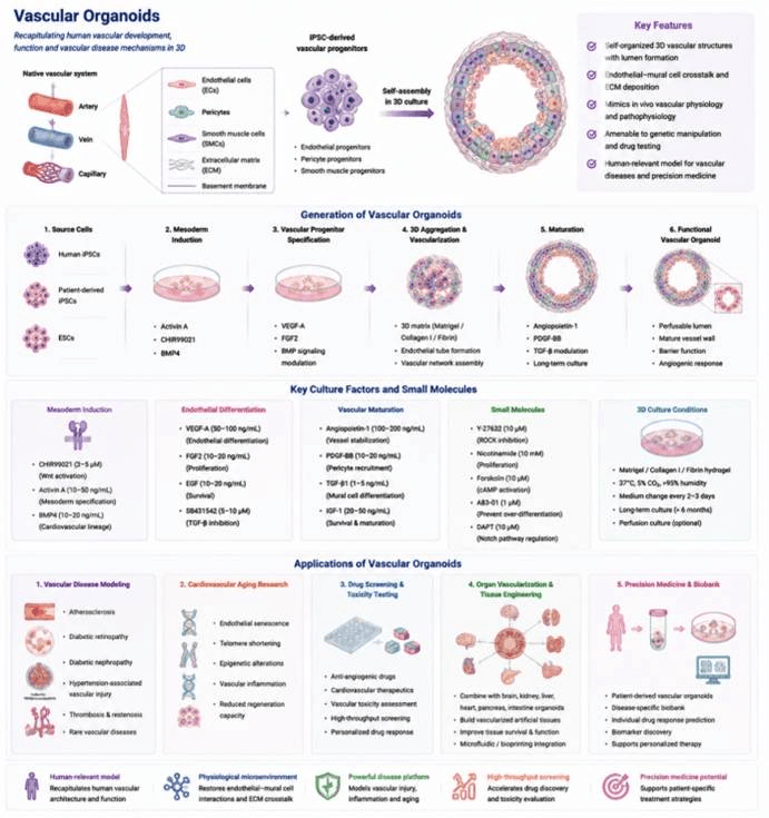
Generation, Culture, and Applications of vascular Organoids.
Data display.
(A) Organoid culture status (bright-field morphology)
(B) Multicolor immunofluorescence staining
(C) Post-translational modification proteomics
(D) Epigenetic profiling (ChIP-seq / ATAC-seq)
(E) Gene editing validation (CRISPR)