
info@ucallmlabs.com
As the colonization site of endometrial progenitor cells, severe injury to the endometrial basal layer acts as the core cause of refractory uterine diseases. Severe endometrial damage triggered by surgical trauma such as dilation and curettage or viral infection completely disrupts the cyclic regeneration rhythm of the functional endometrial layer, thereby inducing various menstrual disorders including amenorrhea and hypomenorrhea. Meanwhile, persistent chronic inflammation mediates aberrant tissue repair processes, drives progressive endometrial fibrosis, and markedly inhibits epithelial regeneration and angiogenesis. Excessive abnormal deposition of fibrous connective tissue gradually replaces the normal extracellular matrix architecture, ultimately leading to irreversible severe impairment of uterine reproductive function.
At present, mainstream clinical interventions include hysteroscopic adhesiolysis, intrauterine balloon or catheter placement, and postoperative adjuvant hormonal therapy. Nevertheless, these therapeutic approaches generally suffer from high recurrence rates and limited clinical efficacy and cannot achieve radical cure. Compared with conventional research models, endometrial organoids support long-term stable in vitro expansion, steadily retain epithelial biological characteristics, and accurately recapitulate the physiological responses of human endometrium to sex hormones. This model has become a central experimental tool for endometrial drug screening, tissue injury repair and regenerative medicine research.
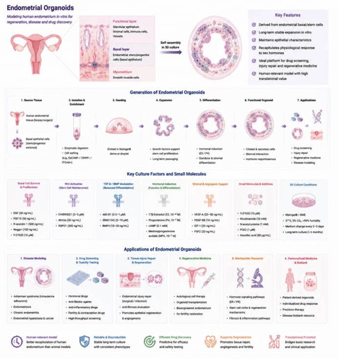
Generation, Culture, and Applications of endometrial Organoids.
Data display.
(A) Organoid culture status (bright-field morphology)
(B) Multicolor immunofluorescence staining
(C) Post-translational modification proteomics
(D) Epigenetic profiling (ChIP-seq / ATAC-seq)
(E) Gene editing validation (CRISPR)