
info@mail.ucallm.com
Comprehensive autophagy research requires integrated analysis of autophagy initiation, autophagic flux, lysosomal function, selective organelle clearance, and metabolic signaling networks to understand cellular homeostasis, aging biology, and disease progression.
We provide an integrated research toolkit — from autophagy core regulators to dynamic flux monitoring assays — enabling accurate investigation of autophagic activity and biological function.
Key Target Highlights
Core Molecular Targets in Autophagy Research
Biological Process | Key Targets | Biological Significance | Research Applications |
Autophagy initiation | ULK1, ULK2, ATG13, FIP200 | Controls autophagy induction | Nutrient stress studies |
Nutrient sensing | mTORC1, AMPK, TSC2 | Integrates energy and growth signals | Metabolic regulation |
Autophagosome nucleation | Beclin-1, VPS34, ATG14 | Regulates phagophore formation | Autophagy initiation studies |
Autophagosome elongation | ATG5, ATG7, ATG12, ATG16L1 | Drives membrane expansion | Autophagy machinery analysis |
LC3 lipidation | LC3-I/LC3-II, GABARAP | Canonical autophagosome marker system | Autophagic flux evaluation |
Cargo recognition | p62/SQSTM1, NBR1 | Selective cargo recruitment | Protein aggregate clearance |
Lysosomal degradation | LAMP1, LAMP2, TFEB | Controls autolysosome function | Lysosome biology |
Mitophagy | PINK1, Parkin, BNIP3, FUNDC1 | Removes damaged mitochondria | Mitochondrial quality control |
Selective autophagy | NCOA4, OPTN, TAX1BP1 | Regulates selective organelle/cargo removal | Disease mechanisms |
Key research trend:
Modern autophagy research is increasingly moving beyond measuring single markers such as LC3-II accumulation toward comprehensive evaluation of autophagic flux, lysosomal competence, selective autophagy pathways, metabolic regulation, and organelle quality control. Emerging studies emphasize that autophagy functions as an adaptive signaling network integrating mTOR/AMPK nutrient sensing, mitochondrial homeostasis, immune regulation, and aging-associated stress responses, providing new opportunities for therapeutic intervention.
Recommended Autophagy Marker Strategy
Multi-Level Validation Framework
Detection Layer | Representative Markers | Scientific Purpose |
Autophagy Initiation | ULK1, phospho-ULK1, Beclin-1 | Determine pathway activation |
Autophagosome Formation | LC3-I/LC3-II, ATG5, ATG7 | Evaluate autophagosome biogenesis |
Autophagic Flux | LC3-II turnover, p62/SQSTM1 degradation | Distinguish induction from blockage |
Lysosomal Function | LAMP1, LAMP2, Cathepsin D | Assess degradation capacity |
mTOR Regulation | mTOR, phospho-S6K, 4E-BP1 | Define nutrient signaling control |
Energy Regulation | AMPK, ACC phosphorylation | Evaluate metabolic stress response |
Selective Autophagy | PINK1, Parkin, NCOA4 | Investigate cargo-specific degradation |
Transcriptional Regulation | TFEB, TFE3 | Analyze lysosome biogenesis |
Core Autophagy Validation Strategy
"Golden Pair" Concept for Pathway Confirmation
Strategy | Marker Combination | Scientific Purpose | Application |
Golden Pair A: Autophagosome Formation | LC3-II + ATG5/ATG7 | Confirm autophagosome generation | General autophagy studies |
Golden Pair B: Autophagic Flux | LC3-II + p62/SQSTM1 | Evaluate autophagy progression | Avoid false-positive activation |
Golden Pair C: mTOR Regulation | p-mTOR/p-S6K + LC3-II | Connect nutrient signaling with autophagy | Metabolic research |
Golden Pair D: Mitophagy Activation | PINK1/Parkin + mitochondrial markers | Assess mitochondrial clearance | Aging and neurobiology |
Competitive Technology Landscape
Autophagy Research Workflow
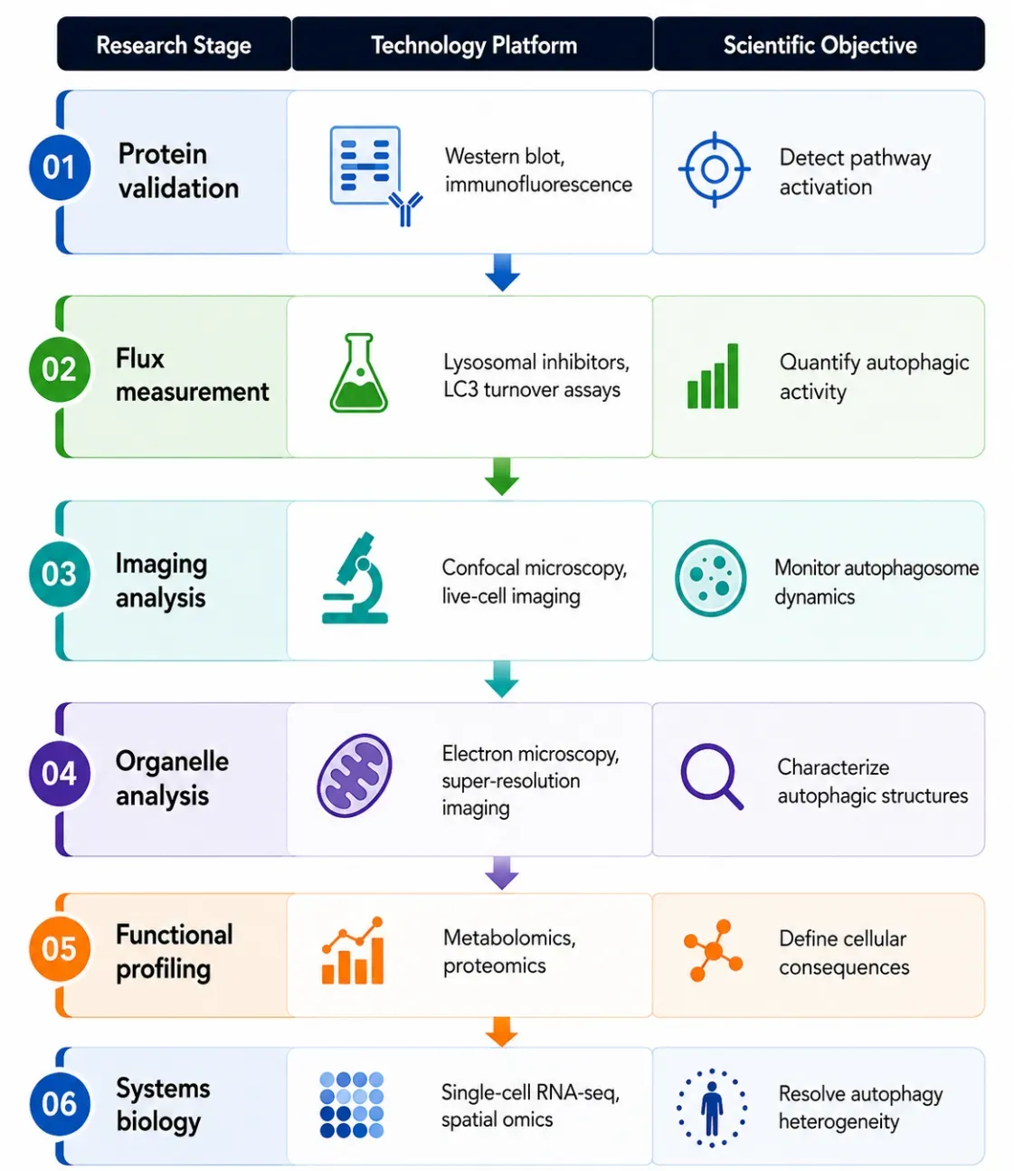
An integrated autophagy research workflow combining molecular validation, functional assays, advanced imaging, multi-omics profiling, and single-cell analysis to comprehensively characterize autophagy dynamics, mechanisms, and biological functions across multiple levels of biological complexity.
Pathway Overview
Autophagy is a conserved cellular recycling process that maintains homeostasis through degradation of damaged proteins and organelles. The ULK1 initiation complex, Beclin-1/VPS34 complex, ATG proteins, and lysosomal pathways coordinate autophagosome formation and cargo degradation. Autophagy regulates metabolism, stress adaptation, immunity, aging, and disease progression.
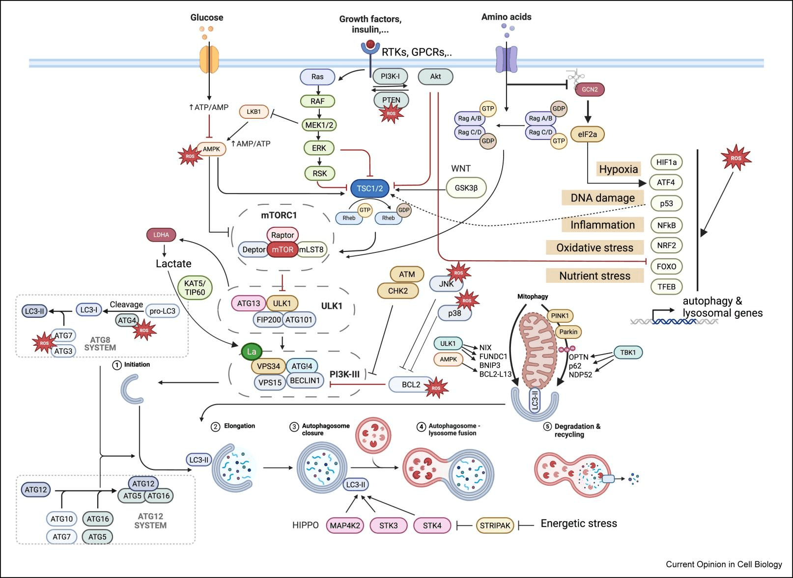
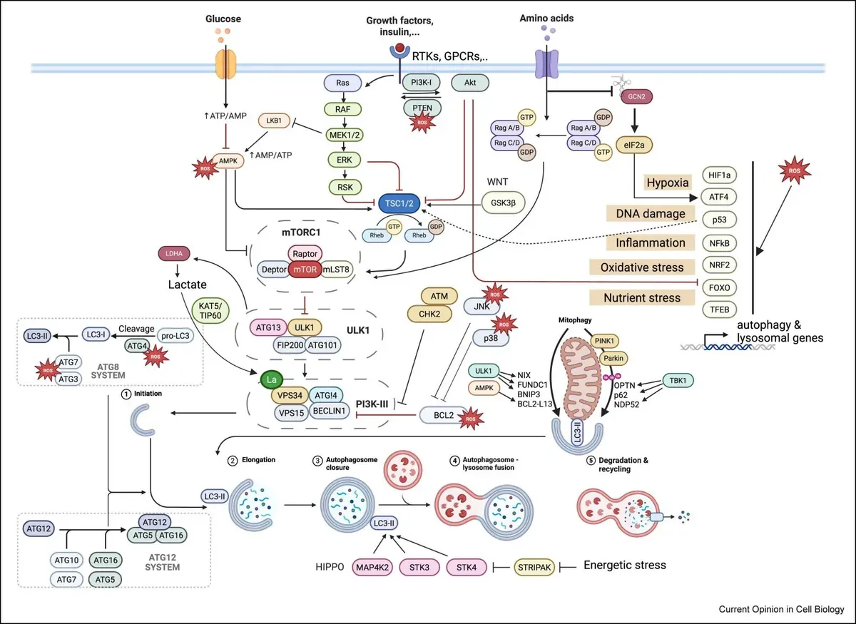
Autophagy control by cellular signaling (PMID: 41086756)
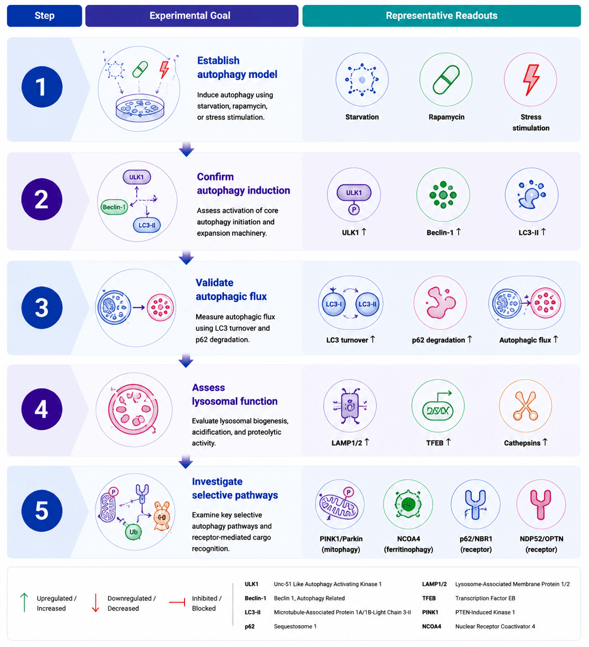
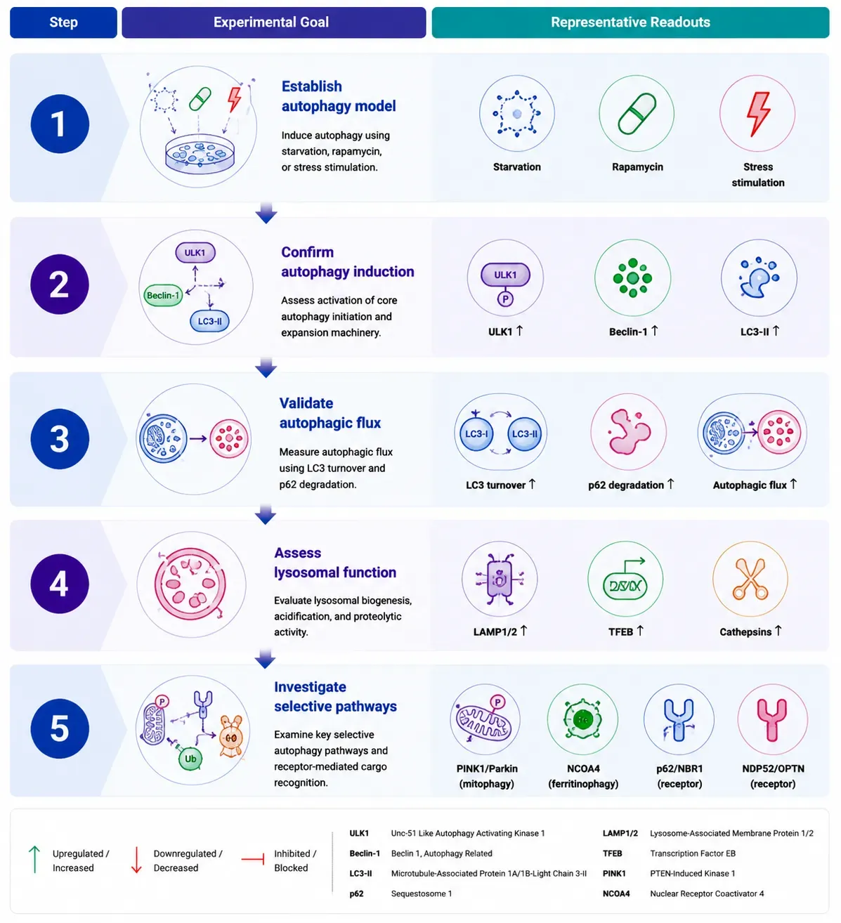
Recommended Experimental Validation Workflow
Step | Experimental Goal | Representative Readouts |
Step 1 | Establish autophagy model | Starvation, rapamycin, stress stimulation |
Step 2 | Confirm autophagy induction | ULK1, Beclin-1, LC3-II |
Step 3 | Validate autophagic flux | LC3 turnover, p62 degradation |
Step 4 | Assess lysosomal function | LAMP1/2, TFEB, Cathepsins |
Step 5 | Investigate selective pathways | PINK1/Parkin, NCOA4, receptors |
Featured Research Application Examples
Research Area | Model System | Key Evaluation Parameters | Scientific Question |
Cancer biology | Tumor models | LC3, p62, mTOR, TFEB | How does autophagy support tumor survival? |
Neurodegeneration | Neuronal models | LC3 flux, lysosomal markers, mitophagy | How does autophagy failure contribute to protein aggregation? |
Aging research | Senescence models | AMPK, mTOR, TFEB | How does autophagy influence cellular aging? |
Metabolic disease | Liver/adipose models | Lipophagy markers | How does autophagy regulate metabolism? |
Immunology | Immune cell models | ATG proteins, selective autophagy markers | How does autophagy regulate immune responses? |
Frequently Asked Questions
Q1. Which biomarkers should I use to accurately assess autophagy?
No single biomarker is sufficient. A comprehensive evaluation should combine LC3-I/II for autophagosome formation, SQSTM1/p62 for cargo degradation, Beclin-1 and ATG proteins for autophagy initiation, together with lysosomal markers such as LAMP1/2 to assess autophagosome maturation.
.Q2. How can I distinguish autophagy induction from impaired autophagic degradation?
Increased LC3-II alone does not necessarily indicate enhanced autophagy. Autophagic flux assays, combining lysosomal inhibitors (e.g., Bafilomycin A1 or Chloroquine) with LC3-II and p62 analysis, are considered the gold standard for differentiating increased autophagy from blocked lysosomal degradation.
Q3. Which experimental approaches provide the most reliable assessment of autophagy?
Robust autophagy studies integrate orthogonal methodologies, including Western blotting, immunofluorescence, autophagic flux assays, live-cell imaging, electron microscopy, and genetic perturbation. Combining complementary approaches minimizes experimental bias and strengthens biological conclusions.
Q4. What are the most common pitfalls in autophagy research?
Common pitfalls include interpreting LC3-II accumulation as increased autophagy without flux analysis, relying on a single biomarker, using insufficient experimental controls, and failing to validate antibody specificity. Multiple independent readouts and appropriate positive and negative controls are essential for accurate interpretation.
Q5. How can autophagy be comprehensively characterized across biological systems?
Modern autophagy research integrates protein validation, functional assays, high-resolution imaging, proteomics, metabolomics, and single-cell/spatial omics to reveal pathway activation, autophagic dynamics, functional remodeling, and cellular heterogeneity from molecular to tissue levels.
Key References
Radulovic M., Yang C., Stenmark H. (2025).Lysosomal membrane homeostasis and its importance in physiology and disease.Nature Reviews Molecular Cell Biology. 27:71–87.
Nixon R.A., Rubinsztein D.C. (2024).Mechanisms of autophagy–lysosome dysfunction in neurodegenerative diseases.Nature Reviews Molecular Cell Biology. 25(12):926–946.
Settembre C., Perera R.M. (2024).Lysosomes as coordinators of cellular catabolism, metabolic signalling and organ physiology.Nature Reviews Molecular Cell Biology. 25:223–245.
Wang L., Klionsky D.J., Shen H.-M. (2023). The emerging mechanisms and functions of microautophagy. Nature Reviews Molecular Cell Biology. 24:186–203
Gross A.S., Ghillebert R., Schuetter M., et al. (2024). A metabolite sensor subunit of the Atg1/ULK complex regulates selective autophagy. Nature Cell Biology. 26.
Nakatogawa H. (2020). Mechanisms governing autophagosome biogenesis. Nature Reviews Molecular Cell Biology. 21:439–458.
Vargas J.N.S., Hamasaki M., Yoshimori T. (2022). The mechanisms and roles of selective autophagy in mammals. Nature Reviews Molecular Cell Biology. 24:167–185.
Kaur J., Debnath J. (2015). Autophagy at the crossroads of catabolism and anabolism. Nature Reviews Molecular Cell Biology. 16:461–472.
Klionsky D.J., Abdel-Aziz A.K., Abdelfatah S., et al. (2021). Guidelines for the use and interpretation of assays for monitoring autophagy (5th Edition). Autophagy.