
info@mail.ucallm.com
1. Overview
Bacillus anthracis is a Gram-positive, facultatively anaerobic, spore-forming bacillus of the family Bacillaceae and genus Bacillus, and is the causative agent of anthrax. It is classified by the U.S. CDC as a Tier 1 biothreat pathogen. The bacterium is a large rod (4–8 × 1–1.5 µm), non-motile, and forms oval, refractile, centrally located spores under certain conditions. Spores are extremely resilient in the environment, surviving in soil, hay, and animal products for decades, and are resistant to extreme temperatures, desiccation, many disinfectants, ultraviolet radiation, and gamma rays.
Anthrax is a major zoonotic disease primarily affecting herbivorous mammals. Humans become infected through contact with infected animals or their products, inhalation of spores, or ingestion of contaminated meat. Human anthrax has four main clinical forms: cutaneous, inhalational, gastrointestinal, and injection anthrax, with inhalational and meningeal anthrax carrying case fatality rates of up to 90% (untreated). In March 2026, China reported an inhalational anthrax case in Guangdong Province linked to environmental exposure in a historically endemic area, highlighting that environmental exposure can pose a significant risk even without direct animal contact.
2. Background Information
2.1 What is Bacillus anthracis?
B. anthracis is a Gram-positive, facultatively anaerobic, spore-forming bacillus of the family Bacillaceae. It is an obligate aerobe or facultative anaerobe with an optimal growth temperature of 35℃, forming characteristic "ground-glass" colonies with wavy margins and a "medusa head" appearance on ordinary media. B. anthracis was the first bacterium proven to cause disease in humans, and the extreme environmental resilience of its spores underpins its long-term persistence in nature and its potential as a bioweapon.
2.2 Genome & Classification
The B. anthracis genome consists of a circular chromosome of approximately 5.2 Mb and two virulence plasmids:
Genome Component | Size | Encoded Products |
Chromosome | ~5.2 Mb | Housekeeping genes, sporulation genes, 24 chromosomal virulence genes |
pXO1 | ~181 kb | Tripartite protein exotoxin complex (PA, LF, EF), 67 virulence-related genes |
pXO2 | ~95 kb | Poly-γ-D-glutamate (PDGA) capsule, synthesized by CapBCAD enzymes |
Wild-type pathogenic strains must carry both pXO1 and pXO2 plasmids for full virulence; loss of either plasmid significantly attenuates virulence. Most chromosomal genes of B. anthracis are also found in B. cereus, with the primary distinction being the carriage of virulence plasmids.
Phylogenomic analyses generally divide B. anthracis into three major clades (A, B, and C), with clade A accounting for the majority of globally sampled isolates. Clade B strains harbor genetic variation in spore germination-related genes, potentially affecting environmental adaptability.
2.3 What Are B. anthracis Virulence Factors?
The pathogenicity of B. anthracis depends primarily on two core virulence factors:
2.3.1 Poly-γ-D-Glutamate Capsule (PDGA Capsule)
The capsule is composed predominantly of poly-γ-D-glutamic acid (PDGA), a homopolymer of D-glutamic acid. The capsule is antiphagocytic, enabling the bacterium to evade host immune cell phagocytosis and killing, which is key to establishing infection and systemic dissemination.
2.3.2 Tripartite Protein Exotoxin
The tripartite toxin encoded by the pXO1 plasmid is the most important virulence factor of B. anthracis, belonging to the AB-type toxin family and comprising three independent proteins:
Toxin Component | Molecular Weight | Function |
Protective Antigen (PA) | 83 kDa | Receptor-binding component (B), mediates LF and EF entry into host cells |
Lethal Factor (LF) | 90 kDa | Zinc metalloprotease, cleaves MAPKK, induces cell death |
Edema Factor (EF) | 89 kDa | Calmodulin-dependent adenylate cyclase, elevates intracellular cAMP |
PA alone is non-toxic, but combines with LF to form Lethal Toxin (LT) and with EF to form Edema Toxin (ET) . AtxA is a multidomain transcription factor that regulates virulence gene expression on both pXO1 and pXO2, with its activity synergistically regulated by glucose and CO₂ through a phosphorelay mechanism involving the glucose-PTS system (PtsG).
2.4 Mechanism of Infection and Pathogenesis
2.4.1 Spore Germination and Early Infection
B. anthracis infection begins when spores enter the host. Spores are phagocytosed by alveolar macrophages and germinate into vegetative cells within the phagolysosome. During germination, spores release large amounts of calcium dipicolinate (Ca-DPA) , a key event in spore germination. A 2025 study found that the SpoVA2 tetrameric membrane complex is required for DPA transport into spores, with SpoVA2 (5A2) being more critical for germination than SpoVA1 (5A1) .
2.4.2 Molecular Mechanism of Toxin Action
Lethal Toxin (LT) : LF acts as a zinc metalloprotease, specifically cleaving the N-terminus of MAPKK (MAP kinase kinase) , blocking MAPK signaling pathways and inducing apoptosis and necrosis of macrophages and dendritic cells.
Edema Toxin (ET) : EF acts as a calmodulin-dependent adenylate cyclase, converting ATP to cAMP and causing a sharp increase in intracellular cAMP levels. A 2025 study published in Cell Reports revealed a novel mechanism by which ET induces endothelial permeability: ET disrupts the endothelial barrier through Rac1- and cofilin-mediated F-actin reorganization, while also activating IGF1R and EGFR in a cAMP-independent manner, together leading to vascular leakage and lethal vascular collapse.
2.4.3 Immune Evasion and Systemic Dissemination
After extensive proliferation in regional lymph nodes, vegetative cells enter the bloodstream, causing massive bacteremia with bacterial concentrations reaching up to 10⁸ CFU/mL. The antiphagocytic action of the capsule and the killing of immune cells by toxins together promote systemic dissemination. A 2025 review noted that post-translational modifications (PTMs) —including phosphorylation, acetylation, glycosylation, and lipidation—play key roles in regulating B. anthracis virulence by influencing toxin enzymatic activity and protein expression.
2.5 Transmission Routes and Process
Transmission of B. anthracis occurs primarily through the following routes:
l Contact transmission (cutaneous anthrax) : Through broken skin contacting infected animals or their products (hides, bone meal, wool), the most common route in natural infection, accounting for approximately 95% of human cases.
l Inhalational transmission (inhalational anthrax) : Inhalation of spore-containing aerosols, the most lethal form, historically associated with occupational exposure in wool mills and tanneries. The 2026 Guangdong case demonstrated that environmental exposure (contaminated soil or dust) during residence in a historically endemic area can also cause inhalational anthrax without direct contact with sick animals.
l Foodborne transmission (gastrointestinal anthrax) : Ingestion of undercooked meat from infected animals.
l Injection transmission (injection anthrax) : Among people who inject drugs, through subcutaneous or intramuscular injection of contaminated substances.
Process: Spores enter host → phagocytosed by macrophages and transported to regional lymph nodes → germinate into vegetative cells in lymph nodes → produce toxins and capsule → massive bacterial proliferation and entry into bloodstream → bacteremia and toxemia → systemic intoxication and organ failure.
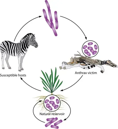
Ecology of B. anthracis. (PMID: 42640059)
2.6 Symptoms and Treatment
2.6.1 Clinical Symptoms
Type | Incubation Period | Main Manifestations | Untreated Case Fatality |
Cutaneous anthrax | 1–7 days | Painless papule → vesicle → black eschar with surrounding edema | ~20% |
Inhalational anthrax | 1–6 days | Influenza-like prodrome → sudden dyspnea, widened mediastinum, pleural effusion, shock | ~90% |
Gastrointestinal anthrax | 2–5 days | Severe abdominal pain, bloody diarrhea, intestinal necrosis | ~40% |
Meningeal anthrax | — | Meningeal signs, altered consciousness | ~90% |
Injection anthrax | — | Severe soft tissue infection at injection site | ~30% |
2.6.2 Treatment
Antimicrobial therapy: CDC recommends combination therapy administered intravenously for ≥2 weeks or until clinically stable, followed by a single oral antibiotic for 60 days to eliminate ungerminated spores in the lungs.
Drug Class | Recommendation Level | Considerations |
Fluoroquinolones | Ciprofloxacin, levofloxacin | Preferred prophylactic agents |
Carbapenems | Meropenem, imipenem/cilastatin | First-line for meningitis (good CNS penetration) |
Penicillins | Penicillin G, ampicillin | Only for penicillin-susceptible strains |
Protein synthesis inhibitors | Minocycline, doxycycline, clindamycin | First-line alternatives |
RNA synthesis inhibitor | Rifampin | Combination therapy |
Antitoxin therapy: Raxibacumab and Obiltoxaximab are two anti-PA monoclonal antibodies that block toxin effects by preventing PA binding to host cell receptors and should be used in combination with antibiotics for inhalational anthrax. A 2025 study reported a bispecific antibody (engineered from two non-protective antibodies 2A6 and 8A7) that achieves full protection against anthrax toxin by inhibiting PA83 cleavage.
Drug resistance: B. anthracis is normally sensitive to penicillin but can induce β-lactamase production, so penicillin or cephalosporin monotherapy is not recommended. The novel bacterial topoisomerase inhibitor OSUAB-0284 retains activity against fluoroquinolone-resistant strains and shows efficacy in a mouse inhalational anthrax model.
2.7 Prevention
l Animal vaccination: The Sterne strain live attenuated vaccine is the primary means of preventing anthrax in livestock, based on a pXO2-deleted attenuated strain.
l Human vaccination: Anthrax Vaccine Adsorbed (AVA, BioThrax™) is the FDA-approved vaccine but requires a prolonged primary series (0, 1, 6 months, then annual boosters). Cyfendus™ is an aluminum-adjuvanted version with a shortened priming schedule, approved for post-exposure prophylaxis. GC1109 is a novel recombinant PA vaccine; a Phase II trial showed 100% of recipients achieved TNA NF50 ≥ 0.56 at week 36.
l Animal carcass disposal: Carcasses of infected animals should be incinerated or deeply buried (with lime) to prevent spore release into the environment.
l Environmental decontamination: Contaminated areas should be disinfected with formaldehyde, glutaraldehyde, or peracetic acid.
l Occupational protection: Workers handling animal hides, wool, and bone meal should wear appropriate personal protective equipment.
l Surveillance and reporting: Anthrax is a notifiable disease in China, with approximately 300–500 cases reported annually, mainly in northern and western provinces.
2.8 Targets for Intervention
2.8.1 Pathogen Targets
Target | Protein/Pathway | Biological Function |
PA | Protective antigen | Receptor binding, toxin translocation |
LF | Lethal factor | MAPKK cleavage |
EF | Edema factor | cAMP elevation, endothelial barrier disruption |
CapBCAD | Capsule synthesis enzymes | PDGA capsule synthesis |
AtxA | Virulence regulator | Toxin and capsule gene expression |
2.8.2 Host Targets
Target | Type | Role in Infection |
Rac1/cofilin | Cytoskeletal regulatory proteins | ET-induced F-actin reorganization and endothelial permeability |
IGF1R/EGFR | Receptor tyrosine kinases | Activated by ET in a cAMP-independent manner, promoting endothelial barrier disruption |
MAPKK | Signaling kinases | Cleavage targets of LF |
2.9 Vaccine Types and Development Progress
Currently, multiple anthrax vaccines are licensed globally, and novel vaccine platforms are rapidly advancing.
2.9.1 Licensed Vaccines
Vaccine | Type | Features |
AVA (BioThraxTM) | Adsorbed vaccine | FDA-approved, prolonged primary series |
CyfendusTM | Aluminum-adjuvanted AVA | Shortened priming schedule, for post-exposure prophylaxis |
BARYTHRAX inj. | Recombinant PA protein | World's first recombinant protein anthrax vaccine, first shipped December 2025 |
2.9.2 Recombinant PA Vaccine (GC1109)
A Phase II clinical trial published in Vaccine in 2025 evaluated GC1109 (a novel recombinant PA vaccine) in healthy adults. All participants in the GC1109 group achieved TNA NF50 ≥ 0.56 at week 36 (100%, 95% CI: 97.6–100.0) , meeting the primary endpoint. Pruritus was the only solicited adverse event significantly more frequent in the GC1109 group; serious adverse events were rare and unrelated to vaccination.
2.9.3 Inhalable Nanoparticle Vaccine (YM1.7)
A 2026 study published in Advanced Materials developed an inhalable nanoparticle anthrax vaccine, YM1.7. The vaccine self-assembles from STING agonist MSA-2 and two disulfide bond-containing polymers, with recombinant PA antigen conjugated to the surface via N─B coordination. After aerosolized intratracheal inoculation, nanoparticles are internalized by antigen-presenting cells, release rPA in the acidic lysosomal environment, and escape into the cytosol via the proton sponge effect, releasing MSA-2. This spatiotemporal delivery mechanism elicits coordinated innate, humoral, mucosal, and cell-mediated immune responses in mice, providing strong protection against inhalational anthrax.
2.9.4 Nasal Booster Vaccine (LP-PA)
A 2025 study published in Journal of Microbiology and Biotechnology developed a nasal booster vaccine displaying PA on the surface of Lactiplantibacillus plantarum (LP-PA) . After intramuscular priming with purified rPA, nasal boosting with LP-PA significantly enhanced germinal center responses and mucosal antibody production, inducing protective mucosal immunity at the pathogen's entry site (respiratory tract).
2.9.5 Lyophilizable Nanoparticle Vaccine
A 2025 study published in Toxins developed a lyophilizable nanoparticle vaccine targeting a novel linear neutralizing epitope in the α2-α3 helices of domain 3 of lethal factor, providing a new strategy for developing cold-chain-independent anthrax vaccines.
2.10 Drugs
2.10.1 Pathogen-Targeted Drugs
Target | Drug/Candidate | Mechanism | Stage |
Type II topoisomerase | OSUAB-0284 | Novel bacterial topoisomerase inhibitor, active against fluoroquinolone-resistant strains | Mouse inhalational anthrax model |
Gepotidacin | Triazaacenaphthylene, targets gyrase and topo IV, efficacious in rabbit inhalational anthrax model | Rabbit model | |
PA | Raxibacumab/Obiltoxaximab | Anti-PA monoclonal antibodies, block toxin receptor binding | FDA-approved |
Bispecific antibody (2A6×8A7) | Inhibits PA83 cleavage, full protection | Preclinical | |
Capsule | Capsule-degrading enzyme (CapD) | Degrades PDGA capsule | Preclinical |
2.10.2 Antimicrobial Agents
Drug | Mechanism | Clinical Status |
Ciprofloxacin | DNA gyrase inhibition | Preferred prophylactic agent |
Meropenem | Cell wall synthesis inhibition | First-line for meningitis |
Doxycycline | Protein synthesis inhibition | First-line alternative |
Rifampin | RNA synthesis inhibition | Combination therapy |
2.11 Recent Research
2.11.1 Molecular Epidemiology and Genomics
A 2025 whole-genome sequencing study of B. anthracis from Ethiopian animal cases confirmed that all strains carried the primary virulence factors: PDGA capsule, lethal toxin (lef), and edema toxin (cya) . A 2026 genomic analysis of clinical and environmental isolates from Peru identified 24 chromosomal virulence genes and 67 pXO1 virulence-related genes.
Anthrax outbreaks continued across Africa in 2025–2026: the Democratic Republic of Congo reported over 50 hippopotamus and buffalo deaths, while Uganda documented 1,165 human anthrax cases between 2017 and 2024. In the 2026 Kazo District outbreak in Uganda, 135 cases (121 suspected, 14 confirmed) were identified between December 2025 and March 2026, with an attack rate of 65 per 100,000 and a case fatality rate of 4%.
2.11.2 Diagnostic Technology Advances
CRISPR/Cas13a field detection technology: A 2025 study published in Microbial Biotechnology established a field detection technology based on MIRA-CRISPR/Cas13a. The CRISPR/Cas13a detection system employing dual crRNAs achieved a detection limit of 1,000 copies/mL for B. anthracis. Combined with a lyophilized reagent system and CRISPR point-of-care device, the detection limit was further improved to 250 copies/mL, with results within 30 minutes, accurately identifying all positive and negative samples.
One-pot RPA-LFD detection: A 2026 study published in International Journal of Biological Macromolecules developed a one-pot recombinase polymerase amplification combined with lateral flow dipstick (RPA-LFD) assay that simultaneously detects B. anthracis and Yersinia pestis in 25 minutes at 42°C, suitable for blood, soil, and water samples, with detection limits comparable to real-time PCR.
CRISPR/Cas12a-functionalized silicon nanowire FET sensor: A 2025 study developed a CRISPR/Cas12a-functionalized silicon nanowire FET sensor capable of quantitatively detecting B. anthracis dsDNA within 10 minutes with a detection limit at the attomolar (aM) level.
Synthetic peptide latex agglutination test: A 2026 study developed an IgY-based latex agglutination test (LAT) for selective detection of B. anthracis spores from soil samples, offering a simple and cost-effective method.
Electrochemical aptasensor: A 2026 study developed a label-free impedimetric electrochemical aptasensor using disposable screen-printed electrodes for detecting B. anthracis spores, with a linear response between 20 and 200 spores/mL.
2.11.3 New Discoveries in Spore Germination Mechanisms
A 2025 study found that dipicolinic acid (DPA) is released in large amounts during spore germination, with calcium-DPA complexes released in the phagolysosome, affecting spore cytotoxicity. A 2026 study further demonstrated that the SpoVA2 tetrameric membrane complex is required for DPA transport into spores, with SpoVA2 being more critical for germination than SpoVA1, and the F1 gene being the most important germination gene in the 5A1 locus of B. anthracis. A 2026 study developed a D-π-A near-infrared fluorescent probe that achieved the first real-time visualization of germination behavior in individual B. anthracis spores.
3. Related Products & Services
l Recombinant Antigens & Receptors
l Antibodies
l Detection Kits
l Recombinant Protein Expression Services
l Antibody Development Services
4. Resources
5. References
1. Burroughs, O. S., Skaar, E. P. (2026). Bacillus anthracis: biological origins and impact on human history. Microbiology and molecular biology reviews : MMBR, 90(3), e0027425.
2. Sangwan, N., Gangwal, A., Jain, P., et al. (2025). Anthrax: Transmission, Pathogenesis, Prevention and Treatment. Toxins, 17(2), 56.
3. Bower, W. A., Yu, Y., Person, M. K., et al. (2023). CDC Guidelines for the Prevention and Treatment of Anthrax, 2023. MMWR. Recommendations and reports : Morbidity and mortality weekly report. Recommendations and reports, 72(6), 1–47.
4. Abdel-Glil, M. Y., Chiaverini, A., Garofolo, G., et al. (2021). A Whole-Genome-Based Gene-by-Gene Typing System for Standardized High-Resolution Strain Typing of Bacillus anthracis. Journal of clinical microbiology, 59(7), e0288920.
5. Moayeri, M., Leppla, S. H., Vrentas, C., et al. (2015). Anthrax Pathogenesis. Annual review of microbiology, 69, 185–208.
6. Jain, P., Guichard, A., Moayeri, M., et al. (2025). Anthrax ET activates Rac1 and RTK signaling to induce F-actin reorganization and endothelial permeability. iScience, 28(11), 113682.
7. Yu, Y., Ramírez-Guadiana, F. H., Gao, Y., et al. (2026). A tetrameric SpoVA2 membrane complex is required for DPA transport into Bacillus anthracis spores. mBio, 17(4), e0377725.
8. Qiao, Z., Wang, S., Shi, Y., et al. (2025). Human anthrax vaccines: Current status and future prospects. Human vaccines & immunotherapeutics, 21(1), 2595774.
9. Kang, C. K., Im, Y. J., Kim, D. Y., et al. (2025). Immunogenicity and safety evaluation of a recombinant protective antigen anthrax vaccine (GC1109): A phase II, dose-confirmed, randomized, double-blind, placebo-controlled study. Vaccine, 65, 127819.
10. Zhang, X., Dai, C., Zhang, H., et al. (2026). Cytosol-Targeting Delivery of Non-Nucleotide STING Agonist Achieves Inhalable Nanoparticle-Based Anthrax Vaccine. Advanced materials (Deerfield Beach, Fla.), 38(28), e12910.
11. Jang, I. H., Jung, I. R., Suh, H., et al. (2025). A Nasal Vaccine Displaying Anthrax Antigen on the Surface of Lactiplantibacillus plantarum Induces Protective Mucosal Immunity against Anthrax Toxin. Journal of microbiology and biotechnology, 35, e2503036.
12. Oscherwitz, J., Cease, K., Milich, D., et al. (2025). A Lyophilizable Nanoparticle Vaccine Specific for a Novel Linear Neutralizing Epitope in the α2-α3 Helices of Domain 3 of Lethal Factor from Bacillus anthracis. Toxins, 17(8), 422.
13. Hilliard, J. J., Jakielaszek, C., Mannino, F., et al. (2024). Efficacy of therapeutically administered gepotidacin in a rabbit model of inhalational anthrax. Antimicrobial agents and chemotherapy, 68(3), e0149723.
14. Fan, J., Huang, L., Chen, J., et al. (2025). Highly Sensitive Field Detection Technology for Anthrax Based on the CRISPR/Cas13a System. Microbial biotechnology, 18(11), e70240.
15. Paul, M., Singh, S., Yadav, P. K., et al. (2026). Development of a rapid and sensitive one-pot recombinase polymerase amplification assay for the simultaneous detection of Bacillus anthracis and Yersinia pestis with naked eye result read-out. International journal of biological macromolecules, 335(Pt 2), 149324.