
info@mail.ucallm.com
1. Overview
Black Creek Canal virus (BCCV) is a member of the genus Orthohantavirus within the family Hantaviridae. It is classified as a New World hantavirus and is one of the hantaviruses associated with hantavirus pulmonary syndrome (HPS) . It is an enveloped, negative-sense, single-stranded, trisegmented RNA virus with spherical or ovoid virions.
BCCV was first discovered in 1993 in Dade County, Florida, USA, following the investigation of a fatal HPS case. The natural reservoir of BCCV is the cotton rat (Sigmodon hispidus) , widely distributed across the southeastern United States. BCCV is transmitted to humans primarily through the inhalation of aerosolized particles from infected rodent excreta (urine, feces, saliva). No licensed BCCV vaccine or specific antiviral drug is currently available, and treatment is supportive.
2. Background Information
2.1 What is Black Creek Canal Virus?
BCCV is an enveloped, negative-sense, single-stranded, trisegmented RNA virus belonging to the family Hantaviridae and genus Orthohantavirus. Its genome consists of small (S), medium (M), and large (L) segments, encoding the nucleoprotein (N), glycoprotein precursor (GPC, cleaved into Gn and Gc), and RNA-dependent RNA polymerase (L), respectively. Complete sequences of the S and M segments and partial sequence of the L segment have been determined for BCCV. Phylogenetic analysis indicates that each of the virus segments is unique, with no evidence of genetic reassortment with other known hantaviruses.
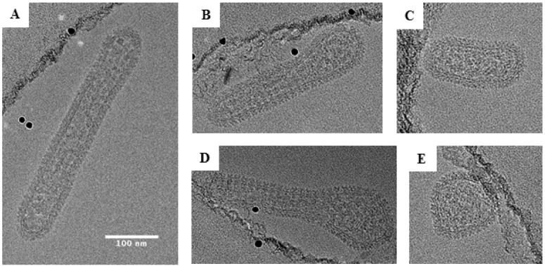
Representative cryo-EM images of Black Creek Canal virus (PMID: 31527500)
2.2 Black Creek Canal Virus Genome & Classification
BCCV belongs to the family Hantaviridae and genus Orthohantavirus, with the following taxonomic classification: Realm Riboviria, Kingdom Orthornavirae, Phylum Negarnaviricota, Class Bunyaviricetes, Order Elliovirales, Family Hantaviridae, Genus Orthohantavirus.
2.3 What Are Black Creek Canal Virus Structural Proteins?
BCCV encodes three major structural proteins:
Protein | Full name | Main Function |
N | Nucleoprotein | Most abundant structural component; encapsidates the trisegmented negative-sense RNA genome to form RNPs; localises to the Golgi and perinuclear regions; involved in viral RNA synthesis |
Gn | Glycoprotein N | Forms spikes on the viral envelope; acts as a chaperone for Gc, facilitating its folding and transport through the Golgi; mediates host cell attachment by binding surface receptors such as β3 integrins; a significant target of the host immune response |
Gc | Glycoprotein C | Together with Gn forms the spike complex; mediates membrane fusion; a key determinant of viral infectivity and tropism |
The BCCV N protein uniquely localises to the Golgi and perinuclear regions, unlike N proteins of other Bunyaviridae family members (which are cytoplasmic), indicating that the morphogenesis mechanisms of New World hantaviruses are distinct
2.4 Mechanism of Cell Entry and Replication
2.4.1 Viral Attachment and Receptor Recognition
BCCV binds to host cell surface receptors via its surface glycoprotein Gn. Gn can bind surface receptors such as β3 integrins, mediating viral attachment.
2.4.2 Viral Entry
BCCV enters host cells through the apical surface of polarized epithelial cells. Studies show that Vero C1008 cells infected through the apical surface demonstrate a high level of susceptibility to BCCV infection, whereas cells infected basolaterally exhibit a barely detectable level of viral protein synthesis.
2.4.3 Viral RNA Replication
After entry, the RNP is released into the cytoplasm, and the L protein (RdRp) initiates genome transcription and replication. The BCCV N protein plays a critical role in viral RNA synthesis, interacting with the viral polymerase (L protein) and host cell factors.
2.4.4 Viral Assembly and Release
BCCV assembly and release exhibit unique polarized characteristics. Unlike most Bunyaviridae family members, which assemble in the endoplasmic reticulum-Golgi intermediate compartment, BCCV assembles and buds at the plasma membrane. The virus is released from the apical surface of polarized cells, with virus titers released from the apical surface approximately 1,200-fold greater than those released into the basolateral media. Actin microfilaments play an important role in BCCV morphogenesis; treatment of infected cells with cytochalasin D decreases the yield of released BCCV by 94%.
2.5 Symptoms and Treatment
2.5.1 Clinical Symptoms
BCCV infection can cause hantavirus pulmonary syndrome (HPS), with an incubation period of approximately several weeks. Clinical manifestations include:
l Influenza-like prodromal symptoms: fever, headache, joint pain
l Progressive pulmonary involvement: can progress to pneumonia, and in severe cases, acute respiratory distress syndrome (ARDS)
Clinical features of HPS include pleural effusions (present in most cases), low cardiac index, low stroke volume index, and high systemic vascular resistance. In survivors, exertional dyspnoea and reduced expiratory flow are common in early convalescence and resolve in most patients, but some patients may manifest long-term pulmonary and cognitive dysfunction.
2.5.2 Antiviral Treatment
No specific antiviral therapy is approved for BCCV. Treatment is primarily supportive, requiring careful management of fluid status to maintain perfusion without exacerbating pulmonary oedema. In severe cases, extracorporeal membrane oxygenation (ECMO) may be beneficial.
2.6 Targets for Intervention
2.6.1 Viral Targets
Target | Protein | Biological Role |
Gn | Glycoprotein N | Receptor binding, Gc chaperone |
Gc | Glycoprotein C | Membrane fusion |
N | Nucleoprotein | RNP formation, RNA synthesis |
L | RNA polymerase | RNA replication |
2.6.2 Host Receptors & Entry Factors
Host Target | Type | Role in Infection |
β3 integrin | Receptor | Viral attachment |
Actin microfilaments | Cytoskeleton | Viral morphogenesis and release |
2.7 Vaccine Types and Development Progress
Currently, no BCCV vaccine is licensed worldwide. Patents exist for BCCV vaccines or purified immunogenic polypeptides. A 2025 patent application relates to a Bunyavirales vaccine, in which BCCV is listed as one of the covered pathogens. In addition, vaccine strategies based on recombinant BCCV Gn protein are under development, as Gn is highly exposed on the virion surface and is a significant target for the host immune response and a key focus in vaccine development.
2.8 Drugs
Currently, no specific small-molecule antiviral is approved for BCCV.
2.8.1 Virus-Targeted Drugs
Target/mechanism | Drug/candidate | Mechanism | Stage |
Viral release | Cytochalasin D | Depolymerizes actin microfilaments, reducing BCCV release by 94% | In vitro |
Viral replication | Ribavirin | Broad-spectrum antiviral, active against hantavirus in vitro | Ineffective in HPS clinical trials |
2.8.2 Host-Targeted Drugs
Target | Drug/candidate | Mechanism | Stage |
Systemic capillary leak | High-dose steroid therapy | Immunosuppression, potentially useful in systemic capillary leak syndrome | Clinical recommendation |
3. Related Products & Services
l Recombinant Antigens & Receptors
l Antibodies
l Detection Kits
l Recombinant Protein Expression Services
l Antibody Development Services
4. Resources
5. References
1. Rollin, P. E., Ksiazek, T. G., Elliott, L. H., et al. (1995). Isolation of black creek canal virus, a new hantavirus from Sigmodon hispidus in Florida. Journal of medical virology, 46(1), 35–39.
2. Ravkov, E. V., Rollin, P. E., Ksiazek, T. G., et al. (1995). Genetic and serologic analysis of Black Creek Canal virus and its association with human disease and Sigmodon hispidus infection. Virology, 210(2), 482–489.
3. Ravkov, E. V., Nichol, S. T., Compans, R. W. (1997). Polarized entry and release in epithelial cells of Black Creek Canal virus, a New World hantavirus. Journal of virology, 71(2), 1147–1154.
4. Ravkov, E. V., Nichol, S. T., Peters, C. J., et al. (1998). Role of actin microfilaments in Black Creek Canal virus morphogenesis. Journal of virology, 72(4), 2865–2870.
5. Ravkov, E. V., Compans, R. W. (2001). Hantavirus nucleocapsid protein is expressed as a membrane-associated protein in the perinuclear region. Journal of virology, 75(4), 1808–1815.
6. Ramanathan, H. N., Jonsson, C. B. (2008). New and Old World hantaviruses differentially utilize host cytoskeletal components during their life cycles. Virology, 374(1), 138–150.
7. Khan, A. S., Gaviria, M., Rollin, P. E., et al. (1996). Hantavirus pulmonary syndrome in Florida: association with the newly identified Black Creek Canal virus. The American journal of medicine, 100(1), 46–48.
8. MacNeil, A., Ksiazek, T. G., Rollin, P. E. (2011). Hantavirus pulmonary syndrome, United States, 1993-2009. Emerging infectious diseases, 17(7), 1195–1201.
9. Mertz, G. J., Miedzinski, L., Goade, D., et al. (2004). Placebo-controlled, double-blind trial of intravenous ribavirin for the treatment of hantavirus cardiopulmonary syndrome in North America. Clinical infectious diseases : an official publication of the Infectious Diseases Society of America, 39(9), 1307–1313.
10. Hooper, J. W., Ferro, A. M., Wahl-Jensen, V. (2008). Immune serum produced by DNA vaccination protects hamsters against lethal respiratory challenge with Andes virus. Journal of virology, 82(3), 1332–1338.
11. Parvate, A., Williams, E. P., Taylor, M. K.,et al. (2019). Diverse Morphology and Structural Features of Old and New World Hantaviruses. Viruses, 11(9), 862.