
info@mail.ucallm.com
Bone diseases involve aberrant changes across multiple layers, including osteogenesis, bone resorption, skeletal stem cell function, cartilage homeostasis, inflammatory responses and bone microenvironment. By establishing disease-specific cellular models, three-dimensional culture models, tissue models and animal disease models, we can recapitulate disease progression at cellular, molecular and tissue levels. These models help dissect complex pathological mechanisms such as bone metabolic imbalance, tissue degeneration, tumor progression and bone aging, and support disease target discovery, investigation of drug action mechanisms and evaluation of candidate therapeutic strategies.
In recent years, with advances in skeletal stem and progenitor cell research, 3D culture and organoid technologies, bone disease models have evolved from conventional monocell culture toward sophisticated models that better mimic physiological and pathological microenvironments. Bone/cartilage organoids and multicellular 3D models can partially reconstruct tissue architecture and cellular crosstalk in vitro, providing novel research tools for bone disease mechanistic studies, drug screening and evaluation of regenerative therapies.
1 Osteoporosis Models
Osteoporosis is a metabolic bone disease characterized by reduced bone mass, deteriorated bone microarchitecture and increased bone fragility. Its core pathological alterations are tightly linked to the imbalance between bone formation and bone resorption. Osteoporosis research models can be built based on osteoblasts, osteoclasts, skeletal stem and progenitor cells and bone marrow microenvironment, to investigate mechanisms including impaired bone-forming capacity, enhanced bone resorption and disrupted bone remodeling.
For in vitro studies, models can be constructed by inducing aberrant osteogenic differentiation of osteoblasts or MSCs, establishing osteoclast differentiation models, and generating osteoblast-osteoclast or skeletal stem cell co-culture systems, to explore the regulation of bone metabolism by signaling pathways including RANK/RANKL/OPG, Wnt/β-catenin and BMP/SMAD. Meanwhile, diverse osteoporosis models are developed by incorporating risk factors such as hormone deficiency, aging, glucocorticoid exposure and metabolic disorders, for evaluating anti-resorptive drugs, bone-anabolic agents and other bone-protective strategies. Skeletal stem and progenitor cells are recognized to be closely associated with metabolic bone diseases such as osteoporosis. Incorporation of SSCs/SSPCs into osteoporosis models can further expand mechanistic investigations of the disease.
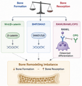
Schematic of the balanced regulation between bone formation and bone resorption. Wnt/β catenin and BMP/SMAD govern osteogenesis, while RANK/RANKL/OPG controls osteoclastogenesis. Decreased bone formation and increased resorption result in bone remodeling imbalance and bone loss.
2 Osteoarthritis Models
Osteoarthritis (OA) is a chronic degenerative disease affecting articular cartilage, subchondral bone, synovium and peri-articular tissues. Its initiation and progression are not simply caused by cartilage wear, but are associated with multiple factors including mechanical stress, extracellular matrix degradation, inflammatory response, chondrocyte dysfunction and subchondral bone remodeling.
Osteoarthritis research models can be established using primary chondrocytes, mesenchymal stem cells, synoviocytes and cartilage tissues to investigate chondrocyte phenotypic shifts, extracellular matrix degradation, inflammatory cytokine stimulation and the mechanisms of cartilage degeneration. Combined with osteochondral co-culture, three-dimensional cartilage models and bone/cartilage organoids, these systems enable investigation of crosstalk between cartilage and subchondral bone, better recapitulating the complex tissue microenvironment of OA. In recent years, mechanisms such as cell-matrix interactions, mechanical signals and ion channels have attracted growing attention and are recognized to participate in OA pathological processes including cartilage degeneration, inflammation, bone remodeling and pain.
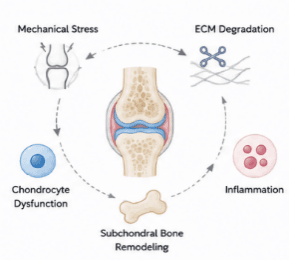
Schematic illustration of the vicious cycle in joint degeneration. Mechanical stress drives chondrocyte dysfunction, subchondral bone remodeling, inflammation and ECM degradation to progressively damage articular tissue.
3 Bone Cancer Models
Bone cancer research mainly focuses on the abnormal proliferation, invasion and metastasis of tumor cells, as well as the crosstalk between tumor cells and the bone microenvironment. Malignant bone tumors represented by osteosarcoma exhibit prominent tissue heterogeneity. Their initiation and progression are jointly regulated by multiple components including tumor cells, osteoblasts, osteoclasts, immune cells, blood vessels and extracellular matrix.
Bone cancer research can be performed using osteosarcoma cell lines, patient-derived tumor cells and three-dimensional tumor models. 3D culture, organoid and co-culture systems are applied to further mimic the bone microenvironment where tumors grow. Research scope covers tumor cell proliferation, migration, invasion, bone destruction, tumor-bone cell interaction and mechanisms of tumor metastasis. These models can also be used for in vitro evaluation of anti-tumor drugs, targeted therapies and combination treatment strategies. There exists complex bidirectional regulation between osteosarcoma and bone microenvironment. The immune and cellular signaling milieu unique to bone tissue also serves as an important foundation for investigating disease progression.
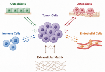
Schematic of cell-cell crosstalk within the bone tumor microenvironment. Tumor cells communicate bidirectionally with osteoblasts, osteoclasts, immune cells, endothelial cells and extracellular matrix to drive tumor progression in bone.
4 Bone Aging Models
With advancing age, bone tissue undergoes changes including reduced bone-forming capacity, imbalance between bone resorption and bone formation, increased bone marrow adiposity and impaired bone regeneration. Research on bone aging focuses on how compromised function of skeletal stem and progenitor cells, cellular senescence, alterations in the bone marrow microenvironment, impaired vascular function and chronic low-grade inflammation affect bone homeostasis.
Senescence of skeletal stem cells and bone marrow stromal cells is regarded as one of the key mechanisms underlying age-related bone loss. Accumulation of senescent cells leads to impaired cell proliferation and osteogenic differentiation. Meanwhile, inflammatory factors and matrix regulators generated by the senescence-associated secretory phenotype (SASP) may further remodel the bone microenvironment, thereby affecting osteogenesis, osteoclastogenesis and tissue repair processes. By establishing replicative senescence, induced senescence and age-related cellular models, we can investigate the relationship between skeletal stem cell senescence, diminished osteogenic capacity and bone loss. These models can also be applied to evaluate anti-aging and bone-protective strategies.
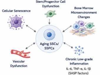
Regulatory mechanisms of aged skeletal stem/progenitor cells (SSCs/SSPCs). Aging SSCs/SSPCs are affected by stem/progenitor cell dysfunction, cellular senescence, vascular dysfunction, bone marrow microenvironment remodeling and chronic low-grade inflammation (SASP factors: IL-6, TNF-α, IL-1β). In turn, aged SSCs/SSPCs drive these pathological alterations, forming an interactive regulatory loop of skeletal aging.