
info@mail.ucallm.com
1 Cell Lines
Bone-related cell lines feature stable culture performance, high reproducibility and convenient experimental operation, making them suitable for research on bone cell differentiation, bone metabolism, signaling pathways, drug screening and disease mechanisms. Depending on research objectives, osteogenic cell lines, osteoclast precursor models, cartilage-related cell lines and bone tumor cell lines can be selected.
Osteogenic cell lines are used to investigate the proliferation, differentiation, maturation, extracellular matrix formation and mineralization of osteoblasts. Commonly used models include MC3T3-E1, SaOS-2 and MG-63. Combined with osteogenic induction culture systems, osteogenic function can be evaluated via ALP, RUNX2, OCN, OPN and mineralization assays.
Osteoclast-related models are mainly applied to study the mechanisms of osteoclast differentiation and bone resorption. Cells such as RAW 264.7 can form multinucleated cells with osteoclast characteristics under appropriate induction conditions, which are used to explore osteoclastogenic pathways including RANK/RANKL, NF-κB and NFATc1.
Cartilage-related cell lines are suitable for studies on chondrocyte function, cartilage matrix metabolism and osteoarthritis. In vitro disease models can be established by inflammatory stimulation, matrix degradation and drug treatment.
Osteosarcoma cell lines such as U2OS, MG-63 and SaOS-2 can be used to examine proliferation, migration, invasion, apoptosis and drug response of bone tumor cells. These cell lines can be further utilized to construct 3D bone tumor models.
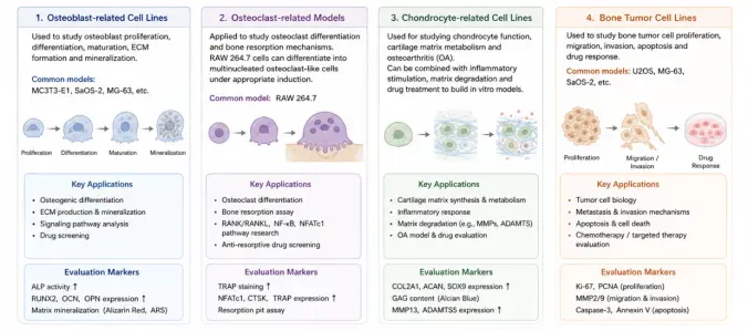
Schematic of in vitro cell models for bone research. Four categories: osteoblast, osteoclast, chondrocyte and bone tumor models, illustrating their applications and evaluation markers.
Products: Culture Media, FBS, Cell Cryopreservation Medium, Cell Culture Dishes/Plates, Antibodies, Assay Kits, Fluorescent Probes
Data display:
RAW264.7
RAW 264.7 is a murine monocyte/macrophage-like cell line. Under appropriate conditions, it can differentiate into osteoclast-like multinucleated cells upon RANKL induction. It is widely used to investigate the mechanisms underlying osteoclast differentiation, bone resorption and bone remodeling.
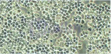
RAW 264.7 is a murine monocyte/macrophage-like cell line cultured in medium supplemented with fetal bovine serum. Under light microscopy, the cells are mostly round or irregular in shape with weak adherence and exhibit semi-adherent growth.
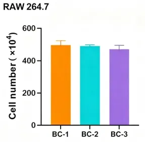
RAW 264.7 cells were cultured with three batches of serum (BC-1, BC-2 and BC-3). The effect of different serum batches on cell expansion performance was evaluated by harvested cell yield, and the consistency of culture performance across serum batches was verified. Cell numbers are expressed as ×10⁴ cells, and error bars represent standard deviation (SD).
DC2.4
DC2.4 is a murine dendritic cell line derived from mouse bone marrow dendritic cell models. It can be applied to studies of bone marrow immune microenvironment, antigen presentation and osteoimmunological regulation.
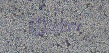
DC2.4 is a murine dendritic cell line cultured in vitro with medium supplemented with fetal bovine serum. The cells exhibit an irregular morphology under light microscopy.
2 Primary Cells
Bone-Related Primary Cells
Primary cells can well retain the biological characteristics and functional phenotypes of their source tissues, making them suitable for research on the physiological functions of bone cells, tissue microenvironment and disease mechanisms. Compared with stable cell lines, primary cells offer advantages in cellular phenotype and tissue relevance, and help improve the physiological relevance of in vitro research models.
Primary Osteoblasts: Used to investigate osteoblast differentiation, bone matrix formation, mineralization and signaling pathways associated with bone formation.
Primary Osteoclasts: Applied to study the mechanisms of osteoclast formation, bone resorption and bone remodeling. They can be co-cultured with osteoblasts to explore the dynamic balance between bone formation and bone resorption.
Primary Chondrocytes: Suitable for research on chondrogenesis, cartilage matrix metabolism, cartilage degeneration and osteoarthritis. They can be used to detect markers including COL2A1, ACAN, SOX9, MMP13 and ADAMTS5.
Primary Mesenchymal Stromal/Stem Cells (Primary MSCs): Possess robust expansion and multipotent differentiation capacity. They are applied in osteogenic, chondrogenic and adipogenic differentiation research, and serve as an important cell source for bone tissue engineering and bone regeneration studies.
Bone Marrow Stromal Cells: Used to investigate the bone marrow microenvironment, functions of skeletal stem and progenitor cells, and interactions between osteoblasts and other bone marrow cells.
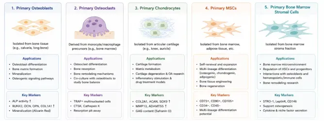
Schematic of primary cell models for bone research. Including primary osteoblasts, osteoclasts, chondrocytes, MSCs and bone marrow stromal cells, illustrating their sources, applications and biomarkers.
Products: Culture Media, FBS, Recombinant Proteins, Cell Cryopreservation Medium, Cell Culture Dishes/Plates, Antibodies, Assay Kits, Fluorescent Probes
3 Stem & Progenitor Cells
Skeletal stem cells and skeletal stem and progenitor cells represent essential cellular sources for bone formation, homeostasis maintenance and injury repair. Investigations into their self-renewal, lineage differentiation and tissue regenerative potential help elucidate the mechanisms of cell fate determination and microenvironmental regulation during bone formation and repair.
Skeletal Stem Cells (SSCs):Used for research on the cellular phenotype, self-renewal, lineage differentiation and bone regenerative potential of skeletal stem cells.
Skeletal Progenitor Cells (SPCs):Applied to study the differentiation process of skeletal progenitor cells toward osteogenic, chondrogenic and other cell lineages.
Mesenchymal Stromal/Stem Cells (MSCs):Widely adopted stromal/stem cell sources in current bone regeneration and tissue engineering research. They can be utilized for osteogenic induction, tissue engineering, cell therapy and cell-biomaterial crosstalk studies.
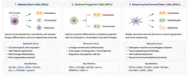
Schematic of skeletal stem cells (SSCs), skeletal progenitor cells (SPCs) and mesenchymal stromal/stem cells (MSCs), showing their differentiation potential, research focuses and key biomarkers.
Products: Culture Media, FBS, Recombinant Proteins, Cell Cryopreservation Medium, Cell Culture Dishes/Plates, Antibodies, Assay Kits, Fluorescent Probes
4 3D Cell Models
3D Bone Cell Models
There are substantial differences between two-dimensional (2D) and three-dimensional (3D) cell culture models. 2D cultures form within minutes to hours, featuring simple operation, good reproducibility and low cost, which facilitates long-term culture and result interpretation. However, they fail to recapitulate the native architecture of in vivo tissues, lacking cell-cell and cell-extracellular matrix interactions as well as in vivo-like microenvironments and niches. This alters cellular morphology, division patterns, phenotypes and polarity. With unlimited nutrient and oxygen supply, cells exhibit deviations in gene expression, splicing and biochemical characteristics compared with in vivo conditions. In contrast, 3D cultures take hours to days to form, with higher operational difficulty, poorer reproducibility, increased costs and fewer commercially available supporting assay reagents. Nevertheless, they can reconstruct the 3D architecture of native tissues, restore interactions between cells and the extracellular microenvironment, and form in vivo-like niches. Cellular morphology, division patterns, phenotypes and polarity are preserved. Nutrients, oxygen and signaling molecules exhibit concentration gradients consistent with in vivo status, and cellular gene expression, splicing, topological structures and biochemical features better resemble the real in vivo state.
MSC Spheroids: Used to investigate 3D aggregation, cell communication, osteogenic differentiation and regenerative potential of MSCs. Combined with osteogenic induction systems, they help explore how 3D environments modulate MSC fate and function.
Osteoblast Spheroids: Applied to study cell-cell crosstalk among osteoblasts, extracellular matrix deposition and mineralization processes.
Co-culture Spheroids: Combinations of osteoblasts, MSCs, osteoclast precursors, endothelial cells or immune cells. They mimic multicellular interactions in bone tissue and examine the relationships among osteogenesis, bone resorption, angiogenesis and immune regulation.
Bone Tumor Spheroids: Deployed to establish 3D models of bone tumors such as osteosarcoma, for research on tumor cell proliferation, invasion, drug penetration and tumor microenvironment.
3D bone cell models can also be combined with hydrogels, ECM and biomaterials to further construct 3D bone tissue models with diverse mechanical and biological properties.
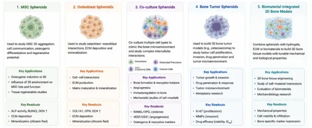
Schematic of 3D spheroids and biomaterial-integrated bone models for bone research, including MSC spheroids, osteoblast spheroids, co-culture spheroids, bone tumor spheroids and biomaterial-integrated bone models, illustrating applications and key readouts.
Products: Culture Media, FBS, Recombinant Proteins, Cell Cryopreservation Medium, U-bottom Cell Culture Dishes/Plates, Antibodies, Assay Kits, Fluorescent Probes
Data display:
hBM-MSCs
Bone marrow is a critical hematopoietic and immune tissue located in the medullary cavity of long bones and the cancellous bone of flat bones. Red bone marrow is responsible for the production of red blood cells, platelets and various immune cells. Therefore, bone marrow functions not only as a hematopoietic organ but also as an essential microenvironment for maintaining immune homeostasis. Bone Marrow Mesenchymal Stem/Stromal Cells (BMSCs) originate from the bone marrow stromal system. Distinct from hematopoietic stem cells, they possess robust self-renewal capacity and multipotent differentiation potential, and can differentiate into multiple cell types including osteoblasts, chondrocytes and adipocytes. They serve as vital seed cells in bone tissue engineering and regenerative medicine research.
BMSCs are present at a low proportion in bone marrow (approximately 0.001%–0.1% of bone marrow nucleated cells). Their in vitro expansion and functional maintenance are affected by multiple factors including adhesion conditions, cell density, medium composition, serum concentration and culture environment. By optimizing the culture system, stable BMSC models can be established to investigate mechanisms of cell differentiation, bone formation, tissue repair and cell-biomaterial crosstalk.
Morphological Observation and Live Cell Station Detection of hBM-MSCs at Different Densities
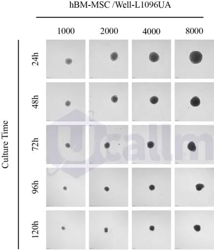
hBM-MSCs cultured on Ucallm® Ultra-Low Attachment Surface forms tumor spheroids. hBM-MSCs were planted in 96-well tissue culture plates and ultra-low attachment plates at concentrations of 500, 1000, 2000, 4000, and 8000 cells per well. Live-cell imaging was conducted at 24, 48, 72, 96, and 120 hours after seeding. Scale bars represent 200 μm.
mBM-MSCs
Murine bone marrow mesenchymal stem/stromal cells (BMSCs) are adult stem cells derived from mouse bone marrow. They possess strong self-renewal capacity and multipotent differentiation potential, and can differentiate into multiple cell types including osteoblasts, chondrocytes and adipocytes. They are widely used in regenerative medicine and stem cell research.
Murine BMSCs are generally isolated from the bone marrow cavity of the tibia or femur of adult mice. Cell suspensions enriched with mesenchymal stem cells are obtained through bone marrow flushing, centrifugation and other procedures. These cells account for a small fraction in bone marrow, approximately 0.001%–0.1% of total bone marrow nucleated cells. When cultured in vitro, murine BMSCs exhibit a fibroblast-like morphology and grow in an adherent manner. Their proliferation and differentiation are susceptible to conditions such as seeding density, medium components, serum concentration, culture temperature and pH. Thanks to their excellent multipotent differentiation capacity, low immunogenicity and favorable expandability, BMSCs hold great research value in tissue repair, cartilage regeneration, nerve injury repair, cell therapy and other research fields.
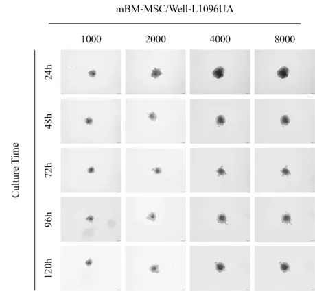
mBM-MSCs cultured on Ucallm® Ultra-Low Attachment Surface forms tumor spheroids. mBM-MSCs were planted in 96-well tissue culture plates and ultra-low attachment plates at concentrations of 500, 1000, 2000, 4000, and 8000 cells per well. Live-cell imaging was conducted at 24, 48, 72, 96, and 120 hours after seeding. Scale bars represent 200 μm.
5 Organoids & Tissue Engineering
Organoids and Bone Tissue Engineering Models | Explore our organoid research portfolio
Bone organoids and bone tissue engineering models integrate stem/progenitor cells, extracellular matrix, biomaterials and bioactive factors within three-dimensional environments, providing sophisticated in vitro models to investigate bone formation, disease onset and tissue regeneration.
Bone Organoids: Used to study bone formation, cellular differentiation and reconstruction of bone microenvironments, and can be further applied for bone disease modeling and evaluation of candidate therapeutics.
Cartilage Organoids: Suitable for exploring chondrogenesis, cartilage maturation, matrix metabolism and pathological processes such as osteoarthritis.
Osteochondral Models: Construct bone- and cartilage-associated tissue structures simultaneously, enabling investigations of the osteochondral interface, subchondral bone alterations and complex pathological events in osteoarthritis.
Biomaterial Scaffolds: Provide 3D support and a permissive microenvironment for tissue reconstruction. Combined with hydrogels, bioceramics, collagen and 3D-printed scaffolds, these systems allow characterization of material properties including pore architecture, degradation profile, mechanical performance and biocompatibility, as well as their regulatory effects on cell differentiation and bone formation.
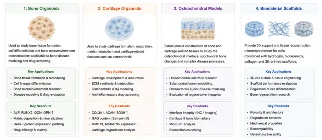
Schematic of bone organoids, cartilage organoids, osteochondral models and biomaterial scaffolds, presenting their research applications and key readouts.
Products: Organoid Culture Systems & Custom Services, Recombinant Proteins, Cell Cryopreservation Medium, Culture Dishes/Plates, Antibodies, Assay Kits, Fluorescent Probes
Data display :
6 3D Bone Marrow Imaging & Spatial Analysis
Bone marrow serves as an essential niche for the maintenance of hematopoietic and immune cells in vivo. It comprises a highly complex three-dimensional network formed by hematopoietic cells, mesenchymal stromal cells, blood vessels and diverse microenvironmental cells. The spatial localization of cells within bone marrow, cell-cell interactions, as well as their relationships with blood vessels and endosteal structures, are critical for understanding hematopoietic homeostasis, immune regulation, bone metabolism and tissue repair. However, the bone marrow inside intact long bones is enclosed within rigid cortical bone, and the marrow tissue itself is soft and prone to deformation. Conventional tissue sectioning and two-dimensional imaging cannot fully preserve its 3D spatial architecture, and continuous high-resolution observation of deep bone marrow regions remains challenging. Therefore, tissue clearing combined with 3D imaging and spatial analysis techniques provides novel tools for dissecting the intact bone marrow microenvironment.
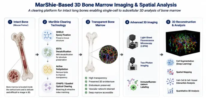
Schematic workflow of MarShie-based 3D bone marrow imaging and spatial analysis, showing intact bone clearing, sample preparation, advanced 3D imaging and spatial quantitative analysis.
MarShie (Marrow Shield) is a tissue-clearing method developed for three-dimensional imaging of bone marrow within intact long bones. This protocol adopts SHIELD multi-epoxy fixation, mild EDTA decalcification, active delipidation, and decolorization-clearing using CHAPS/Quadrol. While enhancing the transparency of bone tissue, it preserves the delicate three-dimensional architecture of bone marrow to the greatest extent possible.
Studies have demonstrated that MarShie enables clearing of bone marrow in intact mouse femurs while retaining endosteum, bone marrow vasculature and deep marrow structures, laying a foundation for subsequent 3D imaging and quantitative spatial analysis. Combined with Light-Sheet Fluorescence Microscopy (LSFM), two-photon imaging, immunofluorescent labeling, as well as image segmentation and machine learning analysis, this technique supports three-dimensional reconstruction of cells, blood vessels and stromal structures inside bone marrow. It permits quantification of cell numbers, spatial distribution, cellular proximity and alterations in tissue architecture. The research also demonstrates the capability of single-cell and even subcellular-level analysis in deep bone marrow regions of intact mouse femurs.
Research Applications
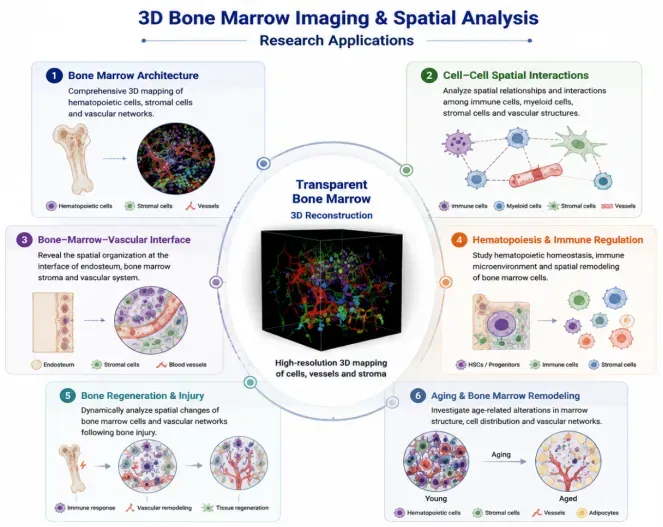
Schematic showing research applications of 3D bone marrow imaging and spatial analysis. Based on transparent bone marrow 3D reconstruction, it covers six research fields including marrow architecture, cell interactions, vascular interface, hematopoiesis, bone injury regeneration and aging-related marrow remodeling.
Representative Technology
MarShie|Bone Marrow Clearing
Mertens et al., Nature Communications, 2024, 15:1764
References
[1] Melis S, et al. Skeletal stem and progenitor cells in bone physiology, ageing and disease. Nature Reviews Endocrinology. 2025;21:135–153.
[2] Miao MZ, et al. Integrin signalling in joint development, homeostasis and osteoarthritis. Nature Reviews Rheumatology. 2024;20:492–509.
[3] Zhou R, et al. Ion channels in osteoarthritis: emerging roles and potential targets. Nature Reviews Rheumatology. 2024;20:545–564.
[4] Bai L, et al. Engineering bone/cartilage organoids: strategy, progress, and application. Bone Research. 2024;12:66.
[5] Wölfel EM, et al. Senescence of skeletal stem cells and their contribution to age-related bone loss. Mechanisms of Ageing and Development. 2024;221:111976.
[6] Mi B, et al. Ageing-related bone and immunity changes: insights into the complex interplay between the skeleton and the immune system. Bone Research. 2024;12:42.