
info@mail.ucallm.com
Tumor-derived Exosomes (TDEs) are critical extracellular vesicles secreted by tumor cells, which play key regulatory roles in tumor initiation, progression and therapeutic response. Exosomes carry characteristic proteins, lipids and nucleic acid cargo from their parent cells and travel across different tissues via body fluid circulation, making them essential messengers bridging tumor cells and their surrounding microenvironment.
Studies have demonstrated that exosomes secreted by tumor cells can be internalized by multiple cell types within the tumor microenvironment, including immune cells, fibroblasts, endothelial cells and other tumor cells. By delivering miRNAs, mRNAs, lncRNAs, proteins and immunomodulatory molecules, they remodel the local microenvironment to promote tumor growth, invasion, metastasis and therapeutic resistance.
1.1 Tumor Microenvironment Remodeling
Tumor-derived exosomes mediate intercellular communication within the tumor microenvironment to support adaptive tumor cell growth. For instance, exosomes released by tumor cells can be taken up by tumor-associated macrophages (TAMs) and induce their polarization toward the immunosuppressive M2-like phenotype, generating an immune microenvironment favorable for tumor progression.
In addition, tumor exosomes act on cancer-associated fibroblasts (CAFs) to stimulate their activation and secretion of extracellular matrix-related factors, enhancing structural remodeling and invasive capacity of tumor tissues. Meanwhile, VEGF-related molecules and pro-angiogenic miRNAs carried by exosomes activate vascular endothelial cells and drive tumor angiogenesis.
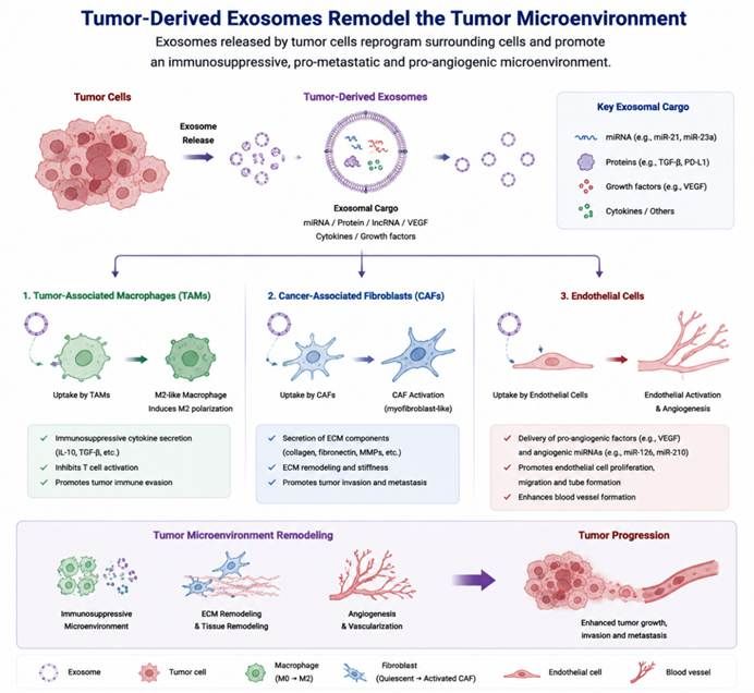
Tumor-derived exosomes remodel the tumor microenvironment.
Tumor cells secrete exosomes loaded with miRNAs, proteins, growth factors and cytokines. These tumor-derived exosomes are internalized by recipient cells within the tumor microenvironment, including tumor-associated macrophages (TAMs), cancer-associated fibroblasts (CAFs), and endothelial cells. Exosomal cargo reprograms these stromal cells: it drives M2 polarization of macrophages, activates CAFs to remodel extracellular matrix, and triggers endothelial activation and angiogenesis. Collectively, these effects shape an immunosuppressive, pro-metastatic and pro-angiogenic tumor microenvironment, which ultimately facilitates tumor progression, invasion and metastasis.
1.2 Immune Evasion
Exosomes play a vital role in tumor immune evasion. Tumor cells transmit immunosuppressive signals to immune cells via exosomes, thereby dampening anti-tumor immune responses.
Tumor-derived exosomes can carry PD-L1 protein. The exosomal surface PD-L1 binds to PD-1 on T cells, inhibiting T cell activation and cytotoxic function and facilitating tumor immune evasion. Exosomal PD-L1 originating from tumor cells can systematically suppress anti-tumor immunity and affect the efficacy of immune checkpoint therapy, revealing the critical function of exosomes in tumor immune regulation.
Furthermore, tumor exosomes may carry immunosuppressive miRNAs such as miR-21 and miR-23a. By modulating signaling pathways in immune cells, they impair T cell function, natural killer (NK) cell activity, and antigen presentation capacity of dendritic cells.
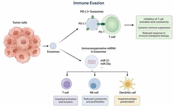
Tumor-derived exosomes mediate the mechanisms of tumor immune evasion. Tumor cells secrete PD-L1-carrying exosomes, which suppress T cell activation and cytotoxicity via PD-L1/PD-1 interaction, leading to systemic immunosuppression and reduced response to immune checkpoint therapy. Meanwhile, exosomes loaded with immunosuppressive miRNAs (such as miR-21 and miR-23a) can inhibit T cell activation, attenuate NK cell killing and proliferation, and block antigen presentation by dendritic cells, enabling multi-pathway immune evasion.
1.3 Metastasis & Pre-metastatic Niche Formation
Exosomes serve as one of the critical mediators facilitating distant tumor metastasis. Exosomes secreted by tumor cells enter the circulatory system and are internalized by cells in specific organs, thereby pre-establishing a “pre-metastatic niche” suitable for tumor cell colonization.
A study published in Nature by Hoshino et al. demonstrated that the integrin combinations on the surface of tumor-derived exosomes determine their organotropism. Exosomes expressing αvβ5 integrin are preferentially taken up by hepatic Kupffer cells, whereas exosomes bearing α6β4 and α6β1 integrins favor lung tissue localization and participate in the formation of the pre-metastatic niche for lung metastasis. This study first uncovered the pivotal role of exosomal surface molecules in organ-specific tumor metastasis.
1.4 Drug Resistance Mechanisms
Exosomes are also key regulators underlying the development of tumor therapeutic resistance. Under therapeutic stress, drug-resistant tumor cells secrete exosomes to transfer drug-resistance-related molecules to sensitive cells, thereby promoting the emergence of population-wide drug resistance.
For example, specific miRNAs, lncRNAs and drug-resistant proteins contained in tumor-cell-derived exosomes modulate pathways associated with drug metabolism, DNA repair and apoptosis, conferring resistance to chemotherapeutics, targeted agents and immunotherapy on tumor cells. Accordingly, profiling exosomal cargo helps dissect tumor drug resistance mechanisms and identify novel therapeutic targets.
1.5 Application Directions of Exosomes in Cancer Research
Ø Tumor-derived Exosome Profiling
Systematic analysis of proteins, RNAs and lipid compositions within tumor-derived exosomes can reveal the status of tumor cells and microenvironmental alterations. Combined with RNA sequencing, proteomics and single-particle exosome analysis, this approach can be applied to dissect tumor heterogeneity and disease progression.
Ø Cancer Biomarker Discovery
Exosomes are widely present in body fluids including blood, urine and saliva, and stably retain molecular information of their parent cells, making them valuable subjects for liquid biopsy research.
Exosomal miRNAs, circRNAs and protein biomarkers have been explored for early cancer detection, disease subtyping and therapeutic response monitoring. For instance, mutant KRAS DNA or characteristic miRNA expression profiles in exosomes can assist the diagnosis of diseases such as pancreatic cancer.
Ø Drug Resistance Mechanism
Exosomal cargo profiling facilitates the discovery of novel drug-resistance-associated molecules and elucidates how tumor cells propagate resistance signals through vesicular communication, providing a foundation for the design of combination therapeutic strategies.
Ø Targeted Exosome Therapy
Leveraging the natural delivery capacity of exosomes, engineered exosomes have emerged as a new platform for precision cancer therapy. Cargo loading technologies enable delivery of siRNA, miRNA and drug molecules into tumor cells; surface engineering modification can enhance the targeting capability of exosomes toward tumor tissues.
For example, Kamerkar et al. used exosomes to deliver KRASG12D siRNA, which effectively downregulated mutant KRAS expression and suppressed tumor progression in pancreatic cancer models, offering compelling evidence for exosome-mediated RNA therapeutics.
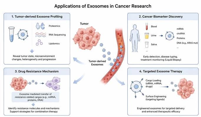
Research applications of exosomes in oncology cover four major directions: multi-omics analysis of tumor-derived exosomes, liquid biopsy biomarker screening, dissection of tumor drug resistance mechanisms, and targeted therapy using engineered exosomes.
Recommended Products:
Category | Product Name | Cat.No. | Specification | Key Application |
EV Reference / Research Control | ContExo® 293F Exosome Reference Material | CTE‑06 | 3E11 Particles/1 mL | Standardization of EV research, experimental control and method validation |
| ContExo® 293F Exosome Standard | CTE‑062 | 3E10 Particles/100 μL | Standard and control for EV research |
EV Isolation from Cell Culture Supernatant | Exosupur® Cell Supernatant Concentration Kit | ES8500 / ES850 | 500 mL / 50 mL | Concentrate EVs from tumor cell culture supernatant |
| Exosupur® Rapid Purification Kit for Cell Supernatant | ESS0500 / ESS050 | 500 mL / 50 mL | Rapid isolation and purification of tumor-cell-derived EVs |
EV Isolation from Tumor Tissue | Exosupur® Tissue Exosome Isolation Reagent | ES9T‑01 / ES9T‑02 | 0.5 mg×1 / 0.5 mg×5 | Isolate EVs derived from tumor tissue |
EV Isolation from Plasma/Serum | Exosupur® EV Purification Consumables — Plasma / Serum Samples | ES9P11e / ES9P14e | 1 mL×1 / 1 mL×4 | Isolation and purification of EVs from plasma and serum for tumor liquid biopsy research |
| Exosupur® EV Purification Consumables — Trace Plasma / Serum | ES9P021e / ES9P024e | 0.2 mL×1 / 0.2 mL×4 | EV isolation from trace clinical plasma and serum samples |
Further EV Purification | Exosupur® Exosome Dilution Column | ES9016 | 0.1 mL×6 | Further purification of EV samples to remove free dyes, antibodies and small molecules |
EV Characterization | ExosuperQC® Exosome 4‑Positive‑1‑Negative WB Identification Kit | EQC‑AP‑01 | 5T | Multi-marker EV characterization with CD9, CD63, CD81, ALIX and Calnexin |
EV Fluorescent Tracing | FluoExo® Exosome Tracing Kit — Green / Red / Near-Infrared | FE‑01‑01 / 02, FE‑02‑01 / 02, FE‑03‑01 / 02 | 1T / 5T | Fluorescent labeling of EVs for cellular uptake and trafficking tracing |
Exosome‑Free Cell Culture | ExoCCM® Exosome‑Free FBS | EDFBS50 / EDFBS5 | 50 mL / 5 mL | Reduce interference of serum-derived EVs in tumor cell experiments |
EV Nucleic Acid Engineering | ExoLoad® Nucleic Acid Loading Kit | ELSR‑06‑02 | 3E11×4T | Load nucleic acids including ASO, miRNA and siRNA into EVs |
| ExoLoad® Nucleic Acid Loading Kit | ELSR‑06‑03 | 1.5E12×6T | Large-scale EV nucleic acid loading and delivery research |
EV Targeting Engineering | ExoBrooch®‑iRGD | EA‑08‑5 | F5000 | Tumor-targeted surface modification of EVs |
EV Surface Modification | ExoCoupl® Exosome Bioorthogonal Conjugation Kit for Surface Modification | EC‑01‑02 | 6T | EV surface functionalization and modification with targeting molecules |