
info@mail.ucallm.com
1. Overview
Chandipura virus (CHPV) is a member of the genus Vesiculovirus within the family Rhabdoviridae and is an important causative agent of acute encephalitis syndrome (AES). It is an enveloped, negative-sense, single-stranded RNA virus with a bullet-shaped virion approximately 70 nm in diameter and 150–180 nm in length.
CHPV was first isolated in 1965 from two patients with dengue-like fever in the village of Chandipura, Nagpur, Maharashtra, India. The virus is transmitted primarily by sandflies, including Phlebotomus and Sergentomyia species. CHPV infection mainly affects children under 15 years of age, presenting as acute encephalitis that progresses rapidly, often leading to coma and death within 24–48 hours after symptom onset. Large outbreaks occurred in Andhra Pradesh and Gujarat, India, in 2003–2004, and in 2024 Gujarat again experienced an outbreak with 245 infections and 82 deaths, the largest outbreak in nearly two decades. CHPV is endemic in India and has also been detected sporadically in Sri Lanka, Nigeria, Kenya, and Senegal. Currently, no specific vaccine or antiviral drug is approved for CHPV.
2. Background Information
2.1 What is Chandipura Virus?
CHPV is an enveloped, negative-sense, single-stranded RNA virus belonging to the family Rhabdoviridae and genus Vesiculovirus. Its genome is approximately 11.1 kb and follows the canonical vesiculovirus genome organization 3′-N-P-M-G-L-5′, encoding five key proteins: nucleoprotein (N), phosphoprotein (P), matrix protein (M), glycoprotein (G), and large RNA-dependent RNA polymerase (L). The ribonucleoprotein (RNP) complex is formed by N protein encapsidating the genomic RNA, with P protein serving as the non-catalytic cofactor of L protein, together forming the viral transcriptase/replicase.
A 2025 phylogenetic study analyzed 23 complete CHPV genome sequences from 1966 to 2024 and found clear lineage separation between Indian and African strains, although overall genomic variability was low. Selection pressure analysis indicated that the coding regions are primarily under purifying selection. In the glycoprotein, all diversification sites (P272H, L424W, K503R) overlapped with predicted B-cell epitopes.
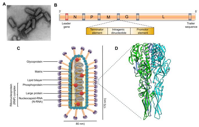
Structure and genome of CHPV (PMID: 39464728)
2.2 Chandipura Virus Genome & Classification
CHPV belongs to the family Rhabdoviridae and genus Vesiculovirus. The genome is a negative-sense single-stranded RNA of approximately 11.1 kb, encoding five structural proteins in the order 3′ to 5′. The International Committee on Taxonomy of Viruses (ICTV) classifies CHPV within the genus Vesiculovirus, which also includes vesicular stomatitis Indiana virus (VSIV) and vesicular stomatitis New Jersey virus (VSNJV).
The genomic RNA contains a 49-nucleotide leader sequence and five transcription units. The N protein encapsidates the genomic RNA to form the nucleocapsid, and the L and P proteins form the ribonucleoprotein complex.
2.3 What Are Chandipura Virus Structural Proteins?
CHPV encodes five structural proteins:
Protein | Full name | Main Function |
N | Nucleoprotein | Encapsidates the viral genomic RNA, forming ribonucleoprotein complexes (RNPs); the most abundantly expressed viral protein in infected cells |
P | Phosphoprotein | Non-catalytic cofactor of L protein, forming the viral transcriptase/replicase; multifunctional |
M | Matrix protein | Links RNPs to the viral envelope, mediates viral assembly and budding; a major driver of host shut-off in VSV |
G | Glycoprotein | Class I transmembrane glycoprotein, mediates receptor binding and low-pH-dependent membrane fusion |
L | Large protein | RNA-dependent RNA polymerase (RdRp), catalyses viral genome replication and transcription |
2.4 Mechanism of Cell Entry and Replication
2.4.1 Viral Attachment and Receptor Recognition
CHPV infection begins with the binding of the viral surface G glycoprotein to specific molecules on the host cell surface. Recent proteomic and biochemical studies suggest that α2-macroglobulin (A2M) and its receptor low-density lipoprotein receptor-related protein 1 (LRP1) may be involved in CHPV cell entry. A2M is a large secreted glycoprotein that is evolutionarily highly conserved, which may explain the broad host range of CHPV. LRP1 is particularly abundant in the brain and plays an important role in maintaining blood-brain barrier integrity, and may serve as an entry portal for CHPV into the central nervous system.
In addition, cell surface vimentin has been identified as participating in CHPV adsorption in Neuro-2a cells; studies show that vimentin interacts with CHPV and promotes virus binding to cells.
2.4.2 Viral Entry
The entry mechanism of CHPV is similar to that of VSV, entering host cells via clathrin-dependent endocytosis. A 2025 study showed that CHPV entry depends on clathrin and intact lipid rafts but not on caveolin-mediated endocytosis. The process is pH-dependent, with endosomal acidification triggering conformational changes in the G protein, exposing the fusion peptide, mediating fusion of the viral envelope with the endosomal membrane, and releasing the viral RNP into the cytoplasm.
2.4.3 Viral RNA Replication
After release of the viral RNP into the cytoplasm, the L protein (RdRp) and its cofactor P protein initiate genome transcription and replication. Transcription starts at the 3′ end of the genome and sequentially synthesizes five mRNAs. During replication, the L protein uses the positive-sense RNA as a template to synthesize full-length negative-sense genomic RNA. CHPV replication occurs entirely in the host cell cytoplasm.
A 2026 time-resolved transcriptomic study revealed the dynamic changes in host responses during CHPV infection and identified host-directed therapeutic targets.
2.4.4 Viral Assembly and Release
Newly synthesized N protein associates with progeny genomic RNA to form RNPs. The M protein connects RNPs to the inner leaflet of the cell membrane and promotes budding. The G protein is enriched at the plasma membrane, and the virus acquires its envelope by budding from the host cell membrane, forming new bullet-shaped virions that are released. The M protein has been demonstrated in VSV to be a major driver of virion assembly, linking the nucleocapsid to the inner leaflet of the plasma membrane through oligomerization.
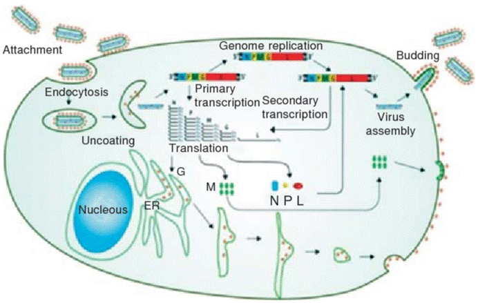
Life cycle of CHPV (PMID: 39464733)
2.5 Symptoms and Treatment
2.5.1 Clinical Symptoms
The incubation period of CHPV infection is not fully defined. The clinical presentation can be divided into two phases:
Prodromal phase: sudden onset of low-grade fever (lasting <3 days), headache, vomiting, diarrhea, and mild influenza-like symptoms with cough.
Central nervous system phase: rapid progression with altered consciousness, convulsions, followed by motor weakness, loss of consciousness, and ultimately coma and death. The case fatality rate ranges from 45% to 70%, with death often occurring within 24–48 hours after hospitalization. Approximately 90% of cases occur in children under 15 years of age.
Other clinical manifestations include tachycardia, tachypnea or dyspnea, irregular respiratory patterns, and auscultatory moist rales. Neurological examination may reveal hypotonia or hypertonia, decreased muscle strength, sluggish pupillary reflexes, papilledema, diminished or absent tendon reflexes, abnormal plantar reflexes, and decerebrate posturing.
2.5.2 Antiviral Treatment
Currently, no specific antiviral therapy is approved for CHPV. Treatment is primarily supportive:
l Hospitalization: intravenous fluids, antipyretics, anticonvulsants
l Management of cerebral edema: including ICU support
l Laboratory confirmation: by RT-PCR and IgM ELISA
2.6 Targets for Intervention
2.6.1 Viral Targets
Target | Protein | Biological Role |
G | Glycoprotein | Receptor binding and membrane fusion |
L | RNA polymerase (RdRp) | RNA transcription and replication |
M | Matrix protein | Viral assembly and budding |
N | Nucleoprotein | RNP formation |
2.6.2 Host Receptors & Entry Factors
Host Target | Type | Role in Infection |
LRP1/A2M | Receptor | Mediates CHPV entry into host cells |
Vimentin | Co-receptor | Promotes viral adsorption |
Clathrin and lipid rafts | Endocytic pathway | Required for viral entry |
GRP78 | Receptor | A2M receptor, interacts with CHPV |
2.6.3 Host Signaling Pathways
Pathway | Key Targets | Biological Role |
RIG-I/MDA5-MAVS | RIG-I, MDA5, MAVS | Viral RNA sensing |
TLR signaling | TLR3, TLR4 | Viral RNA sensing |
JAK–STAT | JAK1, STAT1/2 | IFN signaling |
NF-κB | IKK, p65 | Inflammatory response |
2.6.4 Immune & Inflammatory Targets
Pathway | Key Targets | Major Role |
IFN-α | Cytokine | Antiviral response |
IFN-β | Cytokine | Antiviral response |
IFN-γ | Cytokine | Cellular immunity |
IL-2 | Cytokine | T cell proliferation |
IL-6 | Cytokine | Inflammatory response |
TNF-α | Cytokine | Inflammation |
2.7 Vaccine Types and Development Progress
Currently, no CHPV vaccine is licensed worldwide. Several vaccine candidates are in development.
2.7.1 Recombinant Glycoprotein Vaccine
Early studies developed a CHPV vaccine based on recombinant G protein, which showed immunogenicity in mouse models when co-immunized with DPT.
2.7.2 Vero Cell Inactivated Vaccine
A Vero cell-based inactivated CHPV vaccine has produced adequate seroconversion in mice. Both major vaccine platforms (recombinant and inactivated candidates) have shown robust immunogenicity in mouse models.
2.7.3 Multi-Epitope Vaccine (Immunoinformatics Design)
A 2025 study published in PLoS One used immunoinformatics to design a multi-epitope vaccine combining 11 CTL epitopes, 2 HTL epitopes, and 1 linear B-cell epitope from the G protein, linked to the RS-09 adjuvant. The vaccine had a global population coverage of 99.79%, was soluble, non-allergenic, non-toxic, and highly antigenic. Molecular docking showed a docking score of −1157 kcal/mol with TLR4, and immune simulation showed increased IFN-γ and IL-2 concentrations after each injection.
2.7.4 mRNA Vaccine (Reverse Vaccinology Design)
A 2026 study used a combined structure-guided reverse vaccinology and immunoinformatics approach to develop a multi-epitope mRNA vaccine candidate targeting the G protein. Molecular docking showed a HADDOCK score of −22.6 ± 29.2 with TLR4, and MM-GBSA analysis estimated a binding free energy of −107.28 ± 26.65 kcal/mol.
2.7.5 X-ray Crystallography-Based Epitope Mapping
A 2025 study mapped epitopes of the CHPV G protein and RNA by X-ray crystallography, providing a structural basis for vaccine design.
2.8 Drugs
Currently, no specific small-molecule antiviral is approved for CHPV.
2.8.1 Virus-Targeted Drugs
Target/mechanism | Drug/candidate | Mechanism | Stage |
L protein (RdRp) | Favipiravir | RdRp inhibition | Animal model |
Ribavirin | Broad-spectrum antiviral | In vitro | |
Lycorine | Plant-derived alkaloid; targets the RdRp catalytic domain | In vitro | |
RNA interference | siRNA | Targets viral RNA | Preclinical |
2.8.2 Host-Targeted Drugs
Currently, no host-targeted drugs for CHPV have entered clinical research.
2.8.3 Drugs with Unclear or Non-Specific Targets
Currently, no drugs with unclear or non-specific mechanisms have entered clinical research for CHPV.
2.9 Epidemiology
l First identified: 1965, Chandipura village, Nagpur, Maharashtra, India
l Geographic distribution: endemic in India; sporadic detection in Sri Lanka, Nigeria, Kenya, and Senegal
l Vectors: sandflies, including Phlebotomus and Sergentomyia species
l High-risk populations: children under 15 years of age
l Outbreaks:
Ø 2003–2004: large outbreaks in Andhra Pradesh and Gujarat, India
Ø 2024: Gujarat, India, reported 245 infections and 82 deaths, the largest outbreak in nearly two decades
l Case fatality rate: 45%–70%
l Transmission routes: primarily through sandfly bites
2.10 Diagnosis
Diagnosis f CHPV infection relies on laboratory testing:
l Virus isolation: isolation of CHPV from clinical material (requires BSL-3 laboratory)
l Nucleic acid testing (NAT) : detection of CHPV RNA in clinical material (RT-PCR)
l Serology: detection of CHPV-specific IgM antibodies by ELISA
2.11 Prevention
l Vaccination: no licensed CHPV vaccine is currently available
l Avoid sandfly bites: use insect repellents, wear long-sleeved clothing, and use bed nets
l Vector control: reduce sandfly breeding sites and implement indoor residual spraying
l Surveillance and reporting: strengthen CHPV case surveillance and reporting
l Personal protective measures: wear appropriate personal protective equipment (PPE) when in sandfly-active areas
3. Related Products & Services
l Recombinant Antigens & Receptors
l Antibodies
l Detection Kits
l Recombinant Protein Expression Services
l Antibody Development Services
4. Resources
5. References
1. Bhatt, P. N., Rodrigues, F. M. (1967). Chandipura: a new Arbovirus isolated in India from patients with febrile illness. The Indian journal of medical research, 55(12), 1295–1305.
2. Rao, B. L., Basu, A., Wairagkar, N. S., et al. (2004). A large outbreak of acute encephalitis with high fatality rate in children in Andhra Pradesh, India, in 2003, associated with Chandipura virus. Lancet (London, England), 364(9437), 869–874.
3. Gurav, Y. K., Tandale, B. V., Jadi, R. S., et al. (2010). Chandipura virus encephalitis outbreak among children in Nagpur division, Maharashtra, 2007. Indian Journal of Medical Research, 132, 395–399.
4. Menghani, S., Chikhale, R., Raval, A., et al. (2012). Chandipura virus: An emerging tropical pathogen. Acta Tropica, 124(1), 1–14.
5. World Health Organization. (2024, August 23). Acute encephalitis syndrome due to Chandipura virus – India. Disease Outbreak News.
6. Karanam, S. K., Uppala, P. K., Kandra, N. V., et al. (2024). Chandipura virus: An emerging neurotropic pathogen and its public health implications in India. Discover Public Health, 21, 245.
7. Mallick, D., Yadav, U., Gupta, M., et al. (2025). The evolving landscape of Chandipura virus: A comprehensive account of outbreaks to recent advances. Virology, 608, 110541.
8. Lodha, L., Swaminathan, A., John, A., et al. (2025). Chandipura virus: A comprehensive review. Infectious diseases now, 55(8), 105179.
9. Pandey, N., Singh, S. K. (2025). Biological and pathogenic blueprint of Chandipura virus. Reviews in Medical Virology, 35(2), e70032.
10. Kuri, P. R., Sharma, A. (2026). Unravelling the genomic epidemiology and antigenic evolution of Chandipura virus: A genetically constrained neurotropic threat. PLoS neglected tropical diseases, 20(8), e0014565.
11. Jadi, R. S., Sudeep, A. B., Barde, P. V., et al. (2011). Development of an inactivated candidate vaccine against Chandipura virus (Rhabdoviridae: Vesiculovirus). Vaccine, 29(28), 4613–4617.
12. Padhi, A., Agarwal, A., Mishra, P., et al. (2024). Re-emerging Chandipuravesiculovirus: A cause of concern for global health. Virusdisease, 35(3), 385–399.
13. Basak, S., Mondal, A., Polley, S., et al. (2007). Reviewing Chandipura: a vesiculovirus in human epidemics. Bioscience reports, 27(4-5), 275–298.