
info@mail.ucallm.com
1. Overview
Coxiella burnetii is a Gram-negative, obligate intracellular bacterium of the family Coxiellaceae and genus Coxiella, and is the causative agent of Q fever. The bacterium exhibits a unique phase variation (Phase I / Phase II) and extreme environmental resilience, and is one of the pathogens with the lowest infectious dose known, with a 50% infectious dose (ID₅₀) of approximately 1.18 bacteria. Q fever has a global distribution, reported everywhere except New Zealand and Antarctica, with Germany reporting approximately 50–400 cases annually.
C. burnetii is classified by the U.S. CDC as a Category B biothreat agent. The bacterium primarily infects monocytes/macrophages, establishing a unique replicative niche within host cells—the Coxiella-containing vacuole (CCV)—which is derived from the lysosome but remodeled by the pathogen into a replication-permissive microenvironment. Clinical manifestations vary widely: approximately 50% of infections are asymptomatic, acute disease presents as a self-limiting flu-like illness, and chronic disease can lead to severe complications such as endocarditis, vascular infection, and chronic fatigue syndrome.
2. Background Information
2.1 What is Coxiella burnetii?
C. burnetii is a Gram-negative obligate intracellular bacterium with a unique biphasic life cycle. The small cell variant (SCV, also known as pseudospore) is the environmentally persistent form, extremely resistant to desiccation, heat, and disinfectants; the large cell variant (LCV) is the intracellular replicative form, slowly proliferating within the CCV (generation time approximately 11 hours). The bacterium utilizes a Dot/Icm type IVB secretion system (T4SS) to inject effector proteins into the host cell cytoplasm, manipulating endocytic trafficking and immune evasion to promote intracellular replication.
C. burnetii exists in two antigenic variants: Phase I, which expresses full-length lipopolysaccharide (LPS) and is the virulent form found in nature, and Phase II, which has truncated LPS and is significantly attenuated. LPS is the only defined Coxiella virulence determinant to date.
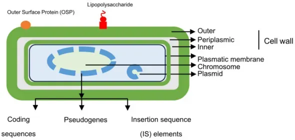
Structural features of Coxiella burnetii. (PMID: 40559597)
2.2 Genome & Classification
The C. burnetii genome is a circular chromosome of approximately 2.0 Mb. A 2026 study published in BMC Microbiology systematically analyzed the genomic characteristics and type IV effector protein repertoire of C. burnetii, finding that genomic features and effector protein repertoires depend primarily on Genomic Groups rather than on host species. Multiple genomic groups exist, with significant differences in effector protein composition between groups.
2.3 What Are C. burnetii Virulence Factors?
The virulence of C. burnetii relies primarily on the Dot/Icm type IVB secretion system (T4SS) and its secreted effector protein repertoire.
2.3.1 Dot/Icm T4SS
The T4SS is an essential virulence factor of C. burnetii, used to deliver a large arsenal of effector proteins into the host cell cytoplasm. A 2025 review published in mLife systematically described the mechanisms by which Dot/Icm effector proteins modulate host cell pathways. A 2024 study published in Pathogens demonstrated the critical role of the IcmE protein in C. burnetii virulence, intracellular replication, and inflammasome activation.
2.3.2 Key Effector Proteins
Effector Protein | Main Function |
EmcB | Deubiquitinase, inhibits RIG-I immune sensor signaling |
CvpB | Selectively removes lysosomal protease cathepsin B, reducing hostility of the intracellular niche |
CYP1B1-AS1/CYP1B1 axis | Regulates mitochondrial homeostasis via AHR signaling, inhibiting ROS and host cell death |
2.4 Mechanism of Infection and Pathogenesis
2.4.1 Invasion and CCV Formation
C. burnetii primarily infects the host through inhalation of contaminated aerosols. After SCVs are phagocytosed by alveolar macrophages, effector proteins secreted via the Dot/Icm T4SS remodel the phagosome into the CCV—a unique replicative niche derived from the lysosome but modified by the bacterium.
2.4.2 Lysosomal Protease Regulation
A 2025 study published in Nature Communications revealed that C. burnetii actively and selectively removes the key lysosomal protease cathepsin B from infected cells through the T4SS effector protein CvpB. Overexpression of cathepsin B leads to defects in CCV biogenesis and impaired bacterial replication, indicating that removal of this protease is a strategy to reduce the hostility of the intracellular niche. Additionally, infection induces the secretion of a broader cohort of lysosomal proteins, including cathepsin B, to the extracellular milieu via retrograde traffic.
2.4.3 Immune Evasion and Mitochondrial Regulation
A 2025 study published in Nature Communications found that C. burnetii infection activates the aryl hydrocarbon receptor (AHR) , driving transcription of the long non-coding RNA CYP1B1-AS1 and CYP1B1. Knockdown of CYP1B1-AS1 or CYP1B1 disrupts mitochondrial homeostasis, increases ROS and mitochondrial dysfunction, and exacerbates apoptosis during infection. The CYP1B1-AS1/CYP1B1 axis acts as a key regulator of mitochondrial homeostasis under AHR signaling, supporting an intracellular environment that benefits C. burnetii replication.
Furthermore, the C. burnetii effector protein EmcB is a deubiquitinase that prevents signaling by the RIG-I immune sensor, defining a broad family of bacterial deubiquitinases.
2.4.4 Immune Mechanisms of Persistent Infection
C. burnetii is described as a "stealth pathogen," and one of its hallmark features is its ability to evade innate immune activation, in contrast to other intracellular pathogens such as Mycobacterium tuberculosis and Legionella pneumophila that actively engage cytosolic sensors such as cGAS, NLRs, and TLRs to trigger robust inflammatory responses.
2.5 Transmission Routes and Process
Transmission of C. burnetii occurs primarily through the following routes:
l Inhalation: The primary route. Inhalation of aerosols containing SCVs, which can be spread by wind up to 10 kilometers from the source, so a clear exposure history is often lacking.
l Contact transmission: Contact with the products of parturition (placenta, amniotic fluid), urine, feces, or milk of infected animals; the primary route for high-risk occupational groups.
l Tick-borne transmission: Ticks are natural reservoirs and vectors of C. burnetii.
l Foodborne transmission: Consumption of unpasteurized dairy products.
Process: Inhalation of SCV aerosols → phagocytosed by alveolar macrophages → remodeling of phagosome into CCV via T4SS → slow replication within CCV → cell lysis and bacterial release → infection of neighboring cells → establishment of local infection or systemic dissemination.
Global outbreak updates 2025–2026: In a 2025 outbreak on a Boer goat farm in South Korea, 60 of 87 goats (69.0%) tested positive; an outbreak among staff at a South African veterinary hospital highlighted the importance of a One Health framework in Africa; Germany's Zollernalbkreis reported several cases of Q fever in 2026.
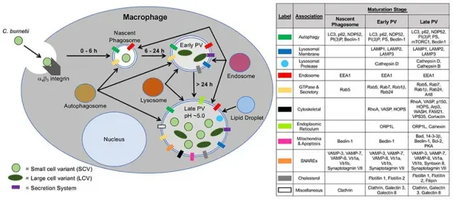
Formation of the C. burnetii parasitophorous vacuole. (PMID: 31574310)
2.6 Symptoms and Treatment
2.6.1 Clinical Symptoms
The incubation period of Q fever is typically 1–3 weeks. Approximately 50% of infected individuals are asymptomatic.
Type | Main Manifestations |
Acute Q fever | Abrupt onset of high fever, severe headache, flu-like illness, cough, pneumonia, hepatitis |
Chronic Q fever | Endocarditis, vascular infection, osteoarticular infection, chronic fatigue syndrome |
Infection during pregnancy | Mostly asymptomatic; may result in obstetric and fetal complications |
Diagnostic factors: Abrupt onset of high fever, flu-like illness, severe headache, cough, inspiratory crackles, rhonchi, or wheezing, hepatomegaly. Risk factors include male sex, age 30-70 years, immunosuppression, preexisting cardiac disease, vasculopathy, and pregnancy.
2.6.2 Treatment
Acute infection: Doxycycline short course (typically 200 mg/day for 14 days).
Chronic/persistent focalized infection: Doxycycline (100 mg twice daily) plus hydroxychloroquine (200 mg three times daily) for at least 18–24 months. Surgical resection of infected vascular tissue or prosthetic material may be required.
Pregnancy: Trimethoprim-sulfamethoxazole is recommended as an alternative to doxycycline.
Treatment limitations: Chronic Q fever requires prolonged antibiotic therapy, and durable cross-strain protection remains elusive.
2.7 Prevention
l Animal vaccination: COXEVAC (Phase I inactivated vaccine) is used in cattle and goats, reducing bacterial shedding, improving fertility, and reducing transmission risk to humans.
l Human vaccination: Q-Vax (formalin-inactivated Phase I whole-cell vaccine derived from the Henzerling strain) is licensed only in Australia and requires pre-screening of antibodies to avoid severe allergic reactions.
l Occupational protection: Abattoir workers, meat handlers, farmers, veterinarians, laboratory personnel, and military personnel are high-risk occupations.
l Environmental management: Strengthen farm biosecurity measures and properly dispose of products of parturition.
l Surveillance and reporting: Implement a One Health strategy integrating human, animal, and environmental surveillance.
2.8 Targets for Intervention
2.8.1 Pathogen Targets
Target | Protein/Pathway | Biological Function |
Dot/Icm T4SS | Type IVB secretion system | Effector protein delivery |
EmcB | Deubiquitinase | Inhibits RIG-I signaling |
CvpB | T4SS effector | Removes cathepsin B |
CYP1B1 | Mitochondrial regulatory enzyme | Maintains mitochondrial homeostasis |
2.8.2 Host Targets
Target | Type | Role in Infection |
AHR | Transcription factor | Drives CYP1B1-AS1/CYP1B1 transcription |
RIG-I | Immune sensor | Inhibited by EmcB deubiquitination |
Cathepsin B | Lysosomal protease | Removed by CvpB to facilitate bacterial replication |
2.9 Vaccine Types and Development Progress
2.9.1 Licensed Vaccines
Vaccine | Type | Features |
Q-Vax | Whole-cell inactivated vaccine | Licensed only in Australia; pre-screening required to avoid allergy |
COXEVAC | Phase I inactivated vaccine | Used in cattle and goats; reduces bacterial shedding |
2.9.2 Single-Dose Nanoparticle-Loaded Hydrogel Vaccine
A 2025 study published in Drug Delivery developed a nanoparticle-loaded hydrogel vaccine that co-delivers CBU1910 protein antigen and CpG1826 adjuvant for single-dose administration. The PLGA-PEG-PLGA (PPP) hydrogel forms a gel at physiological temperature and continuously releases protein for 8 weeks in vivo. The single-dose vaccine elicited stronger and more durable humoral immune responses in mice, yielding a balanced IgG2c/IgG1 response.
2.9.3 LEEI-Inactivated Vaccine
A 2026 study published in IJID One Health used low-energy electron irradiation (LEEI) to inactivate C. burnetii Phase II as a novel vaccine candidate. LEEI destroys nucleic acids while preserving immunogenic surface structures (LPS and proteins), achieving inactivation within minutes to hours without toxic chemicals. Preliminary results showed that LEEI-inactivated bacteria were efficiently taken up by primary bovine macrophages and induced MHC-II expression, indicating macrophage activation.
2.9.4 Computational Multi-Epitope Vaccine
A 2025 study published in ScienceDirect constructed a multi-epitope vaccine based on C. burnetii metabolic pathways, with the potential to elicit immune responses against C. burnetii.
2.10 Drugs
2.10.1 First-Line Treatment Regimens
Drug | Indication | Regimen |
Doxycycline | Acute infection | 200 mg/day, 14 days |
Doxycycline + hydroxychloroquine | Chronic/persistent infection | 100 mg bid + 200 mg tid, ≥18–24 months |
Trimethoprim-sulfamethoxazole | Pregnancy | Alternative regimen |
2.10.2 Drug Resistance
C. burnetii is naturally resistant to β-lactam antibiotics (due to its obligate intracellular nature), and aminoglycoside activity is reduced in the acidic CCV environment. No approved drugs exist to shorten the treatment course for chronic Q fever, and durable cross-strain protective vaccines remain elusive.
2.11 Recent Research
2.11.1 Molecular Epidemiology and Genomics
A 2026 study published in BMC Microbiology revealed that genomic characteristics and T4SS effector protein repertoires depend primarily on Genomic Groups rather than host species, which is of great significance for understanding the host adaptability and pathogenicity evolution of C. burnetii.
A 2025 systematic review published in Transboundary and Emerging Diseases integrated six key domains: ecological reservoirs, diagnostic strategies, molecular epidemiology, therapeutic challenges, vaccine development, and the One Health approach.
2.11.2 Diagnostic Technology Advances
Technology | Target | Features |
Real-time PCR (IS1111) | Multi-copy insertion sequence | Environmental dust sample detection, Ct<40 considered positive |
qPCR (com1) | Single-copy gene | Quantification of bacterial load |
Multiplex/digital PCR | Multiple targets | Improved sensitivity and specificity |
LAMP | Isothermal amplification | Suitable for point-of-care testing |
mNGS | Metagenomics | Identification of rare pathogens |
2.11.3 Environmental Surveillance
A 2025 study published in Applied and Environmental Microbiology established a serology combined with PCR analysis of environmental dust samples method to assess C. burnetii infection status on small ruminant farms. Farms were categorized into four levels based on bacterial load in dust: high (>1,000 GE/mg), moderate (100–1,000 GE/mg), low (<100 GE/mg), and negative.
3. Related Products & Services
l Recombinant Antigens & Receptors
l Antibodies
l Detection Kits
l Recombinant Protein Expression Services
l Antibody Development Services
4. Resources
5. References
1. Brangsch, H., Berens, C., Fuchs, S., et al. (2026). Genome characteristics and type IV effector protein repertoire of Coxiella burnetii depend rather on Genomic Groups than on host species. BMC microbiology, 26(1), 393.
2. Yuan, J., Zhang, Y., Song, L., et al. (2025). Modulation of host cell pathways by Coxiella burnetii Dot/Icm effectors. mLife, 4(6), 579–598.
3. Bird, L. E., Xu, B., Hobbs, A. D., et al. (2025). Coxiella burnetii manipulates the lysosomal protease cathepsin B to facilitate intracellular success. Nature communications, 16(1), 3844.
4. Arunima, A., Niyakan, S., Butler, S. M., et al. (2025). CYP1B1-AS1 regulates CYP1B1 to promote Coxiella burnetii pathogenesis by inhibiting ROS and host cell death. Nature communications, 16(1), 7493.
5. Palanisamy, R., Zhang, Y., Zhang, G. (2024). Role of Type 4B Secretion System Protein, IcmE, in the Pathogenesis of Coxiella burnetii. Pathogens (Basel, Switzerland), 13(5), 405.
6. Mezouar, S., Desnues, B., Bechah, Y., et al. (2025). Pathogenicity and Virulence of Coxiella burnetii: Focus on Q fever. Virulence, 16(1), 2495842.
7. Christodoulou, M., Papagiannis, D. (2025). Q Fever Vaccines: Unveiling the Historical Journey and Contemporary Innovations in Vaccine Development. Vaccines, 13(2), 151.
8. Gisbert, P., Hurtado, A., Guatteo, R. (2024). Efficacy and Safety of an Inactivated Phase I Coxiella burnetii Vaccine to Control Q Fever in Ruminants: A Systematic Review. Animals : an open access journal from MDPI, 14(10), 1484.
9. Wang, L., Ramirez, A., Felgner, J., et al. (2025). Development of a single-dose Q fever vaccine with an injectable nanoparticle-loaded hydrogel: effect of sustained co-delivery of antigen and adjuvant. Drug delivery, 32(1), 2476144.
10. García-Pérez, A. L., Zendoia, I. I., Ferrer, D., et al. (2025). Combination of serology and PCR analysis of environmental samples to assess Coxiella burnetii infection status in small ruminant farms. Applied and environmental microbiology, 91(10), e0093125.
11. Mohammadi, M. R., Moradkasani, S., Latifian, M., et al. (2025). Coxiella burnetii: Emerging threats, molecular insights, and advances in diagnosis and control measures. Journal of microbiological methods, 237, 107213.
12. Khademi, P., Tukmechi, A., Sgroi, G., et al. (2024). Molecular and genotyping techniques in diagnosis of Coxiella burnetii: An overview. Infection, genetics and evolution : journal of molecular epidemiology and evolutionary genetics in infectious diseases, 123, 105655.
13. Seshadri, R., Paulsen, I. T., Eisen, J. A., et al. (2003). Complete genome sequence of the Q-fever pathogen Coxiella burnetii. Proceedings of the National Academy of Sciences of the United States of America, 100(9), 5455–5460.
14. Toledo-Perona, R., Contreras, A., Gomis, J., et al. (2024). Controlling Coxiella burnetii in naturally infected sheep, goats and cows, and public health implications: a scoping review. Frontiers in veterinary science, 11, 1321553.
15. Centers for Disease Control and Prevention. (2025). Clinical Guidance for Q fever. CDC Q Fever Clinical Guidance.