
info@mail.ucallm.com
```
```
Disulfidptosis
1.1 Mechanism Overview
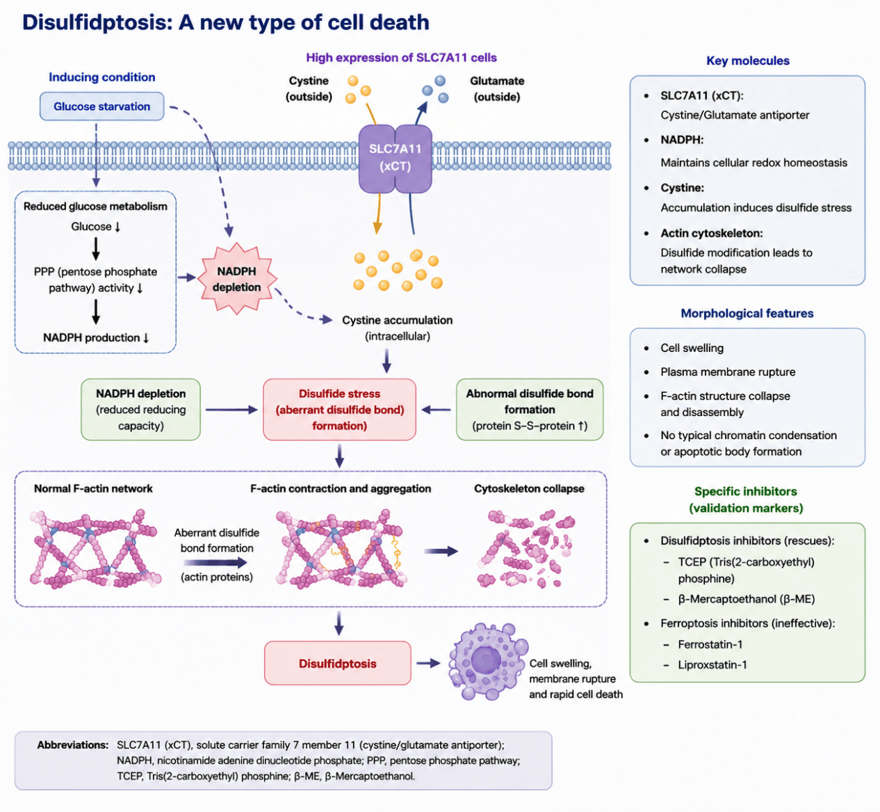
Disulfidptosis, a newly identified type of cell death, has attracted growing attention. Although its regulatory mechanisms have not been fully elucidated, current studies indicate that upregulation of SLC7A11 serves as a key trigger for disulfidptosis. As a transporter protein, SLC7A11 mediates the cellular uptake of extracellular cystine. Under glucose deprivation, elevated SLC7A11 leads to massive intracellular cystine accumulation, which induces disulfide stress and ultimately causes cell death. Accumulating evidence suggests that disulfidptosis plays vital roles in the initiation and progression of numerous diseases. It is closely linked to tumors, neurodegenerative disorders, cardiovascular diseases and liver diseases, and is also associated with many other illnesses. Accordingly, systematic research on disulfidptosis mechanisms bears prominent clinical significance. It not only helps uncover fundamental biological principles, but also offers novel strategies for disease prevention and treatment.
Disulfidptosis Model Diagram. Glucose deprivation induces NADPH depletion. In cells with high SLC7A11 expression, this triggers cystine accumulation and aberrant disulfide bond formation, provokes actin cytoskeleton collapse, and ultimately initiates disulfidptosis.
1.2 Target List
| Mechanism Module | Target (Gene/Protein) | Full Name | Function | Role in Disulfidptosis |
|---|---|---|---|---|
| Cystine Uptake and Transport System | SLC7A11 (xCT) | Solute carrier family 7 member 11 | Cystine/glutamate antiporter that mediates cystine uptake | Core determinant of disulfidptosis; high SLC7A11 expression promotes excessive cystine import and serves as an essential prerequisite for disulfidptosis |
| SLC3A2 (4F2hc) | Solute carrier family 3 member 2 | Chaperone subunit of System Xc⁻ | Forms the System Xc⁻ complex with SLC7A11 to facilitate cystine transport | |
| Glucose Metabolism and NADPH Generation | Glucose | Glucose | Primary cellular carbon source | Glucose deprivation reduces cellular reducing capacity and triggers disulfidptosis |
| PPP Pathway | Pentose phosphate pathway | Generates NADPH to maintain cellular reductive capacity | Provides NADPH to sustain intracellular redox balance and protect against disulfide stress | |
| G6PD | Glucose‑6‑phosphate dehydrogenase | Rate‑limiting enzyme of the pentose phosphate pathway | Controls PPP flux and NADPH production, influencing cellular sensitivity to disulfidptosis | |
| PGD | 6‑Phosphogluconate dehydrogenase | Key enzyme involved in PPP‑mediated NADPH synthesis | Contributes to NADPH generation and maintenance of cellular antioxidant capacity | |
| Cellular Redox Homeostasis | NADPH | Nicotinamide adenine dinucleotide phosphate | Major source of cellular reducing power | NADPH depletion drives thiol oxidation and aberrant disulfide bond formation, serving as a critical trigger of disulfidptosis |
| GSH | Glutathione | Major intracellular antioxidant maintaining thiol reduction state | Maintains protein thiols in a reduced state and buffers disulfide stress | |
| GCLC/GCLM | Glutamate‑cysteine ligase catalytic subunit / modifier subunit | Rate‑limiting enzyme complex for glutathione biosynthesis | Regulates GSH synthesis and determines cellular antioxidant capacity | |
| TXN | Thioredoxin | Component of the thioredoxin redox system | Mediates disulfide bond reduction and maintains protein thiol homeostasis | |
| Disulfide Bond Accumulation and Protein Crosslinking | Cystine | Cystine | Oxidized dimer of cysteine containing a disulfide bond | Accumulates after SLC7A11‑mediated import and increases intracellular disulfide burden |
| Cysteine | Cysteine | Reduced form of cystine and precursor for GSH synthesis | Normally utilized for glutathione synthesis after cystine reduction | |
| Protein disulfide bonds (S–S) | Protein disulfide bonds | Covalent bonds formed between cysteine residues | Aberrant accumulation induces protein crosslinking, protein dysfunction, and proteotoxic stress | |
| Cytoskeletal Collapse and Actin Remodeling | ACTB (β‑actin) | Actin beta | Major cytoskeletal actin isoform | Primary execution target of disulfidptosis; abnormal disulfide crosslinking disrupts cytoskeletal integrity |
| ACTG1 (γ‑actin) | Actin gamma 1 | Actin isoform involved in F‑actin network organization and stabilization | Maintains F‑actin network stability and cytoskeletal structure | |
| F‑actin | Filamentous actin | Polymerized actin cytoskeletal structure | Undergoes contraction, aggregation, and collapse, representing the major morphological hallmark of disulfidptosis | |
| PFN1 | Profilin 1 | Regulates actin polymerization dynamics | Modulates actin filament assembly and remodeling | |
| FLNA | Filamin A | Cytoskeletal crosslinking protein | Maintains cytoskeletal connectivity and mechanical stability |
1.3 Application Scheme
| Purpose | Markers/Assays |
|---|---|
| Target verification | SLC7A11 |
| Metabolic stress evaluation | NADPH/NADP⁺ |
| Disulfide stress detection | Protein‑S‑S |
| Phenotypic identification | ACTB + F‑actin |
| Cell death confirmation | PI / LDH |
| Exclusion of ferroptosis | GPX4 + Ferrostatin‑1 |
1.4 References
- Liu X, Nie L, Zhang Y, et al. Actin cytoskeleton vulnerability to disulfide stress mediates disulfidptosis[J]. Nature Cell Biology, 2023, 25(3): 404‑414. DOI: 10.1038/s41556‑023‑01091‑2.
- Zheng T, Li X, Chen Y, et al. Disulfidptosis: A new form of programmed cell death[J]. Journal of Experimental & Clinical Cancer Research, 2023, 42: 137.
- Machesky LM. Deadly actin collapse by disulfidptosis[J]. Nature Cell Biology, 2023, 25: 353‑354.
- Koppula P, Zhuang L.Disulfidptosis: Disulfide stress‑induced cell death[J]. Trends in Cancer, 2023, 9(10): 857‑860.