
info@mail.ucallm.com
1. Overview
Eastern Equine Encephalitis Virus (EEEV) is a member of the genus Alphavirus within the family Togaviridae and is the causative agent of Eastern Equine Encephalitis (EEE). It is an enveloped, positive-sense, single-stranded RNA virus with a spherical virion approximately 60–65 nm in diameter.
EEEV is the most deadly encephalitic arbovirus infection in North America, with a case fatality rate of approximately 30%. Approximately 99% of human infections are asymptomatic or mild, but about 5% of cases progress to severe neuroinvasive disease. EEEV is maintained in a natural cycle primarily between Culiseta melanura mosquitoes and passerine birds in freshwater hardwood swamps. Humans and horses serve as dead-end hosts, and transmission to humans depends on "bridge" mosquitoes—including Aedes, Culex, and Coquillettidia species—that feed on both birds and mammals.
2. Background Information
2.1 What is EEEV?
EEEV is an enveloped, positive-sense, single-stranded RNA virus belonging to the family Togaviridae and genus Alphavirus. The genome is a positive-sense single-stranded RNA of approximately 11.7 kb, containing a 5′ non-structural open reading frame and a 3′ structural open reading frame. The 5′ ORF is translated directly into a polyprotein that is cleaved into four non-structural proteins (nsP1–nsP4), which together form the viral replication complex, mediating RNA synthesis and contributing to evasion of host antiviral responses. nsP1 possesses capping activity, nsP2 possesses RNA triphosphatase and protease activities, nsP3 participates in replication complex assembly, and nsP4 is the RNA-dependent RNA polymerase. The 3′ ORF is translated from the 26S subgenomic RNA into five structural proteins (C, E3, E2, 6K, E1). The EEEV complex includes lineage I (EEEV) and lineages II–IV, which have been reclassified as Madariaga virus (MADV).
In 2025, Bandyopadhyay et al. resolved the asymmetric structural transitions of EEEV at 40°C and pH 5.6 by cryo-EM at 3.8 Å resolution, revealing the conformational changes in the glycoproteins and nucleocapsid core of a prototypical alphavirus prior to membrane fusion.
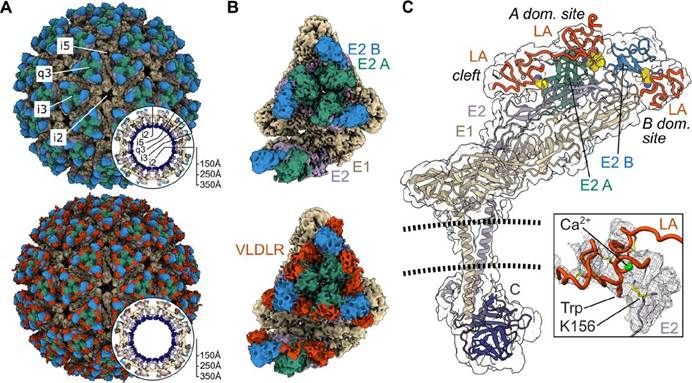
Cryo-EM structure of EEEV PE–6 in complex with VLDLR (PMID: 38176410)
2.2 EEEV Genome & Classification
The EEEV genome is approximately 11.7 kb, encoding a non-structural polyprotein p1234 and a structural polyprotein. The non-structural polyprotein is cleaved into four non-structural proteins: nsP1, nsP2, nsP3, and nsP4. The structural polyprotein is translated from the 26S subgenomic RNA into five structural proteins: capsid (C), E3, E2, 6K, and E1.
EEEV belongs to the genus Alphavirus. The EEEV antigenic complex includes four groups: Group I consists of only EEEV, endemic in North America and the Caribbean and causing most human disease cases; Groups IIA, IIB, and III are caused by the related Madariaga virus (MADV, formerly known as South American eastern equine encephalitis virus) and cause primarily equine illness in Central and South America.
2.3 What Are EEEV Structural Proteins?
EEEV encodes five structural proteins:
Protein | Full name | Main Function |
C | Capsid | Helps pack and protect the RNA genome; functions as a potent IFN antagonist protein by globally inhibiting host cell gene expression |
E3 | Envelope protein 3 | Signal peptide; involved in viral assembly |
E2 | Envelope glycoprotein 2 | Contains the binding site for the cellular attachment factor heparan sulfate; mediates viral binding to host cell receptors |
6K | 6K protein | Plays important roles in infection |
E1 | Envelope glycoprotein 1 | Class II fusion protein; mediates fusion of the viral membrane with the host cell membrane |
TF | TF protein | Plays important roles in infection |
2.4 Mechanism of Cell Entry and Replication
2.4.1 Viral Attachment and Receptor Recognition
EEEV infects cells through adhesive interactions between its surface glycoproteins and membrane proteins on the cell surface. The virus can bind several different molecules, including the very low density lipoprotein receptor (VLDLR) , apolipoprotein E receptor 2 (ApoER2/LRP8) , and heparan sulfate.
A 2024 study published in Cell resolved multiple cryo-EM structures of EEEV-VLDLR complexes, showing that EEEV uses multiple sites (E1/E2 cleft and E2 A domain) to engage more than one LA domain simultaneously. However, no single LA domain is necessary or sufficient to support efficient EEEV infection. All EEEV strains show conservation of two VLDLR binding sites, while the EEEV PE-6 strain and a few other EEE complex members feature a single amino acid substitution that enables binding of LA domains to an additional site on the E2 B domain. This multi-site, multivalent binding mode allows EEEV to adapt to diverse hosts and is one of the molecular bases for its high pathogenicity.
2.4.2 Viral Entry
EEEV enters host cells via endocytosis. After the viral E2 protein binds to host receptors, the virus particle is internalized. Within the endosome, the decrease in pH triggers conformational changes in the envelope proteins, exposing the fusion protein E1, which inserts into the endosomal membrane, creating a pore and releasing the viral capsid into the cytosol. The capsid is then disassembled by host ribosomes, releasing the positive-sense viral RNA.
A 2025 structural study published in Nature Communications provided snapshots of the conformational changes in EEEV at 40°C or pH 5.6, revealing the structural transitions in the viral glycoproteins and nucleocapsid core prior to fusion. The study revealed transitional epitopes that are inaccessible at room temperature or neutral pH through antibody binding studies, providing new structural insights into the EEEV entry mechanism.
2.4.3 Viral RNA Replication
After release into the cytoplasm, the positive-sense viral RNA can be directly read by ribosomes, leading to the production of the non-structural polyprotein. The non-structural polyprotein is cleaved into nsP1–nsP4, which assemble into the replication complex. The replication complex forms a replication spherule in ER-derived membrane vesicles.
The EEEV replication cycle is extremely rapid. A 2025 rule-based kinetic model predicted that EEEV replicates efficiently and produces progeny viral particles as soon as 3–4 hours post-infection. The model revealed a key characteristic: further cleavage of the replicase complex signals a shift from negative-sense to positive-sense synthesis—first nsP1 is cleaved from nsPs2-3, and second, the junction between nsP2 and nsP3 is cleaved, leading to four individual proteins. This process is defined by the rate constant r_replicase_cleave and yields the replicase form responsible for positive-sense synthesis, C1234.
Within the replication spherule, free genome (posRNA) binds with the complementary template strand (negRNA) forming a double-stranded RNA intermediate (dsRNA), governed by the rate constant r_rna_bind. nsP2 in the replicase complex has helicase activity, and the negative strand (free or in-duplex) can be used as a template to synthesize positive-sense RNA products. C1234 can initiate transcription through four mechanisms, based on the form of the negative-sense template (free negRNA vs in-duplex dsRNA) and which of the two promoters it binds to (full-length genome and subgenomic). The model includes two parameters—ka_transcribe_neg_4pos and ka_transcribe_neg_4sg—defining genome and subgenome promoter affinities. The relative subgenome and genome production of EEEV is asymmetric in favour of subgenome as the respective promoters are bound at distinct sites on the RNA-dependent RNA polymerase.
2.4.4 Viral Assembly and Release
Newly synthesized structural proteins and genomic RNA assemble and bud at the peripheral membrane to form progeny virions. The cytoplasmic domain of the E2 glycoprotein interacts with the nucleocapsid, while its ectodomain is responsible for binding a cellular receptor.
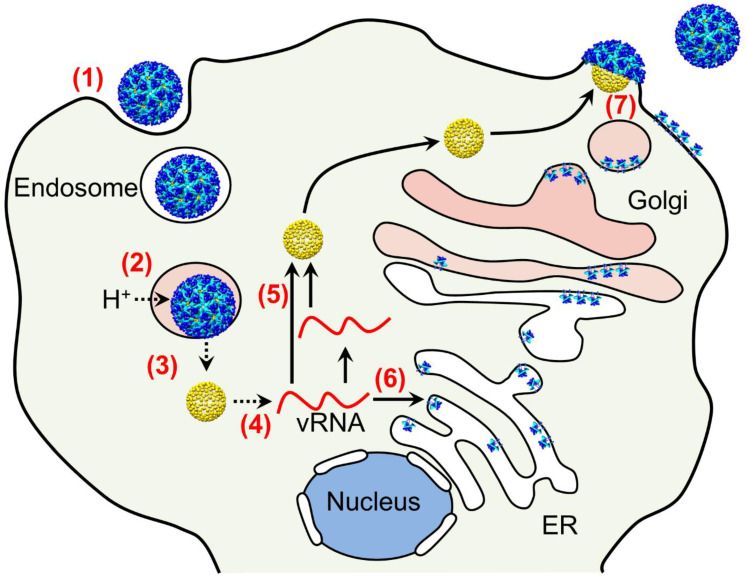
Entry and replication cycle of alphaviruses. (PMID: 34451437)
2.5 Symptoms and Treatment
2.5.1 Clinical Symptoms
The incubation period for EEEV infection is typically 4–10 days. Most infections are asymptomatic or present as a mild febrile illness. Symptomatic patients may develop fever, chills, malaise, arthralgia, and myalgia. Approximately 5% of cases progress to severe neuroinvasive disease, presenting with encephalitis, with symptoms including high fever, headache, altered mental status, seizures, and focal neurological deficits.
The case fatality rate is approximately 30%, and a high proportion of survivors experience long-term neurological sequelae. In the 2019 U.S. EEE outbreak, 19 of 38 confirmed cases died (case fatality rate 50%), and survivors experienced neurological sequelae. US median data from 2014–2018 showed a neuroinvasive rate of approximately 99%, with 41% of these patients dying.
People at highest risk are those living in or visiting endemic forested areas near swamps or marshes and those whose occupations (e.g., agricultural work) or hobbies (e.g., camping, gardening) involve outdoor exposure.
2.5.2 Antiviral Treatment
Currently, no specific antiviral therapy is approved for EEEV, and clinical management is supportive. Patients with severe meningeal symptoms often require pain control for headaches and antiemetic therapy, as well as rehydration for associated nausea and vomiting. Patients with encephalitis require close monitoring for the development of elevated intracranial pressure, seizures, and inability to protect their airway.
2.6 Targets for Intervention
2.6.1 Viral Targets
Target | Protein | Biological Role |
E1/E2 | nvelope glycoproteins | Receptor binding & membrane fusion |
nsP2 | on-structural protein 2 | Protease, helicase, host immune antagonism |
nsP4 | Non-structural protein 4 | RNA-dependent RNA polymerase |
nsP1 | Non-structural protein 1 | RNA capping, membrane association |
C | Capsid | Nucleocapsid formation, IFN antagonism |
2.6.2 Host Receptors & Entry Factors
Host Target | Type | Role in Infection |
VLDLR | Receptor | Primary entry mediator, engaging multiple LA domains at multiple sites |
ApoER2/LRP8 | Receptor | Accessory entry receptor |
Heparan sulfate | Attachment factor | Viral attachment, promotes neurovirulence |
2.6.3 Host Signaling Pathways
Pathway | Key targets | Biological role |
TLRs/RLRs pathways | TLRs, RIG-I-like receptors | Viral RNA sensing, activating innate immune responses of dendritic cells and fibroblasts |
Inflammatory signaling | IL-6, TNF-α, CCL2, CXCL10 | Synthesis of pro-inflammatory cytokines and chemokines |
IFN signaling | IFN-α/β | Antiviral response; EEEV almost completely avoids serum IFN-α/β induction through altered myeloid cell tropism |
miR-142-3p pathway | miR-142-3p | Binds to EEEV 3′ non-translated region, suppressing virus replication and systemic IFN-α/β induction |
2.6.4 Immune & Inflammatory Targets
Target | Category | Major role |
IFN-α | Cytokine | Antiviral response; EEEV evades its induction via altered myeloid cell tropism |
IFN-β | Cytokine | Antiviral response |
IL-6 | Cytokine | Inflammatory response; lower levels correlated with BDGR-164 protection |
IL-1β | Cytokine | Inflammasome signaling; released by infected neurons, activating glial cells |
TNF-α | Cytokine | Inflammation; lower levels correlated with BDGR-164 protection |
CCL2 | Chemokine | Immune-cell recruitment |
CXCL10 | Chemokine | Immune-cell recruitment; released by astrocytes, promoting neuroinflammation |
RANTES | Chemokine | Immune-cell recruitment; lower levels correlated with BDGR-164 protection |
2.7 Vaccine Types and Development Progress
Currently, no publicly licensed EEEV vaccine is available, and at-risk individuals are restricted to superseded vaccines. Equine EEEV vaccines are licensed and widely used, but no approved human vaccines exist.
2.7.1 MVA-BN-WEV (Recombinant Live-Attenuated Vaccine)
MVA-BN-WEV is a recombinant multivalent vaccine based on the Modified Vaccinia Ankara vector, encoding the envelope surface proteins of three equine encephalitis viruses (WEEV, EEEV, VEEV), intended for the prevention of disease induced by these three viruses.
l Phase 1 clinical trial (NCT04131595): 45 healthy adults were enrolled in 3 dose groups (1×10⁷, 1×10⁸, 2×10⁸ Inf.U), received 2 doses 4 weeks apart, and were monitored for 6 months. The safety profile was acceptable at all doses. The highest dose resulted in seroconversion of all (100%) participants for WEEV and VEEV and 92.9% for EEEV, 2 weeks following the second vaccination, with durability observed for 6 months.
l Phase 2 clinical trial (NCT06899802): initiated in March 2025, planning to enroll 411 healthy adults aged 18–50 to evaluate optimal dose, dosing regimen, reactogenicity, safety, and immunogenicity.
2.7.2 Self-Replicating RNA Platform
In 2025, researchers developed a self-replicating RNA platform demonstrating enhanced efficacy for EEEV vaccines in low-dose or single-dose applications, with potential for combination cancer immunotherapies.
2.7.3 Trivalent Virion-Based Vaccine
A 2025 study published in Science Translational Medicine developed an inactivated trivalent virion-based vaccine against VEEV, EEEV, and WEEV. The vaccine uses a combination of attenuated chimeric Sindbis-VEEV/EEEV/WEEV viruses inactivated with H₂O₂. The alum-adjuvanted vaccine conferred complete protection in mice with two doses, and substantial or complete protection in nonhuman primates at low and high doses, respectively.
2.7.4 mRNA Vaccine
The mRNA platform is an emerging direction for EEEV vaccine development. A 2026 review published in Vaccines noted that global climate change and ecological disruption drive a need for public access to safe and effective vaccines against EEEV, and novel platforms such as mRNA and viral vectors may be able to achieve this.
2.8 Drugs
Currently, no specific small-molecule antiviral is approved for EEEV. Several antiviral candidates have shown significant progress.
2.8.1 Virus-Targeted Drugs
Target | Drug/candidate | Mechanism | Stage |
Broad-spectrum antiviral | BDGR-164 | Novel quinazolinone with nanomolar potency against VEEV, EEEV, WEEV | Mouse model |
BDGR-251 | Novel directly acting antiviral with outstanding efficacy in both pre-exposure prophylaxis and post-exposure therapy | Mouse model | |
nsP2 protease | nsP2 protease inhibitors | Block New World alphavirus replication | Mouse model |
Viral late stage | Fluticasone propionate | Inhibits late stage of EEEV viral life cycle, blocking viral RNA replication and protein synthesis | In vitro |
Ubiquitin-proteasome pathway | NSC697923, BARM, OMA | Ubiquitin-mediated signaling pathway inhibitors, broad-spectrum against VEEV and EEEV | In vitro |
2.8.2 Host-Targeted Drugs
Target | Drug/candidate | Mechanism | Stage |
Pyrimidine synthesis | A771726 | Leflunomide active metabolite | In vitro |
Interferon pathway | Type I interferon | Antiviral immune modulation | Basic research |
2.9 Epidemiology
l First identified: 1930s, Americas
l Geographic distribution: EEEV is primarily found in the eastern and southeastern coastal regions of the United States and the Caribbean. In recent years, cases have expanded westward, with cases reported in Arizona in 2023 and North Dakota in 2020
l Vectors: Culiseta melanura is the principal maintenance vector, transmitting the virus among birds in freshwater hardwood swamps; Aedes, Culex, and Coquillettidia species serve as bridge vectors
l Natural cycle: mosquitoes ↔ passerine birds; humans and horses are dead-end hosts
l 2025 US outbreak: CDC ArboNET reported 3 human EEE cases (as of 16 December 2025), including 1 in Maine (death), 1 in New York (death), and 1 in South Carolina (death), all neuroinvasive cases, with a total of 3 deaths
l 2025 Ontario, Canada: reported its first human EEE case
l Historic data (2003–2024) : 215 cumulative human EEE cases, including 84 deaths
l 2019 US outbreak: 38 confirmed cases and 19 deaths, the largest outbreak in recent years
l Cyclicity: EEEV exhibits pronounced 2–3 year cyclical outbreak patterns in the northeastern United States, linked to shifts in mosquito feeding preferences between hatch-year and adult avians
l Organ transplant transmission: EEEV has been documented to be transmitted through solid organ transplantation, with one organ donor transmitting the infection to three organ transplant recipients
2.10 Diagnosis
Diagnosis of EEEV infection relies on laboratory testing:
l Virus isolation: isolation of EEEV from clinical material (requires high-containment laboratory)
l Nucleic acid testing (NAT) : detection of EEEV RNA in clinical material (RT-PCR), appropriate for immunocompromised patients
l Serology: detection of EEEV-specific IgM antibodies in serum or CSF, the front-line diagnostic assay; positive IgM results should be confirmed by neutralizing antibody testing
CDC recommends that combined with a consistent clinical presentation in an endemic area, a rapid and accurate diagnosis of EEE can be made by the detection of EEEV-specific IgM antibodies in serum or CSF. EEEV IgM testing is available commercially, at some state health departments, and at CDC. Some patients who are severely immunocompromised might require molecular (e.g., RT-PCR) testing for diagnosis. EEE is a nationally notifiable condition in the United States, and all cases should be reported to state or local public health authorities.
2.11 Prevention
l Vaccination: No licensed human EEEV vaccine is currently available; equine EEEV vaccines are licensed and widely used for horses
l Avoid mosquito bites: in the absence of a vaccine, prevention of EEE depends on personal protective measures to decrease exposure to infected mosquitoes
Ø Use insect repellent
Ø Wear long-sleeved shirts and pants
Ø Treat clothing and gear with 0.5% permethrin
Ø Take steps to control mosquitoes indoors and outdoors, including emptying water-holding containers
l Personal protective measures: wear appropriate personal protective equipment (PPE) when in mosquito-active areas
l Blood safety: people recently diagnosed with EEE should not donate blood for 120 days (4 months) after their illness; EEEV infections temporally associated with organ transplantation or blood transfusion should be reported promptly
l Surveillance and reporting: EEEV is a notifiable disease in the United States; strengthen surveillance of human cases, mosquito vectors, and avian hosts
3. Related Products & Services
l Recombinant Antigens & Receptors
l Antibodies
l Detection Kits
l Recombinant Protein Expression Services
l Antibody Development Services
4. Resources
5. References
1. Bandyopadhyay, A., Williamson, L. E., Buchman, C. D., et al. (2026). Asymmetric structural transitions in the icosahedral organization of Eastern equine encephalitis virus. Nature communications, 17(1), 8230.
2. Adams, L. J., Raju, S., Ma, H., et al. (2024). Structural and functional basis of VLDLR usage by Eastern equine encephalitis virus. Cell, 187(2), 360–374.e19.
3. Yang, P., Li, W., Fan, X., et al. (2024). Structural basis for VLDLR recognition by eastern equine encephalitis virus. Nature communications, 15(1), 6548.
4. Bowers, D. F., Hightower, L. E. (2022). Eastern Equine Encephalitis Virus Taxonomy, Genomics, and Evolution. Journal of Medical Entomology, 59(1), 14–23.
5. Zubair, A. S., McAlpine, L. S., Gobeske, K. T. (2024). Virology, ecology, epidemiology, pathology, and treatment of eastern equine encephalitis. Journal of the neurological sciences, 457, 122886.
6. Aguilar, P. V., Weaver, S. C., Basler, C. F. (2007). Capsid protein of eastern equine encephalitis virus inhibits host cell gene expression. Journal of virology, 81(8), 3866–3876.
7. Gardner, C. L., Burke, C. W., Tesfay, M. Z.,et al. (2008). Eastern and Venezuelan equine encephalitis viruses differ in their ability to infect dendritic cells and macrophages: impact of altered cell tropism on pathogenesis. Journal of virology, 82(21), 10634–10646.
8. Trobaugh, D. W., Sun, C., Bhalla, N., et al. (2019). Cooperativity between the 3' untranslated region microRNA binding sites is critical for the virulence of eastern equine encephalitis virus. PLoS pathogens, 15(10), e1007867.
9. Fierro, C., Weidenthaler, H., Vidojkovic, S., et al. (2024). Safety and immunogenicity of a novel trivalent recombinant MVA-based equine encephalitis virus vaccine: A Phase 1 clinical trial. Vaccine, 42(10), 2695–2706.
10. Lam, L. K. M., Gilliland, T., Jr, Dunn, M., et al. (2025). An inactivated trivalent virion-based vaccine protects against aerosol challenge with encephalitic alphaviruses in mice and macaques. Science translational medicine, 17(818), eadv7079.
11. Williams, E. P., Xue, Y., Yang, D., et al. (2025). Broad spectrum antiviral BDGR-164 provides protection against lethal neurotropic alphavirus infection in mice. Antiviral research, 240, 106206.
12. Lindsey, N. P., Martin, S. W., Staples, J. E., et al. (2020). Notes from the Field: Multistate Outbreak of Eastern Equine Encephalitis Virus - United States, 2019. MMWR. Morbidity and mortality weekly report, 69(2), 50–51.
13. Brown, S. C., Cormier, J., Tuan, J., et al. (2021). Four Human Cases of Eastern Equine Encephalitis in Connecticut, USA, during a Larger Regional Outbreak, 2019. Emerging infectious diseases, 27(8), 2042–2051.
14. Armstrong, P. M., Andreadis, T. G. (2010). Eastern equine encephalitis virus in mosquitoes and their role as bridge vectors. Emerging infectious diseases, 16(12), 1869–1874.
15. Lambert, A. J., Martin, D. A., Lanciotti, R. S. (2003). Detection of North American eastern and western equine encephalitis viruses by nucleic acid amplification assays. Journal of clinical microbiology, 41(1), 379–385.
16. CDC. (2025). Clinical Testing and Diagnosis for Eastern Equine Encephalitis.
17. CDC. (2024). Treatment and Prevention of Eastern Equine Encephalitis.