
info@mail.ucallm.com
Exosomes are a subset of Extracellular Vesicles (EVs). Classically, exosomes originate from the endosomal system and are released into the extracellular environment through the formation, trafficking and exocytosis of Multivesicular Bodies (MVBs). Together with other types of EVs including microvesicles (ectosomes) and apoptotic bodies, they constitute the complex EV repertoire.
With advances in high‑throughput sequencing, proteomics, lipidomics and other technologies, accumulating studies have revealed that EVs carry a wide range of biologically active molecules such as proteins, lipids, mRNAs, miRNAs, lncRNAs and DNA. Via cellular uptake and molecular delivery, EVs participate in intercellular communication, immune regulation, tissue repair, as well as the onset and progression of diseases.
Benefiting from favorable biocompatibility, intrinsic cellular‑uptake properties and amenability to engineering modification, EVs have been widely explored in disease‑mechanism investigation, biomarker discovery, drug delivery and precision therapeutics. Characterization and modulation of EV sources, isolation‑purification workflows, surface properties and cargo contents further enable the construction of EV research systems tailored to diverse experimental requirements.
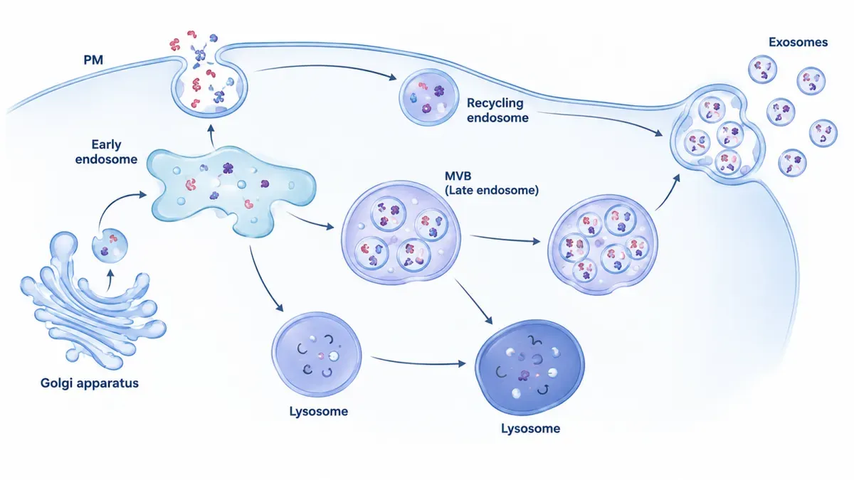
Biogenesis, cargo composition and intercellular‑communication mechanisms of exosomes. Donor cells generate multivesicular bodies (MVBs) via early and late endosomes. Exosomes (30–150 nm) are released upon fusion of MVBs with the plasma membrane. Exosomes are characterized by membrane marker proteins including CD9, CD63 and CD81, and encapsulate biologically active cargos such as proteins, lipids, DNA, mRNA, miRNA and lncRNA. Exosomes act on recipient cells through receptor‑ligand binding, endocytosis or membrane fusion, and mediate diverse biological effects including immune regulation, tissue repair and regeneration, disease progression and metastasis, signal transduction, as well as gene regulation.
To meet end‑to‑end research requirements, we have built a one‑stop total‑solution for exosome studies. It covers eight product portfolios: reference standards, multi‑sample EV isolation and purification, standardized characterization and validation, in‑vivo/cellular fluorescent tracing, exosome‑free cell‑culture systems, nucleic‑acid loading, targeting modification, and surface engineering. It delivers a full‑loop closed‑service workflow spanning sample processing, quality‑control validation, functional assays, and engineered drug‑loading modification. The solution is suitable for basic research, mechanistic investigation, vector development and pre‑clinical studies.
Exosome Research Product Portfolio:
Research Stage | Core Research Content | Key Products |
Cell Culture and Exosome Biogenesis | Donor cell culture, exosome biogenesis and release | Cell culture media, FBS / exosome‑depleted FBS, cytokines, cell‑culture consumables, transfection reagents |
Exosome Isolation and Purification | Exosome extraction, purification and impurity removal | Exosome extraction kits, ultrafiltration tubes, SEC columns, centrifugation consumables, low‑binding consumables |
Exosome Identification and Characterization | Detection of particle size, concentration, morphology and specific biomarkers | CD9/CD63/CD81 antibodies, ELISA kits, Western Blot reagents, flow‑cytometry antibodies, NTA‑related products, TEM sample‑preparation reagents |
Exosome Labeling and Cellular Uptake | Exosome tracing, target‑cell uptake and intracellular trafficking | Fluorescent probes, membrane dyes, cell‑tracing dyes, fluorescent labeling reagents, cell‑imaging products |
Cargo Analysis and Functional Validation | Analysis of protein, RNA, miRNA and other cargos; biological‑function studies | RNA‑isolation reagents, qPCR reagents, ELISA kits, antibodies, cell‑viability assay reagents, reporter‑gene assay kits, cytokines |
Disease Model and Applied Research | Research on oncology, immunology, bone metabolism, tissue repair and drug development | Organoids, stem cells, immune cells, tumour‑cell models, cell‑death detection reagents, transfection reagents, imaging products |