
info@mail.ucallm.com
1 Exosome Biogenesis
Exosomes are mainly generated via the canonical endosomal pathway, starting with plasma membrane endocytosis. First, the plasma membrane invaginates to form early endosomes. This process not only mediates the uptake of extracellular substances, but also provides the initial membrane structure for exosome biogenesis. As endosomes mature, early endosomes gradually transform into late endosomes, which further develop into multivesicular bodies (MVBs).
During MVB formation, the endosomal membrane invaginates inward to generate numerous small vesicles loaded with specific biomolecules, known as intraluminal vesicles (ILVs). This process is regulated by multiple molecular machineries, including the ESCRT (Endosomal Sorting Complex Required for Transport) complex, tetraspanins (such as CD9, CD63, CD81), lipid rearrangement and Rab GTPases. Meanwhile, cells selectively package specific functional molecules into ILVs, including proteins, lipids, mRNAs, miRNAs, lncRNAs and other nucleic acid molecules, thereby endowing exosomes with distinct biological functions.
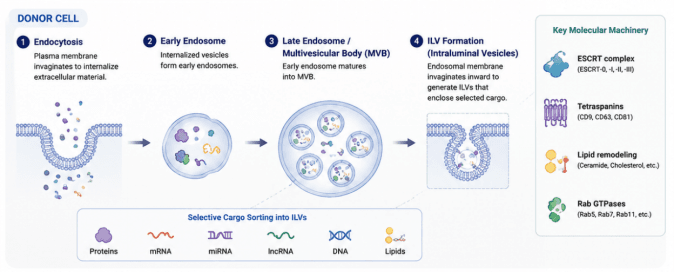
Endosomal pathway-mediated exosome biogenesis. The donor cell first internalizes extracellular substances through endocytosis to form early endosomes, which further mature into late endosomes/multivesicular bodies (MVBs). During inward budding of the endosomal membrane to form intraluminal vesicles (ILVs), bioactive components including proteins, mRNAs, miRNAs, lncRNAs, DNAs and lipids are selectively sorted into ILVs. This process relies on core molecular machineries such as the ESCRT complex, tetraspanins (CD9, CD63, CD81), lipid remodeling and Rab GTPases. After MVBs fuse with the plasma membrane, ILVs are released extracellularly as mature exosomes.
Mature multivesicular bodies can subsequently fuse with the plasma membrane and release internal ILVs into the extracellular environment via exocytosis. Once ILVs are secreted from the parent cell, they become mature exosomes. Released exosomes can travel between cells, be taken up by target cells through receptor binding, membrane fusion or endocytosis, and deliver their bioactive cargo to recipient cells, thereby regulating gene expression, signal transduction, immune responses and tissue microenvironment.
Therefore, exosome biogenesis arises not only from dynamic regulation of the cellular endomembrane system, but also serves as a critical foundation for intercellular communication. In-depth investigation of exosome biogenesis helps further dissect their functional mechanisms in tumor progression, immune regulation, tissue repair and disease diagnosis.
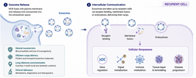
Mechanisms of exosome release, cellular uptake and exosome-mediated intercellular communication. Multivesicular bodies (MVBs) fuse with the plasma membrane of donor cells and release intraluminal vesicles (i.e., exosomes) into the extracellular space. Exosomes can be internalized by recipient cells via three pathways: receptor binding, membrane fusion or endocytosis. They then release internal bioactive cargo to regulate gene expression, signal transduction, immune response and tissue remodeling in target cells, and also participate in disease progression. As natural nanocarriers, exosomes feature favorable biocompatibility, low immunogenicity, effective protection and delivery of bioactive molecules, and support long-distance signal transmission. Hence, they hold great clinical value in the fields of disease biomarker discovery, diagnosis and therapy.
Exosome Product
2 Core Components of Exosomes
2.1 Membrane Proteins
Exosomes possess a typical lipid bilayer membrane structure. Their surface and membrane compartment are enriched with a variety of characteristic protein molecules. These membrane proteins not only participate in exosome biogenesis, trafficking and target cell recognition, but also serve as critical molecular markers for exosome purification and quality identification. Among them, tetraspanin family members CD9, CD63 and CD81 are the most widely used canonical marker proteins for exosomes.
CD9, CD63 and CD81 all belong to the tetraspanin family. They form protein complexes to stabilize exosome membrane structure, facilitate vesicle biogenesis and mediate intercellular interactions. CD63 is mainly localized on the membrane of late endosomes and multivesicular bodies (MVBs), and is closely associated with exosome biogenesis and intraluminal vesicle (ILV) formation. CD9 and CD81 are abundantly present on the exosome surface, participating in recognition, adhesion and uptake between exosomes and recipient cells. Therefore, CD9/CD63/CD81 are commonly used as positive markers in exosome identification to verify whether isolated products exhibit typical exosome characteristics.
In addition to tetraspanins, exosomes are enriched with multiple cytoplasmic proteins related to vesicle biogenesis and endosomal sorting. For instance, Alix (ALG-2-interacting protein X) is one of the ESCRT-associated regulatory proteins involved in endosomal membrane remodeling, ILV formation and exosome cargo sorting. TSG101 (Tumor Susceptibility Gene 101) is a key component of the ESCRT-I complex and plays an essential role in MVB formation, membrane protein trafficking and exosome secretion. Accordingly, Alix and TSG101 are generally regarded as structural marker proteins inside exosomes for further validation of exosome origin and biogenesis mechanisms.
In exosome research, proteins including CD9, CD63, CD81, Alix and TSG101 are commonly detected by Western blot, flow cytometry and immuno-electron microscopy to assess the purity, integrity and quality consistency of exosome samples. Combined with nanoparticle tracking analysis (NTA), transmission electron microscopy (TEM) and proteomic analysis, a more comprehensive exosome characterization system can be established.
Therefore, exosome membrane proteins constitute a vital part of exosome biological functions, and act as core indicators for identification, quality control and functional characterization in exosome research.
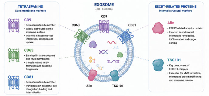
Schematic diagram of canonical exosome markers. Exosomes range from 30–150 nm in diameter. Tetraspanin signature proteins on the membrane surface include CD9, CD63 and CD81. CD9 and CD81 mediate the recognition, adhesion and uptake between exosomes and target cells; CD63 is enriched on the membranes of late endosomes and multivesicular bodies and participates in intraluminal vesicle generation and exosome biogenesis. Alix and TSG101 are ESCRT-associated structural markers. Alix participates in endosomal membrane remodeling, intraluminal vesicle formation and cargo sorting; TSG101 is a core component of the ESCRT-I complex, regulating multivesicular body formation, membrane protein trafficking and exosome release.
2.2 Nucleic Acid Cargo
In addition to proteins, exosomes are rich in a variety of bioactive nucleic acid molecules, serving as critical carriers for intercellular transmission of genetic information. Studies have shown that exosomes selectively sort specific nucleic acids from donor cells and package them inside vesicles. This protects the nucleic acids from degradation by extracellular nucleases and enables their stable transport across different cells and tissues. Nucleic acids carried by exosomes mainly include mRNA, microRNA (miRNA), long non-coding RNA (lncRNA), circular RNA (circRNA) and DNA fragments. These molecules can be taken up by recipient cells and further regulate gene expression, signal transduction and cellular functional status.
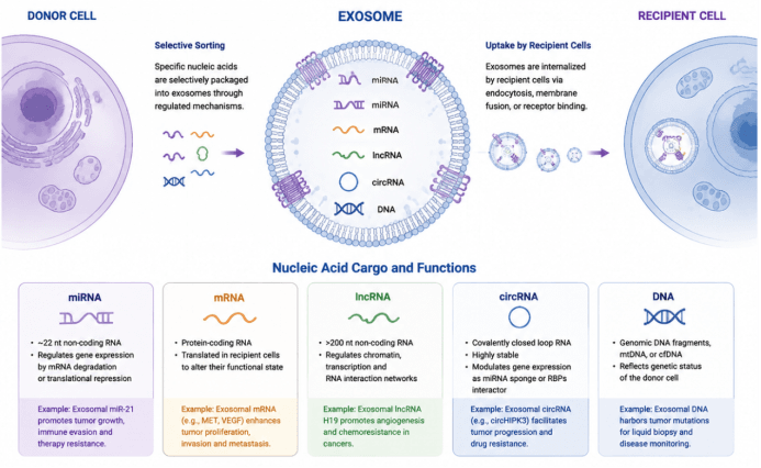
Mechanisms of sorting, delivery and biological functions of diverse nucleic acid cargos in exosomes. Donor cells selectively sort and package specific nucleic acids into exosomes via regulatory machineries. Exosomes carry nucleic acid components including miRNA, mRNA, lncRNA, circRNA and DNA, and are internalized by recipient cells through endocytosis, membrane fusion or receptor binding. miRNA are ~22 nt non-coding RNAs that regulate gene expression by degrading mRNA or repressing translation; mRNA can be translated inside recipient cells to alter cellular functions; lncRNAs are non-coding RNAs longer than 200 nt, modulating chromatin, transcription and RNA interaction networks; circRNAs are covalently closed circular RNAs with high stability, acting as miRNA sponges or interactors of RNA-binding proteins. Exosomal DNA includes genomic fragments, mitochondrial DNA or circulating cell-free DNA, which can reflect the genetic status of donor cells. The figure illustrates typical biological roles of various exosomal nucleic acids in tumors.
Among these, microRNA (miRNA) is one of the most extensively studied types of exosomal nucleic acid cargo. miRNAs are a class of ~22-nucleotide non-coding RNAs. By binding to target gene mRNAs, they promote mRNA degradation or translational repression, thereby regulating cell proliferation, differentiation, immune responses and disease progression. In 2007, Valadi et al. first reported the presence of functional mRNA and miRNA in exosomes, and proposed that exosomes serve as critical mediators for intercellular RNA transfer. This discovery transformed our understanding of exosome functions. Subsequent numerous studies have revealed that specific miRNAs contained in exosomes derived from tumor cells, immune cells and stem cells participate in tumor microenvironment modulation, inflammatory responses and tissue repair.
Beyond miRNAs, exosomes can also carry characteristic mRNA molecules from donor cells. Once delivered into recipient cells, exosomal mRNA can act as templates for protein translation and thereby modify the functional status of recipient cells. For example, tumor-derived exosomes can facilitate the proliferation, invasion and metastasis of tumor cells by delivering specific mRNAs; functional mRNAs within stem cell-derived exosomes are thought to regulate tissue repair and regeneration. Protected by the exosomal membrane, RNAs remain stable in blood, urine and other body fluids, making exosomal mRNA an important research focus for liquid biopsy and disease biomarker discovery.
In recent years, increasing attention has been paid to the roles of lncRNA (long non-coding RNA) and circRNA (circular RNA) in exosome research. lncRNAs are generally longer than 200 nucleotides and affect cellular functions by regulating chromatin architecture, transcription and RNA interaction networks. Thanks to their closed-loop structure, circRNAs possess high stability, can be preserved in exosomes for extended periods, and participate in the regulation of gene expression. Studies have demonstrated that specific lncRNAs and circRNAs enriched in tumor-derived exosomes promote tumor immune escape, angiogenesis and drug resistance, and hold potential as diagnostic biomarkers for diseases.
In addition, DNA components can be detected in exosomes, including genomic DNA fragments, mitochondrial DNA and circulating DNA. Although the origin and loading mechanisms of exosomal DNA remain somewhat controversial, accumulating evidence indicates that exosomal DNA can reflect the genetic status of source cells and shows promising applications in tumor mutation detection, disease monitoring and liquid biopsy.
Overall, nucleic acid cargo endows exosomes with powerful capacity for information transmission. Exosomes therefore act not only as key mediators of intercellular communication, but also as an important research platform for investigating disease mechanisms, discovering biomarkers and delivering nucleic acid therapeutics. Characterization of exosomal molecules such as miRNA, mRNA, lncRNA, circRNA and DNA enables in-depth understanding of the rules governing intercellular information exchange, and promotes the application of exosomes in precision medicine and regenerative medicine.
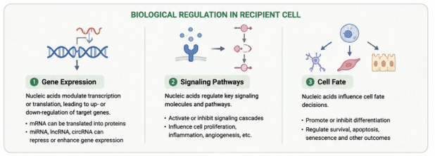
Biological regulatory effects of exosome-derived nucleic acids in recipient cells. After entering recipient cells, nucleic acids carried by exosomes regulate cellular biological behaviors at three levels: ① Gene expression: Nucleic acids modulate transcription and translation to upregulate or suppress target genes. mRNA can be directly translated into proteins, while miRNA, lncRNA and circRNA participate in the repression or enhancement of gene expression; ② Signaling pathways: Regulate key signaling molecules and pathways, activate or inhibit signaling cascades, and affect cell proliferation, inflammation, angiogenesis, etc.; ③ Cell fate: Govern cell fate decisions such as cell differentiation, survival, apoptosis and senescence.
2.3 Lipid Components
Exosomes possess a membrane structure composed of lipid bilayers, and their lipid composition is closely related to the plasma membrane and endosomal membrane system of donor cells. Compared with conventional cell membranes, exosomal membranes are enriched with specific lipid molecules, including cholesterol, sphingomyelin, ceramide and phosphatidylserine (PS). These lipids not only form the basic skeleton of the exosome membrane to maintain the structural stability and integrity of vesicles, but also participate in exosome biogenesis, cargo sorting, secretion and interactions with recipient cells.
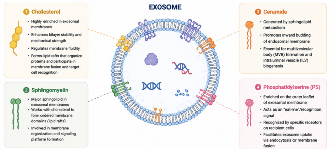
Major lipid components of exosomes and their biological functions. The exosomal membrane is enriched with four characteristic lipids: ① Cholesterol, highly enriched in the exosomal membrane. It strengthens the stability and mechanical strength of the lipid bilayer, modulates membrane fluidity, participates in lipid raft assembly, and mediates membrane fusion and target cell recognition; ② Sphingomyelin, the principal sphingolipid in exosomal membranes. It cooperates with cholesterol to form ordered membrane structures (lipid rafts) and contributes to membrane organization and signaling platform construction; ③ Ceramide, generated from sphingolipid metabolism. It promotes inward budding of the endosomal membrane and acts as a key component for the formation of multivesicular bodies and intraluminal vesicles; ④ Phosphatidylserine (PS), located on the outer leaflet of the exosomal membrane. It serves as an “eat-me” recognition signal, which is recognized by specific receptors on recipient cells and facilitates exosome entry into target cells via endocytosis or membrane fusion.
Cholesterol is one of the abundant lipid components in exosomal membranes. It enhances lipid bilayer stability and tunes membrane fluidity and mechanical strength, enabling exosomes to maintain structural integrity in the extracellular environment. In addition, membrane microdomains formed by cholesterol enrichment, known as lipid rafts, are implicated in exosomal protein clustering, membrane fusion and target cell recognition.
Sphingomyelin and ceramide play vital roles during exosome biogenesis. Studies have demonstrated that ceramide promotes inward budding of the endosomal membrane and participates in the formation of intraluminal vesicles (ILVs) within multivesicular bodies (MVBs). By regulating membrane curvature and lipid arrangement, the sphingolipid metabolic pathway affects exosome production efficiency and cargo loading.
Phosphatidylserine (PS) is an important phospholipid on the exosome surface and acts as a cellular recognition signal for binding and uptake between exosomes and recipient cells. Surface PS on exosomes can be recognized by specific receptors to facilitate vesicle entry into target cells through endocytosis, membrane fusion and other pathways, thereby delivering bioactive molecules.
In recent years, lipidomic analysis has further revealed that exosomes possess distinctive lipid profiles, and exosomes secreted by different cell types may exhibit divergent lipid signatures. These lipid characteristics not only affect exosome stability and biodistribution, but also modulate diverse biological processes including inflammatory responses, immune regulation, tumor microenvironment and tissue repair. Therefore, exosomal lipids are not only essential for maintaining vesicle structure, but also critical regulators governing exosome biogenesis, functional performance and the efficiency of intercellular communication. Research on exosomal lipid composition and metabolic mechanisms can deepen our understanding of exosome biology and advance their applications in disease diagnosis, drug delivery and regenerative medicine.
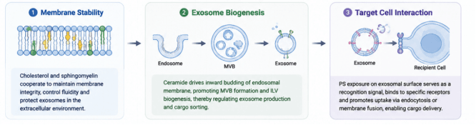
Functions of exosomal lipids in membrane stabilization, vesicle biogenesis and target cell interaction. Lipids regulate the exosome life cycle at three core levels: ① Membrane stability: Cholesterol and sphingomyelin synergistically maintain membrane integrity and modulate membrane fluidity, preserving the structural stability of exosomes in the extracellular environment; ② Exosome biogenesis: Ceramide drives inward budding of the endosomal membrane, promotes the generation of multivesicular bodies (MVBs) and intraluminal vesicles (ILVs), and controls exosome production and cargo sorting; ③ Target cell interaction: Phosphatidylserine (PS) exposed on the exosome surface acts as a recognition signal, binding to specific receptors on recipient cells and facilitating exosome uptake via endocytosis or membrane fusion to deliver cargo.