
info@mail.ucallm.com
1. Overview
Hendra virus (HeV) is the prototype species of the genus Henipavirus within the family Paramyxoviridae and is closely related to Nipah virus (NiV). It is an enveloped, negative‑sense, single‑stranded, non‑segmented RNA virus with pleiomorphic, roughly spherical virions.
HeV was first identified in 1994 in the Brisbane suburb of Hendra, Queensland, Australia, after which it was named. The virus is transmitted primarily through fruit bats (Pteropus species, also known as flying foxes) to horses, and then from horses to humans. To date, 7 human cases have been reported, with 4 fatalities; equine outbreaks total 89 spillover events with over 100 horse deaths. Although human cases are exceedingly rare, Hendra virus has an extremely high case fatality rate (approximately 57%) and has been designated by the WHO as a priority pathogen. Currently, no approved vaccines or antiviral therapies exist for human use.
2. Background Information
2.1 What is Hendra Virus?
Hendra virus is an enveloped, negative‑sense, single‑stranded, non‑segmented RNA virus belonging to the family Paramyxoviridae and genus Henipavirus. The virion is pleiomorphic, roughly spherical, composed of helical nucleocapsids enclosed within an envelope. The virus was originally named equine morbillivirus but was renamed Hendra virus (HeV) following genetic analysis confirming its distinctness from other paramyxoviruses.
The HeV genome is a negative‑sense single‑stranded RNA of approximately 18,324 nucleotides, one of the largest genomes among paramyxoviruses. The genome contains six transcriptional units encoding six structural proteins in the order 3′ to 5′.
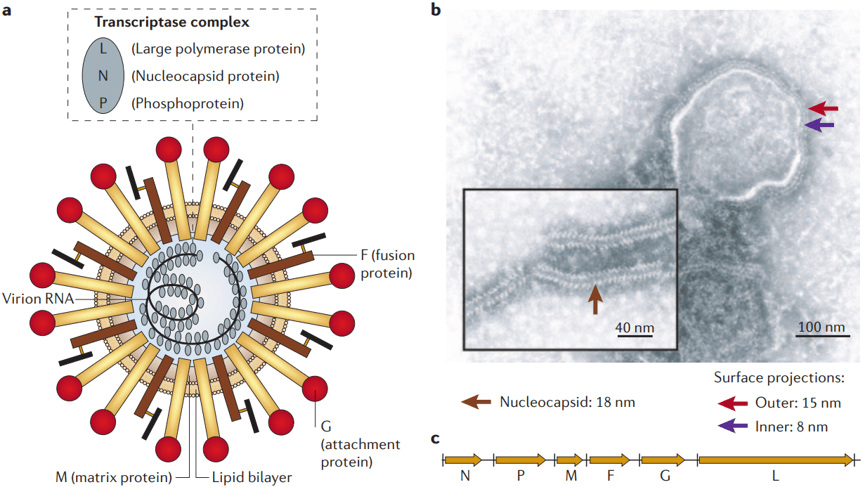
Structure of henipaviruses and their genomes (PMID: 16357858)
2.2 Hendra Virus Genome & Classification
HeV belongs to the genus Henipavirus in the family Paramyxoviridae. The genus Henipavirus also includes Nipah virus (NiV) , with which HeV shares high structural, sequence, and antigenic similarity. HeV and NiV share 78% nucleotide homology in their N genes. HeV currently exists in two genotypes:
l Genotype 1 (g1) : the original classical genotype
l Genotype 2 (g2) : a novel genotype discovered near Newcastle, Australia, in 2021
2.3 What Are Hendra Virus Structural Proteins?
HeV encodes six major structural proteins and three non‑structural proteins:
Protein | Gene | Main Function |
N (Nucleoprotein) | N gene | Binds tightly to viral RNA, forms the nucleocapsid core |
P (Phosphoprotein) | P gene | Polymerase cofactor, involved in transcription and replication |
M (Matrix protein) | M gene | Inner envelope protein, determines virion architecture |
F (Fusion protein) | F gene | Class I fusion protein, mediates membrane fusion |
G (Attachment protein) | G gene | Surface attachment glycoprotein, recognizes host receptors |
L (Large polymerase) | L gene | RNA‑dependent RNA polymerase (RdRp), catalyzes transcription and replication |
In addition, similar to Nipah virus, the P gene of Hendra virus encodes three non-structural proteins-C, V, and W-through RNA editing.
2.4 Mechanism of Cell Entry and Replication
2.4.1 Viral Attachment and Receptor Recognition
HeV infection begins with the binding of the viral G protein to Ephrin-B2 or Ephrin-B3 receptors on host cells. Henipaviruses use tryptophan as a "latch" to facilitate G‑receptor association.
2.4.2 Viral Entry
Following G protein–Ephrin receptor binding, conformational changes activate F protein. F protein mediates fusion of the viral envelope with the host cell membrane. HeV membrane fusion is pH‑independent, occurring either at the endosome or at the cell surface. F protein rearranges from prefusion to postfusion conformation, inserting the fusion peptide into the host membrane and releasing the viral nucleocapsid into the cytoplasm.
2.4.3 Viral RNA Replication
After nucleocapsid release into the cytoplasm, the L protein (RdRp) together with its cofactor P protein initiates transcription and replication:
l Transcription: negative‑sense RNA genome is transcribed into mRNAs, which are translated into viral proteins
l Replication: positive‑sense RNA intermediates are synthesized using negative‑sense RNA as template
l Progeny negative‑sense genomic RNAs are synthesized using positive‑sense RNA as template
2.4.4 Viral Assembly and Release
Newly synthesized N protein associates with progeny genomic RNA and L protein to form new ribonucleoprotein complexes (RNPs). M protein recruits RNPs to the cell membrane. The virus acquires its envelope by budding from the host cell membrane, releasing new virions.
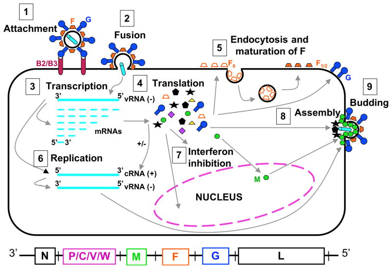
The life cycle of Henipavirus. (PMID: 21345285)
2.5 Symptoms and Treatment
2.5.1 Clinical Symptoms
The incubation period for HeV infection is not fully defined. Clinical manifestations in humans are diverse:
l Influenza‑like illness: some patients experience self‑limited influenza‑like illness
l Acute pneumonitis and multi‑organ failure: can present as fatal acute pneumonitis and multi‑organ failure
l Encephalitis: some patients present with acute encephalitis (partially fatal)
l Relapsed infection: relapsed infection has been reported
Equine symptoms include high fever, acute respiratory distress, and influenza‑like signs such as nasal discharge.
2.5.2 Treatment
Currently, no specific antiviral therapy is approved for HeV infection. Treatment is primarily early intensive supportive care:
l Intensive care support
l Respiratory support
l Management of neurologic complications
2.6 Targets for Intervention
2.6.1 Viral Targets
Target | Protein | Biological Role |
G | Attachment protein | Ephrin receptor binding |
F | Fusion protein | Membrane fusion |
L (RdRp) | RNA polymerase | RNA transcription & replication |
N | Nucleoprotein | RNP formation |
M | Matrix protein | Assembly & budding |
2.6.2 Host Receptors & Entry Factors
Host Target | Type | Role in Infection |
Ephrin-B2 | Cell surface receptor | G protein‑mediated viral attachment |
Ephrin-B3 | Cell surface receptor | G protein‑mediated viral attachment |
2.6.3 Host Signaling Pathways
Pathway | Key Targets | Biological Role |
Interferon pathway | IFN‑α/β | Antiviral response, suppressed by V protein |
2.7 Vaccine Types and Development Progress
Currently, no human Hendra virus vaccine is licensed worldwide. However, several candidates have entered clinical trials or preclinical development.
2.7.1 HeV-sG-V (Soluble G Glycoprotein Subunit Vaccine)
HeV-sG-V is a subunit vaccine candidate based on the Hendra virus soluble G glycoprotein (HeV sG). Due to the structural and sequence similarities between HeV and NiV G glycoproteins, HeV‑sG provides heterologous cross‑protective immunity.
2.7.2 rVSVΔG‑HeV‑G (Single‑Cycle Recombinant VSV‑Vectored Vaccine)
The single‑cycle recombinant vesicular stomatitis virus (rVSV)‑vectored vaccine (G*rVSVΔG‑HeV‑G) displays the HeV G glycoprotein.
2.7.3 Other Vaccine Candidates
According to 2026 reports, Phase 2 clinical trials for two human Hendra virus vaccines were set to begin in 2026.
2.8 Drugs
Currently, no specific small‑molecule antiviral is approved for HeV infection. Drug development efforts focus on the following areas:
2.8.1 Virus-Targeted Drugs
Target | Drug/candidate | Mechanism | Stage |
L protein (RdRp) | Remdesivir | RdRp inhibition | Shown antiviral activity against HeV |
Menadiol diphosphate | RdRp inhibition | In silico drug repurposing | |
Masoprocol | RdRp inhibition | In silico drug repurposing | |
Dinoprostone | RdRp inhibition | In silico drug repurposing | |
Pamidronic acid | RdRp inhibition | In silico drug repurposing | |
F protein | Rhesus‑derived mAbs | Target prefusion F conformation | Preclinical |
| Nanobody DS90 | Binds conserved F glycoprotein site | Preclinical |
Multiple targets | mRNA‑encoded antibody (mRNA‑1E5‑LNPs) |
| Preclinical |
2.9 Epidemiology
l First identified: 1994, Hendra suburb of Brisbane, Queensland, Australia
l Natural reservoir: fruit bats (Pteropus species, flying foxes), seroprevalence 20%–50%
l Transmission routes:
Ø Bat → horse: contamination of pastures or feed with infected bat fluids or tissues (urine, saliva, gestational products)
Ø Horse → horse: relatively inefficient, but can occur in stabled environments
Ø Horse → human: 7 cases, each involving heavy exposure to infected horse secretions, tissues, or body fluids
Ø Bat → human: no direct transmission evidence
Ø Human → human: no cases reported
l Geographic distribution: all known cases have occurred in Queensland and northern New South Wales, Australia. As of December 2025, 89 outbreak events recorded. The most southerly cases are near Scone in the Upper Hunter Valley (2019) and near Newcastle (2021 and 2023)
2.10 Diagnosis
Diagnosis of HeV infection relies on laboratory testing:
l Virus isolation: isolation of HeV from clinical material (requires PC4 laboratory)
l Nucleic acid testing (NAT) : detection of HeV RNA in clinical material
l Serology: detection of seroconversion or four‑fold or greater rise in HeV‑specific IgM or IgG titres
2.11 Prevention
l Equine vaccination: Equivac® HeV vaccine has been registered in Australia since 2012 and is a key measure for preventing HeV infection in horses
l Avoid contact: avoid contact with fruit bats and potentially contaminated food and water sources
l Horse management: place horse feed and water sources in areas inaccessible to fruit bats
l Personal protective measures: veterinarians and handlers should wear appropriate personal protective equipment (PPE) when handling suspected or confirmed HeV‑infected horses
l Surveillance and reporting: HeV is a notifiable disease and Emergency Animal Disease (EAD) in Australia
3. Related Products & Services
l Recombinant Antigens & Receptors
l Antibodies
l Detection Kits
l Recombinant Protein Expression Services
l Antibody Development Services
4. Resources
5. References
1. Spengler, J. R., Lo, M. K., Welch, S. R., et al. (2025). Henipaviruses: epidemiology, ecology, disease, and the development of vaccines and therapeutics. Clinical Microbiology Reviews, 38(1), e00128-23.
2. Eaton, B. T., Broder, C. C., Middleton, D., et al. (2006). Hendra and Nipah viruses: different and dangerous. Nature Reviews Microbiology, 4, 23–35.
3. Wang, L. F., Harcourt, B. H., Yu, M., et al. (2001). Molecular biology of Hendra and Nipah viruses. Microbes and Infection, 3, 279–287.
4. Bowden, T. A., Aricescu, A. R., Gilbert, R. J. C., et al. (2008). Structural basis of Nipah and Hendra virus attachment to their cell-surface receptor ephrin-B2. Nature Structural & Molecular Biology, 15, 567–572.
5. Xu, K., Rajashankar, K. R., Chan, Y. P., et al. (2012). New insights into the Hendra virus attachment and entry process from structures of the virus G glycoprotein and its complex with ephrin-B2. PLoS Pathogens, 8, e1002890.
6. Murray, K., Selleck, P., Hooper, P., et al. (1995). A morbillivirus that caused fatal disease in horses and humans. Science, 268, 94–97.
7. Pallister, J., Middleton, D., Wang, L. F., et al. (2013). A recombinant Hendra virus G glycoprotein-based subunit vaccine protects ferrets against Hendra and Nipah viruses. PLoS ONE, 8, e70383.
8. Middleton, D., Pallister, J., Klein, R., et al. (2012). Hendra virus vaccine, a one health approach to protecting horse, human and environmental health. Emerging Infectious Diseases, 18, 10–14.
9. Rodrigue, V., Gravagna, K., Yao, J., Nafade, V., & Basta, N. E. (2024). Current progress towards prevention of Nipah and Hendra disease in humans: A scoping review of vaccine and monoclonal antibody candidates being evaluated in clinical trials. Tropical Medicine & International Health, 29(5), 354–364.
10. Bossart, K. N., Geisbert, T. W., Feldmann, H., et al. (2009). A neutralizing human monoclonal antibody protects against lethal disease in a new model of acute Nipah virus infection. PLoS Pathogens, 5, e1000642.
11. Lo, M. K., Feldmann, F., Gary, J. M., et al. (2019). Remdesivir (GS-5734) protects African green monkeys from Nipah virus challenge. Science Translational Medicine, 11, eaau9242.
12. Plowright, R. K., Foley, P., Field, H. E., et al. (2011). Urban habituation, ecological connectivity and epidemic dampening: the emergence of Hendra virus from flying foxes. Proceedings of the Royal Society B, 278, 370–377.