
info@mail.ucallm.com
1. Overview
Japanese encephalitis virus (JEV) is a major member of the genus Orthoflavivirus within the family Flaviviridae and is the causative agent of Japanese encephalitis (JE). It is an enveloped, positive‑sense, single‑stranded RNA virus with a spherical virion approximately 40–60 nm in diameter.
JEV was first isolated in 1935 from the brain tissue of a fatal human case in Japan. The virus is transmitted primarily by Culex tritaeniorhynchus mosquitoes and is maintained in a natural cycle between pigs and water birds. Humans are incidental hosts; most infections are asymptomatic, but severe cases can develop fatal encephalitis. Globally, an estimated 50,000–175,000 human infections occur annually, with 30%–50% of survivors left with permanent neuropsychiatric sequelae.
JEV is endemic in East Asia, South Asia, Southeast Asia, the Western Pacific coastal regions, and eastern‑central Australia. In recent years, climate change and land‑use alterations have facilitated its spread to new geographic areas, including Australia, Africa, and Russia. In the absence of specific antiviral drugs, vaccination and mosquito control remain the mainstays of prevention and control.
2. Background Information
2.1 What is Japanese Encephalitis Virus?
Japanese encephalitis virus is an enveloped, positive‑sense, single‑stranded RNA virus belonging to the family Flaviviridae and genus Orthoflavivirus. The virion is spherical, approximately 40–60 nm in diameter, composed of an internal nucleocapsid and an external lipid envelope. The nucleocapsid consists of genomic RNA and capsid protein (C); the envelope is derived from the host cell membrane and is studded with membrane (prM/M) and envelope (E) proteins.
The JEV genome is a positive‑sense single‑stranded RNA of approximately 11 kb. It possesses a methylated cap at the 5′ end but lacks a poly(A) tail at the 3′ end, instead containing a conserved CU region. The genome contains a single open reading frame (ORF) encoding a polyprotein precursor, which is cleaved by host and viral proteases into 10 mature proteins – three structural proteins (C, prM/M, E) and seven non‑structural proteins (NS1, NS2A, NS2B, NS3, NS4A, NS4B, NS5).
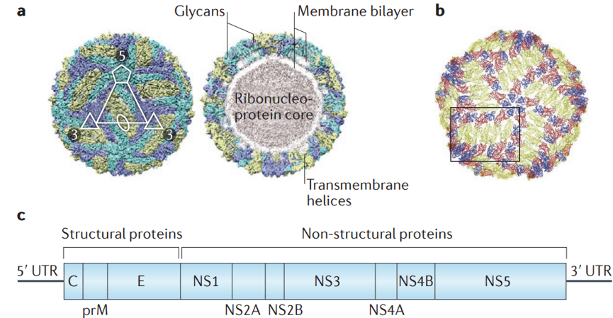
Japanese Encephalitis Virus Structure (PMID: 29697099)
2.2 Japanese Encephalitis Virus Genome & Classification
JEV exists in multiple genotypes. Currently, genotype I (GI) predominates globally, while the classical live‑attenuated vaccine SA14‑14‑2 is based on genotype III (GIII). In recent years, genotype V (GV) has re‑emerged, attracting public health attention.
2.3 What Are Japanese Encephalitis Virus Structural Proteins?
JEV encodes 3 structural proteins:
Protein | Full name | Main Function |
C | Capsid | Encapsidates the viral genomic RNA, forming the nucleocapsid core |
prM/M | pre‑Membrane/Membrane | prM is cleaved by furin during maturation to produce M; involved in assembly and envelope formation |
E | Envelope | Major surface protein; mediates receptor binding and membrane fusion; primary target of neutralizing antibodies; amino acids 107, 138, and 315 are key determinants of neurovirulence |
2.4 Mechanism of Cell Entry and Replication
The JEV replication cycle resembles that of other flaviviruses and occurs entirely within the host cell cytoplasm.
2.4.1 Viral Attachment and Receptor Recognition
Infection begins with the binding of the viral E protein to one or more attachment factors on the host cell surface. Identified receptors and entry factors include:
Host receptor/entry factor | Type | Role in infection |
TfR1 (Transferrin receptor 1) | Receptor | JEV E protein interacts via its protease‑like domain (residues 384–606); TfR1 knockdown significantly inhibits viral entry |
EGFR (Epidermal growth factor receptor) | Receptor | Specifically interacts with the ED3 domain of JEV E in neuronal cells; EGFR inhibitors reduce entry and replication |
GRP78 | Receptor | Involved in JEV binding to neurons |
CD4 | Receptor | Mediates entry in porcine kidney cells via lipid‑raft‑localised CD4 |
2.4.2 Viral Entry
JEV enters host cells via receptor‑mediated endocytosis. The endocytic pathway varies by cell type, involving either clathrin‑dependent endocytosis or clathrin‑independent endocytosis (the latter mediated by Endophilin in neuronal cells).
After internalisation, endosomal acidification triggers low‑pH‑induced conformational changes in E protein, mediating fusion of the viral envelope with the endosomal membrane. Following fusion, the viral genomic RNA is released into the cytoplasm.
2.4.3 Viral RNA Replication
Upon release, the viral RNA associates with ribosomes on the rough endoplasmic reticulum (ER) membrane, initiating translation. The polyprotein precursor is co‑translationally and post‑translationally cleaved to produce structural and non‑structural proteins.
The non‑structural proteins assemble into replication complexes on ER‑derived membrane vesicles. NS5 (RdRp) catalyses viral RNA replication.
2.4.4 Viral Assembly and Maturation
Newly synthesised genomic RNA interacts with structural proteins C, prM, and E, and buds into the ER lumen to form immature virions. During transport through the Golgi apparatus, the prM protein is cleaved by host furin protease to generate M protein. This cleavage triggers rearrangement of the envelope proteins, maturating the virions. Mature and partially mature virions are finally released via exocytosis.
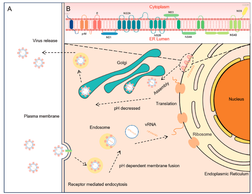
The life cycle of Japanese Encephalitis Virus. (PMID: 39983879)
2.5 Symptoms and Treatment
2.5.1 Clinical Symptoms
The incubation period for JEV infection is typically 5–15 days. Approximately 99% of infections are asymptomatic or present with only mild symptoms. Symptomatic patients usually present with an acute encephalitis syndrome:
l Fever
l Headache
l Vomiting
l Altered consciousness
l Seizures
l Neck stiffness
Severe cases may progress to coma, paralysis, and death. Case fatality rates can be as high as 30%. Among survivors, 30%–50% experience permanent neuropsychiatric sequelae, including intellectual disability, movement disorders, and hearing loss.
High‑risk populations: children and rural residents are the main affected groups. However, with increasing childhood vaccination coverage, the proportion of adult cases has risen significantly. In the 2025 Nepal outbreak, approximately 76% of deaths occurred in adults aged over 40 years.
2.5.2 Treatment
Currently, no specific antiviral therapy is approved for JEV infection. Treatment is primarily supportive:
l Intensive care support
l Intracranial pressure control
l Anticonvulsant therapy
l Respiratory support
2.6 Targets for Intervention
2.6.1 Viral Targets
Target | Protein | Biological Role |
E | Envelope protein | Receptor binding & membrane fusion |
NS3 | Helicase/protease | RNA unwinding, polyprotein processing |
NS5 | RNA polymerase (RdRp) | RNA replication |
NS1 | Non‑structural protein 1 | Membrane curvature, replication complex formation |
2.6.2 Host Targets
Host Target | Type | Role in Infection |
TfR1 | Receptor | Viral entry |
EGFR | Receptor | Viral entry (neuronal cells) |
AT1 receptor | GPCR | Mediates angiotensin II‑induced inflammatory cytokine release |
GSPT1 | Host protein | Interacts with viral NS5 to support viral translation and replication |
2.7 Vaccine Types and Development Progress
Vaccination is the most effective measure for preventing JEV infection. Several JEV vaccines are currently licensed, and new platforms are rapidly advancing.
2.7.1 Licensed Vaccines
l SA14‑14‑2 (live‑attenuated) : a live‑attenuated vaccine based on genotype III, widely used in China. However, studies indicate that GIII‑based vaccines elicit suboptimal neutralising responses against emerging genotype I isolates.
l Inactivated vaccines: include mouse brain‑derived and cell culture‑derived inactivated vaccines.
l Chimeric live‑attenuated vaccine (JE/YF17D, IMOJEV®) : a chimeric vaccine constructed by inserting JEV prM and E genes into the yellow fever vaccine YF17D backbone.
2.7.2 mRNA Vaccine (GBP560)
In 2025, SK company initiated a Phase I/II clinical trial (NCT06680128) for the JEV mRNA vaccine GBP560 in healthy adults aged ≥18 years, evaluating safety, reactogenicity, and immunogenicity.
2.7.3 Liposome-Based Vaccines
A 2025 study published in Clinical and Experimental Vaccine Research evaluated liposome‑based oral and injectable JEV vaccines. The injectable formulation induced stronger IgG responses, while the oral formulation, though less immunogenic, showed promise as a non‑invasive delivery route.
2.7.4 Chimeric Virus-Vectored Vaccine (CYV-JEV)
In December 2025, a study in Vaccines reported the construction of a chimeric JEV genotype I prME protein vaccine CYV‑JEV using Chaoyang Virus (CYV) as the backbone. This vaccine replicated efficiently in mosquito cells but was replication‑defective in mammalian cells. In IFNAR−/− mice, two immunisations provided 100% complete protection.
2.7.5 Other Vaccine Directions
l JEV virus-like particle (VLP)-based nanoparticle vaccines: using JEV-VLPs as carriers to induce neutralising antibodies and Th1 responses
l NS1-based vaccines: truncated NS1 fused with E. coli heat-labile enterotoxin B subunit
2.8 Drugs
Currently, no specific antiviral drug is approved for JEV. Several novel antiviral candidates have shown promise:
2.8.1 Virus-Targeted Drugs
Target | Drug/candidate | Mechanism | Stage |
Viral replication | Nitazoxanide (NTZ) | Antiviral via SUCLG2‑ and PRKAG1‑mediated autophagy pathways | In vitro & animal |
NS5 (RdRp) | CC‑90009 | Cereblon E3 ligase modulator; induces proteasomal degradation of GSPT1/NS5 complexes | In vitro & animal |
NS3 protease | Novel thiazole derivatives | Flaviviral protease inhibitors, active against both DENV2 and JEV | In vitro |
Multiple targets | JEV‑GFP replicon system | Screening tool for antivirals targeting JEV non‑structural proteins | Drug screening |
2.8.2 Host-Targeted Drugs
Target | Drug/candidate | Mechanism | Stage |
AT1 receptor/PPARγ | Telmisartan | Antihypertensive; AT1 antagonist and PPARγ agonist; anti‑inflammatory and antiviral via AT1/PPARγ axis | In vitro & animal |
EGFR | Erlotinib/Gefitinib | EGFR kinase inhibitors; reduce viral entry and replication | In vitro & primary mouse neurons |
2.9 Epidemiology
l First identified: 1935, Japan
l Global disease burden: 50,000–175,000 annual infections, 10,000–20,000 deaths
l Geographic distribution: East Asia, South Asia, Southeast Asia, Western Pacific coastal regions, and eastern‑central Australia; has recently spread to Australia, Africa, and Russia
l Natural cycle: Culex tritaeniorhynchus ↔ pigs/water birds
l Seasonality: incidence is 20%–30% higher from July to September than other months
Major outbreaks in 2025:
l Assam, India: 389 laboratory‑confirmed cases, 72 deaths, affecting 33/35 districts
l Nepal: 175 confirmed cases, 34 deaths; ~76% of deaths occurred in adults >40 years
l Australia: 2 locally acquired confirmed cases reported in Victoria and Queensland in January 2025
Epidemiological shift: With large‑scale childhood vaccination, the burden of JEV disease is shifting from children to adults. This “adult immunity gap” has become a new public health challenge.
2.10 Diagnosis
Diagnosis of JEV infection relies on laboratory testing:
l Serology: detection of anti‑JEV IgM antibodies in CSF or serum (MAC‑ELISA)
l RT‑PCR: detection of viral RNA in blood or CSF
l Virus isolation: from clinical specimens
Due to serological cross‑reactivity with other flaviviruses (e.g., dengue, West Nile), diagnosis should be interpreted alongside clinical and epidemiological information.
2.11 Prevention
l Vaccination: the most effective preventive measure (see Section 2.6)
l Mosquito control: reduce breeding sites, use bed nets and repellents
l Pig management: pigs are the main amplifying hosts; pig farms should be located away from residential areas
l Traveller precautions: travellers to endemic areas should be vaccinated and take anti‑mosquito measures
l Surveillance: strengthen monitoring of human, pig, and mosquito populations
3. Related Products & Services
l Recombinant Antigens & Receptors
l Antibodies
l Detection Kits
l Recombinant Protein Expression Services
l Antibody Development Services
4. Resources
5. References
1. Turtle, L., Solomon, T. (2018). Japanese encephalitis — the prospects for new treatments. Nature Reviews Neurology, 14, 298–313.
2. Park, J. Y., Lee, H. M. (2025). Advances and Challenges in Vaccination and Therapeutic Strategies Against Japanese Encephalitis Virus. Pathogens, 14(12), 1204.
3. Gong, J., Duan, X., Ge, Z. (2025). Molecular mechanisms of Japanese encephalitis virus infection and advances in vaccine research. Microbial pathogenesis, 201, 107397.
4. Kumar, S., Verma, A., Yadav, P., et al. (2022). Molecular pathogenesis of Japanese encephalitis and possible therapeutic strategies. Archives of virology, 167(9), 1739–1762.
5. Turtle, L., Solomon, T. (2018). Japanese encephalitis - the prospects for new treatments. Nature reviews. Neurology, 14(5), 298–313.
6. Campbell, G. L., Hills, S. L., Fischer, M., et al. (2011). Estimated global incidence of Japanese encephalitis: a systematic review. Bulletin of the World Health Organization, 89(10), 766–774E.
7. van den Hurk, A. F., Ritchie, S. A., Mackenzie, J. S. (2009). Ecology and geographical expansion of Japanese encephalitis virus. Annual review of entomology, 54, 17–35.
8. Yun, S. I., Lee, Y. M. (2014). Japanese encephalitis: the virus and vaccines. Human vaccines & immunotherapeutics, 10(2), 263–279.
9. Lee, A. R., Kim, W. J., Choi, H., et al. (2025). Genotype III-Based Japanese Encephalitis Vaccines Exhibit Diminished Neutralizing Response to Reemerging Genotype V. The Journal of infectious diseases, 231(5), 1281–1289.
10. Halstead, S. B., Thomas, S. J. (2011). New Japanese encephalitis vaccines: alternatives to production in mouse brain. Expert review of vaccines, 10(3), 355–364.
11. Barzon, L., Palù, G. (2018). Recent developments in vaccines and biological therapies against Japanese encephalitis virus. Expert opinion on biological therapy, 18(8), 851–864.
12. Hegde, N. R., Gore, M. M. (2017). Japanese encephalitis vaccines: Immunogenicity, protective efficacy, effectiveness, and impact on the burden of disease. Human vaccines & immunotherapeutics, 13(6), 1–18.
13. Nain, M., Mukherjee, S., Karmakar, S. P., et al. (2017). GRP78 Is an Important Host Factor for Japanese Encephalitis Virus Entry and Replication in Mammalian Cells. Journal of virology, 91(6), e02274-16.
14. Wang, Q., Yang, S., Yang, K., et al. (2023). CD4 is an important host factor for Japanese encephalitis virus entry and replication in PK-15 cells. Veterinary microbiology, 287, 109913.
15. Wei, M., Ma, R., Wang, X., et al. (2025). SUCLG2 plays a crucial role in the activity of Nitazoxanide against Japanese encephalitis virus. Antiviral Research, 106301.
16. Yang, Q., Xie, S., Zhao, Y., et al. (2025). Transferrin receptor 1 is an entry factor for Japanese encephalitis virus. Veterinary microbiology, 309, 110698.
17. Cui, J., Huang, X., Li, Y., et al. (2025). Construction and Evaluation of a Chimeric Japanese Encephalitis Virus Vaccine Candidate Strain with Chaoyang Virus as the Backbone. Vaccines, 14(1), 30.
18. He, Z., Chen, Y., Xia, B., et al. (2025). CC-90009, a Cereblon E3 Ligase Modulator, Exhibits Antiviral Efficacy Against JEV In Vitro and In Vivo via Targeted Degradation of GSPT1 and Viral NS5 Protein. Pharmaceutics, 17(12), 1524.