
info@mail.ucallm.com
1. Overview
Listeria monocytogenes is a Gram-positive, facultative anaerobic, intracellular bacillus of the family Listeriaceae and genus Listeria, and is the causative agent of listeriosis. The bacterium is recognized by the World Health Organization (WHO) as one of the most important foodborne pathogens, with a case fatality rate of 20–30%, among the highest of all foodborne pathogens.
L. monocytogenes is a facultative intracellular pathogen capable of surviving and replicating within a variety of host cells, including macrophages, epithelial cells, and hepatocytes. The bacterium is highly adaptable to environmental stresses, capable of growing at 4°C, tolerating high salt concentrations and a wide pH range, thus posing a persistent threat in refrigerated and ready-to-eat foods. Pregnant women, neonates, the elderly, and immunocompromised individuals are at high risk for listeriosis, which can cause meningitis, sepsis, abortion, and neonatal infection.
2. Background Information
2.1 What is Listeria monocytogenes?
L. monocytogenes is a Gram-positive, facultative anaerobic, intracellular bacillus belonging to the family Listeriaceae and genus Listeria. The bacterium was first discovered in 1926, and its name derives from "monocytosis," referring to the marked increase in monocytes in the blood of infected animals. L. monocytogenes possesses a unique intracellular parasitic capacity, employing a series of virulence factors to achieve phagosome escape, intracellular replication, and cell-to-cell spread.
2.2 Genome & Classification
The L. monocytogenes genome is a circular chromosome of approximately 2.9 Mb, containing approximately 2,800–2,900 genes. The bacterium is divided into four phylogenetic lineages, with lineages I and II being the primary sources of clinical infection. Lineage I includes serovars 1/2b, 3b, and 4b, with serovar 4b associated with most invasive listeriosis outbreaks; lineage II includes serovars 1/2a, 1/2c, and 3a.
The pathogenicity of L. monocytogenes is primarily attributed to the presence of Listeria pathogenicity islands (LIPIs) . LIPI-1 encodes core virulence factors including listeriolysin O (LLO); LIPI-3 encodes listeriolysin S; LIPI-4 is associated with hypervirulent clones (e.g., ST87) and participates in bacterial invasion and intracellular proliferation.
2.3 What Are L. monocytogenes Virulence Factors?
Virulence Factor | Encoding Location | Main Function |
Listeriolysin O (LLO) | LIPI-1 (hly gene) | Cholesterol-dependent pore-forming toxin, mediates phagosome escape |
nternalins (InlA/InlB) | LIPI-1 | Mediate host cell adhesion and invasion |
ActA | LIPI-1 | Mediates actin aggregation and cell-to-cell spread |
Phospholipases (PlcA/PlcB) | LIPI-1 | Synergize with LLO in phagosome membrane disruption |
PrfA | Chromosome | Positive regulator of virulence gene expression |
LLS (Listeriolysin S) | LIPI-3 | Cytotoxicity, associated with hypervirulence |
LIPI-4 | LIPI-4 | Promotes flagellum formation, enhances colonization and cellular invasion |
2.3 Mechanism of Infection and Pathogenesis
2.3.1 Adhesion and Invasion
L. monocytogenes infection begins when the bacterium enters the host via the faecal-oral route. Upon reaching the small intestine, the bacterium uses surface proteins internalin A (InlA) and internalin B (InlB) to bind host cell surface receptors, mediating bacterial invasion of intestinal epithelial cells. InlA interacts with host E-cadherin, while InlB binds the c-Met receptor, together promoting bacterial internalization.
2.3.2 Phagosome Escape
After entering host cells, L. monocytogenes is encapsulated within a phagosome. Listeriolysin O (LLO) is the key virulence factor mediating phagosome escape. LLO is a cholesterol-dependent pore-forming toxin with maximal activity at acidic pH. A 2025 study published in Virulence identified critical residues within the amino acid 251–255 domain of LLO: F251, Q253, and Y255 are essential for LLO's hemolytic activity and cytotoxicity; alanine mutations at these sites significantly impair bacterial proliferation in mouse organs and disruption of late phagosome membranes. Notably, F251A and Y255A mutations completely abolish late phagosome escape capacity.
2.3.3 Intracellular Replication and Cell-to-Cell Spread
After escaping into the cytoplasm, L. monocytogenes exploits host cell nutrients for rapid replication. The surface protein ActA mediates host actin aggregation, forming an "actin tail" that propels the bacterium through the cytoplasm and into adjacent cells, achieving cell-to-cell spread without exposure to the extracellular environment.
2.3.4 Central Nervous System Infection Mechanisms
A 2025 study published in Acta Neuropathologica revealed divergent host-pathogen interactions in L. monocytogenes CNS infection:
l Microglia: L. monocytogenes thrives in the cytosol of microglia, driving productive infection and facilitating bacterial spread
l Monocyte-derived macrophages (MDM) : MDMs effectively sequester L. monocytogenes within the phagolysosomal system, limiting its replication and inducing a viable but non-culturable (VBNC) state without completely eliminating the pathogen
2.3.5 Immune Evasion Mechanisms
L. monocytogenes evades host immune recognition through multiple mechanisms. A 2025 study published in Autophagy found that the host protein RNF144A inhibits autophagy by targeting BECN1 for degradation, thereby promoting L. monocytogenes infection, revealing a novel regulatory mechanism of host factors in bacterial infection.
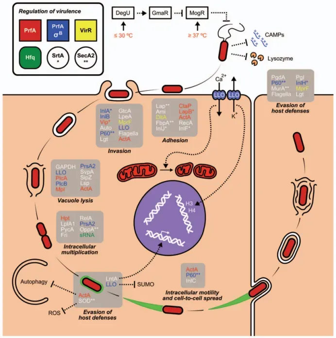
The Listeria monocytogenes cell infection cycle and the virulence factors involved in the successive steps (PMID: 21921683)
2.4 Transmission Routes and Process
Transmission of L. monocytogenes occurs primarily via the foodborne route:
l Ready-to-eat foods: refrigerated ready-to-eat meat products, soft cheeses, unpasteurized dairy products, smoked seafood
l Fresh produce: unwashed vegetables and fruits
l Food processing environments: L. monocytogenes can form persistent biofilms in food processing facilities, leading to recurrent contamination
Process: Ingestion of contaminated food → bacteria reach the small intestine → invade intestinal epithelial cells via InlA/InlB → escape into the cytosol via LLO → replicate intracellularly → disseminate via blood and lymph to liver, spleen, and CNS → establish infection in target organs.
2.5 Symptoms and Treatment
2.5.1 Clinical Symptoms
Non-invasive listeriosis: febrile gastroenteritis, with symptoms including diarrhoea, fever, headache, and myalgia.
Invasive listeriosis: severe clinical manifestations, including:
l Bloodstream infection: the most common presentation, accounting for 42.86% of clinical infections
l Central nervous system infection: meningitis, encephalitis, accounting for 26.19%
l Perinatal infection: miscarriage, stillbirth, premature birth, and neonatal infection, accounting for 26.19%
2.5.2 Treatment
Drug | Recommendation Level | Considerations |
Ampicillin | First-line | Often combined with gentamicin |
Penicillin G | First-line | Alternative to ampicillin |
Trimethoprim-sulfamethoxazole (TMP/SMX) | Alternative | For penicillin-allergic patients |
Neonatal treatment: ampicillin plus an aminoglycoside (e.g., gentamicin).
ICU patients: gentamicin combined with ampicillin remains the standard and effective treatment for Listeria meningitis.
2.6 Prevention
l Food safety: thoroughly cook meat and eggs; avoid unpasteurized dairy products and soft cheeses; thoroughly wash produce; separate raw and cooked foods
l High-risk populations: pregnant women, the elderly, and immunocompromised individuals should avoid high-risk foods (e.g., refrigerated ready-to-eat meat products, smoked seafood)
l Food processing environment surveillance: strengthen L. monocytogenes monitoring and biofilm control in food processing facilities
l Hand hygiene: thoroughly wash hands after handling raw food
l Vaccination: currently no licensed human Listeria vaccine is available
2.7 Targets for Intervention
2.7.1 Pathogen Targets
Target | Protein/Pathway | Biological Function |
LLO | Listeriolysin O | Phagosome escape |
InlA/InlB | Internalins | Host cell invasion |
ActA | Actin aggregation protein | Cell-to-cell spread |
PrfA | Virulence regulator | Virulence gene expression |
2.7.2 Host Targets
Target | Type | Role in Infection |
E-cadherin | Cell adhesion molecule | InlA receptor |
c-Met | Receptor tyrosine kinase | InlB receptor |
BECN1 | Autophagy protein | Targeted for degradation by RNF144A, regulates autophagy |
2.8 Vaccine Types and Development Progress
Currently, no licensed human L. monocytogenes vaccine is available, but several candidates are in development.
2.8.1 Oral Cancer Vaccine (Colorectal Cancer)
A 2026 study published in Journal for Immunotherapy of Cancer developed an oral L. monocytogenes cancer vaccine. By deleting the ActA and InlB virulence genes, a vaccine strain capable of oral administration in mice was constructed. Oral immunization induced CD8 T cell responses comparable in magnitude to fully virulent L. monocytogenes; immunized mice did not lose weight, and L. monocytogenes was contained to intestinal tissues. Prophylactic immunization protected mice from colorectal cancer tumour challenge; therapeutic immunization combined with immune checkpoint inhibitors (anti-PD-1 and anti-CTLA-4) controlled tumour growth.
2.8.2 HPV Multivalent E6E7 Recombinant Listeria Vaccine
A 2025 study published in BMC Cancer constructed recombinant Listeria vaccines expressing HPV6/11/16/18 E6E7 proteins using a LM prime-LI boost-LM final heterologous immunization strategy. In HPV16/18 E6/E7-expressing tumour models, this vaccination protocol achieved complete tumour cure rates of 30% and 40%, respectively, and significantly extended median survival time.
2.8.3 Inactivated Vaccine (ISA 61 VG Adjuvant)
A 2025 study published in Veterinary Research developed inactivated L. monocytogenes vaccines using the ISA 61 VG water-in-oil adjuvant. 61 VG-AIV induced higher levels of specific antibodies and pro-inflammatory cytokines than the alum-adjuvanted vaccine in mice, conferring 83.4% protection against serotype 4h challenge (vs. 50% for alum-adjuvanted vaccine), and provided cross-protection against serotypes 1/2a, 1/2b, and 4b. High immunoprotection (83.4%) was also conferred in sheep.
2.8.4 Multi-Epitope mRNA Vaccine
A 2025 study published in International Journal of Biological Macromolecules computationally designed a multi-epitope mRNA vaccine targeting E. coli K1, group B Streptococcus, L. monocytogenes, Neisseria meningitidis, and Streptococcus pneumoniae, targeting outer membrane porins (OMPs).
2.8.5 Immunoinformatics Epitope Vaccine
A 2025 study published in Springer used immunoinformatics approaches to design a multi-epitope subunit vaccine targeting the LLO exotoxin. The final vaccine construct comprised 315 amino acids; immune simulation predicted robust cellular and humoral responses within five days of administration, and protein-protein docking analysis predicted significant binding affinity to MHC-II receptors.
2.9 Drugs
2.9.1 Pathogen-Targeted Drugs
Target | Drug/candidate | Mechanism | Stage |
LLO | Tanshinone IIA | Inhibits LLO pore-forming activity, forming a stable LLO-Tan IIA complex | Animal model |
Curcumin/nisin dual-loaded liposomes | Photodynamic combination therapy, synergistic bactericidal and antibiofilm effects | In vitro |
Tanshinone IIA: A 2025 study published in Scientific Reports demonstrated that Tanshinone IIA (Tan IIA) significantly inhibits LLO pore-forming activity by forming a stable LLO-Tan IIA complex. Tan IIA concentration-dependently decreased L. monocytogenes cytotoxicity toward J774 and BeWo cells, with inhibitory rates of 41.3% and 54.5%, respectively (8 µg/mL). In mouse models, Tan IIA (80 mg/kg) increased survival rate by 26.7% after L. monocytogenes infection, reduced bacterial loads, alleviated tissue damage, and diminished inflammation.
2.9.2 Antimicrobial Agents
Drug | Mechanism | Clinical Status |
Ampicillin | Cell wall synthesis inhibition | First-line |
Penicillin G | Cell wall synthesis inhibition | First-line |
Gentamicin | Protein synthesis inhibition | Combination therapy |
Trimethoprim-sulfamethoxazole | Folate synthesis inhibition | Alternative |
2.10 Recent Research
2.10.1 Molecular Epidemiology and Genomics
A 2026 study published in International Journal of Food Microbiology analyzed the prevalence and genomic characteristics of L. monocytogenes in the Korean pork production chain. L. monocytogenes was recovered from 19.0% of retail meat samples but not from farms or slaughterhouses, suggesting that contamination occurs mainly during downstream processing and distribution. Whole-genome sequencing identified nine sequence types across two lineages, with lineage II-ST9 being most prevalent. ST9 isolates carried the repUS43 plasmid replicon and the tetM resistance gene. ST87 (a hypervirulent clone) carried LIPI-4 and was detected in multiple provinces.
A 2026 study published in the Chinese Journal of Nosocomiology analyzed 42 clinical L. monocytogenes isolates from a hospital between 2010 and 2025. Clinical infections were dominated by bloodstream infections (42.86%) , central nervous system infections (26.19%) , and perinatal infections (26.19%) . 75.61% of non-perinatal cases had underlying diseases causing immunosuppression. All strains were 100% susceptible to ampicillin, penicillin, meropenem, and erythromycin. Clinical strains predominantly belonged to lineage I (54.76%) , with the most common serovar being 1/2a (45.24%) and predominant sequence types ST87 and ST8. All strains carried LIPI-1 and LIPI-2 virulence islands.
2.10.2 Diagnostic Technology Advances
One-pot LAMP-CRISPR/Cas12b-LFA assay: A 2026 study published in Food Microbiology developed a one-pot LAMP-CRISPR/Cas12b detection system based on the lmo0753 gene, integrated with a lateral flow assay (LFA) for naked-eye result reading. The limits of detection were 10 CFU/mL (pure culture) and 20 CFU/g (pork samples). Enrichment time for pork samples was substantially shortened to 3 hours (single strain) and 4–5 hours (mixed bacteria). Results from 66 fresh pork, 20 ready-to-eat food, and 24 raw milk samples were completely consistent with the culture-based standard method.
Direct qPCR detection: A 2025 study published in Scientific Reports developed qPCR primers targeting the LPXTG cell wall anchor domain and a leucine-rich repeat hypothetical protein, enabling direct detection of L. monocytogenes and L. innocua within 3 hours without DNA extraction, significantly reducing contamination risk and cost per reaction.
Dual-signal amplified LFIA platform: A 2025-developed dual-signal amplified lateral flow immunoassay (D-LFIA) platform integrating nanobody-engineered magnetofluorescent nanoprobes enabled simultaneous ultrasensitive detection of Salmonella Enteritidis, L. monocytogenes, and Campylobacter jejuni.
Recombinant p60 antigen sandwich ELISA: A 2025 study published in PubMed developed and validated a sandwich ELISA targeting recombinant p60 antigen for rapid detection of L. monocytogenes in food samples, with high sensitivity, specificity, and regional adaptability.
Activity-based ratiometric electrochemical probe: A 2026 study published in PubMed developed a novel activity-based ratiometric electrochemical probe for real-time detection of L. monocytogenes in human blood and milk samples.
2.10.3 New Pathogenesis Discoveries
Novel LLO amino acid residues: A 2025 study published in Virulence identified critical residues within the LLO amino acid 251–255 domain, expanding understanding of LLO's role in L. monocytogenes pathogenicity and phagosome escape.
LIPI-4 and flagellum formation: A 2025 study published in PubMed found that LIPI-4 promotes flagellum formation through metabolic regulation, thereby enhancing L. monocytogenes colonization and cellular invasion. LIPI-4 is involved in the invasion and intracellular proliferation of Caco-2 and RAW264.7 cells and facilitates intercellular migration.
Neurolisteriosis mechanisms: A 2025 study published in Acta Neuropathologica revealed the distinct roles of microglia and monocyte-derived macrophages in neurolisteriosis, providing new perspectives for innovative therapeutic approaches.
2.10.4 Epidemiological Updates
A 2026 comprehensive review published in ScienceDirect noted that L. monocytogenes remains a major threat to global postharvest food safety, with mortality rates of 20–30% among vulnerable populations.
In 2025, Denmark reported an outbreak of invasive listeriosis caused by fish cakes, with sequence type 7 (ST7) registered in 11 cases between August and October 2025.
In 2025, Austria reported 32 cases of invasive listeriosis, with an incidence of 0.35 per 100,000 inhabitants.
A 2025 study published in PubMed examined the gene profiles and serotypes of multidrug-resistant L. monocytogenes: Listeria spp. were detected in 18.5% of food products, 4.75% of animal faecal samples, 18.0% of aborted fetuses, and 17.0% of human samples.
3. Related Products & Services
l Recombinant Antigens & Receptors
l Antibodies
l Detection Kits
l Recombinant Protein Expression Services
l Antibody Development Services
4. Resources
5. References
1. Radoshevich, L., Cossart, P. (2018). Listeria monocytogenes: towards a complete picture of its physiology and pathogenesis. Nature reviews. Microbiology, 16(1), 32–46.
2. Hamon, M., Bierne, H., Cossart, P. (2006). Listeria monocytogenes: a multifaceted model. Nature reviews. Microbiology, 4(6), 423–434.
3. Quereda, J. J., Nahori, M. A., Meza-Torres, J., et al. (2017). Listeriolysin S Is a Streptolysin S-Like Virulence Factor That Targets Exclusively Prokaryotic Cells In Vivo. mBio, 8(2), e00259-17.
4. Camejo, A., Carvalho, F., Reis, O., et al. (2011). The arsenal of virulence factors deployed by Listeria monocytogenes to promote its cell infection cycle. Virulence, 2(5), 379–394.
5. Pizarro-Cerdá, J., Kühbacher, A., Cossart, P. (2012). Entry of Listeria monocytogenes in mammalian epithelial cells: an updated view. Cold Spring Harbor perspectives in medicine, 2(11), a010009.
6. Glaser, P., Frangeul, L., Buchrieser, C., et al. (2001). Comparative genomics of Listeria species. Science (New York, N.Y.), 294(5543), 849–852.
7. Maury, M. M., Tsai, Y. H., Charlier, C., et al. (2016). Uncovering Listeria monocytogenes hypervirulence by harnessing its biodiversity. Nature genetics, 48(3), 308–313.
8. Maury, M. M., Bracq-Dieye, H., Huang, L., et al. (2019). Author Correction: Hypervirulent Listeria monocytogenes clones' adaptation to mammalian gut accounts for their association with dairy products. Nature communications, 10(1), 3619.
9. Tavares-Gomes, L., Polidori, M., Monney, C.,et al. (2025). Divergent host-pathogen interactions in neurolisteriosis: cytosolic replication vs. phagosomal dormancy of Listeria monocytogenes in CNS macrophages. Acta neuropathologica, 149(1), 63.
10. Charlier, C., Perrodeau, É., Leclercq, A., et al. (2017). Clinical features and prognostic factors of listeriosis: the MONALISA national prospective cohort study. The Lancet. Infectious diseases, 17(5), 510–519.
11. Wang, H., Hu, S., Feng, H., et al. (2025). Protective effect of tanshinone IIA against Listeria monocytogenes infection in vitro and in vivo. Scientific reports, 15(1), 36051.
12. Xia, J., Feng, Y., Xu, J., et al. (2025). Novel amino acid residues in listerolysin O drive phagosome escape and pathogenicity in Listeria monocytogenes. Virulence, 16(1), 2580098.
13. Disson, O., Grayo, S., Huillet, E., et al. (2008). Conjugated action of two species-specific invasion proteins for fetoplacental listeriosis. Nature, 455(7216), 1114–1118.
14. Cossart, P., Lebreton, A. (2014). A trip in the "New Microbiology" with the bacterial pathogen Listeria monocytogenes. FEBS letters, 588(15), 2437–2445.
15. World Health Organization. (2022). Listeriosis. World Health Organization.