
info@mail.ucallm.com
1. Overview
Marburg virus (MARV) is the prototype species of the genus Orthomarburgvirus within the family Filoviridae and is the causative agent of Marburg virus disease (MVD). It is an enveloped, negative‑sense, single‑stranded, non‑segmented RNA virus with filamentous virions, approximately 80 nm in diameter and variable in length.
MARV was first identified in 1967 simultaneously in Marburg and Frankfurt, Germany, and Belgrade, former Yugoslavia, among laboratory workers handling African green monkeys imported from Uganda. Along with Ebola virus, MARV belongs to the family Filoviridae and is among the most pathogenic viruses known to infect humans, with case fatality rates ranging from 23% to 90%. The natural reservoir of MARV is the Egyptian fruit bat (Rousettus aegyptiacus) .
Between 2021 and 2025, multiple Marburg virus outbreaks occurred across Africa, including Guinea (2021), Ghana (2022), Equatorial Guinea and Tanzania (2023), Rwanda (2024), and Tanzania and Ethiopia (2025). In November 2025, Ethiopia confirmed its first-ever Marburg virus outbreak. The World Health Organization (WHO) has designated Marburg virus as a priority pathogen. Currently, no specific vaccines or antiviral therapies are approved for MARV infection.
2. Background Information
2.1 What is Marburg virus?
Marburg virus is an enveloped, negative‑sense, single‑stranded, non‑segmented RNA virus belonging to the family Filoviridae and genus Orthomarburgvirus. The virion is pleiomorphic, mostly filamentous, approximately 80 nm in diameter and variable in length. The viral envelope is derived from the host cell membrane and contains trimeric glycoprotein (GP) spikes responsible for receptor recognition and membrane fusion. The inner side of the envelope is lined by the matrix protein VP40. The MARV genome is a negative‑sense single‑stranded RNA of approximately 19 kb. Marburg virus exists in two known variants: Marburg virus (MARV) and Ravn virus (RAVV) .
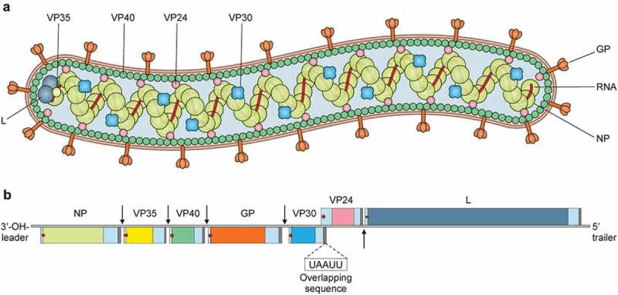
Virion structure and genome organization of Marburg virus (PMID: 35363588)
2.2 Marburg Virus Genome & Classification
MARV belongs to the genus Orthomarburgvirus in the family Filoviridae. Its genome is a negative‑sense single‑stranded RNA of approximately 19 kb. The genome contains seven genes in the order: 3′-UTR-NP-VP35-VP40-GP-VP30-VP24-L-5′-UTR.
Among the seven structural proteins, NP, VP35, VP30, and L form the helical nucleocapsid, which is surrounded by a matrix composed of VP40 and VP24. The glycoprotein GP is the sole target of neutralizing antibodies on the viral surface.
2.3 What Are Marburg Virus Structural Proteins?
Marburg virus encodes seven major structural proteins:
Protein | Full name | Main Function |
NP | Nucleoprotein | Encapsidates viral RNA, forms the nucleocapsid core, protects the genome |
VP35 | Viral protein 35 | Polymerase cofactor, involved in transcription/replication; immune suppression; antagonizes host interferon responses |
VP40 | Viral protein 40 | Matrix protein, mediates viral assembly and budding |
GP | Glycoprotein | Surface spike protein, mediates host cell entry and membrane fusion |
VP30 | Viral protein 30 | Transcriptional activator |
VP24 | Viral protein 24 | Nucleocapsid component; interferon signaling suppression; immune evasion |
L | Large Protein | RNA‑dependent RNA polymerase (RdRp); responsible for viral genome replication and transcription |
2.4 Mechanism of Cell Entry and Replication
MARV entry and replication occur entirely within the host cell cytoplasm, and its life cycle is highly similar to that of Ebola virus.
2.4.1 Viral Attachment and Receptor Recognition
MARV infection begins with the binding of the viral GP protein to host cell surface receptors. GP is the sole mediator of viral entry.
2.4.2 Viral Entry
The virus enters host cells via receptor‑mediated endocytosis. After internalization, the virus is trafficked through endosomes to late endosomes/lysosomes, where host proteases cleave GP. The cleaved GP then binds to endosomal receptors, triggering membrane fusion and release of the viral nucleocapsid into the cytoplasm.
2.4.3 Viral RNA Replication
After nucleocapsid release into the cytoplasm, the L protein (RdRp) together with its cofactor VP35 initiates transcription and replication:
l Transcription: the negative‑sense RNA genome is transcribed into mRNAs, which are translated into viral proteins
l Replication: positive‑sense RNA intermediates are synthesized using negative‑sense RNA as template
l Progeny negative‑sense genomic RNAs are synthesized using positive‑sense RNA as template
VP24 helps the virus evade host immune responses by suppressing interferon signaling pathways.
2.4.4 Viral Assembly and Release
Newly synthesized NP protein associates with progeny genomic RNA and L protein to form new ribonucleoprotein complexes. VP40 recruits nucleocapsids to the cell membrane. The virus acquires its envelope by budding from the host cell membrane, releasing new virions.
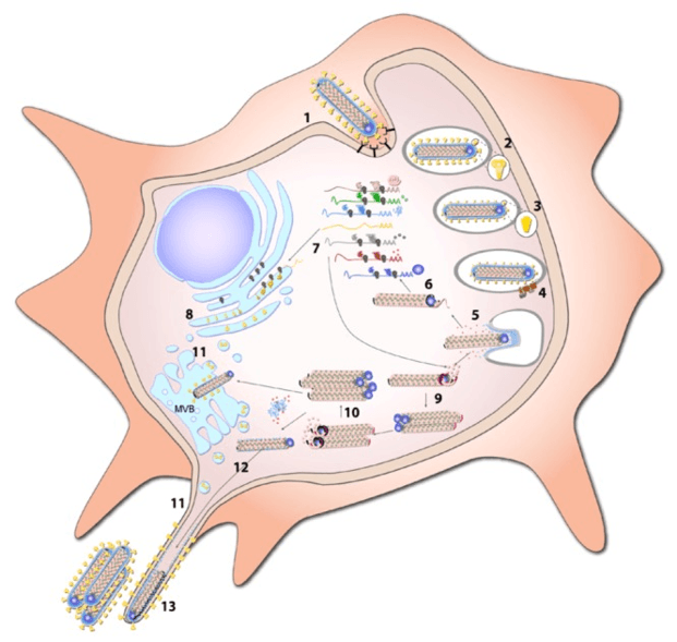
Replication cycle of Marburg virus. (PMID: 23202446)
2.5 Symptoms and Treatment
2.5.1 Clinical Symptoms
The incubation period for Marburg virus disease is typically 2–21 days. Clinical presentation ranges from asymptomatic infection to severe hemorrhagic fever. Common symptoms include:
l Acute onset of fever
l Severe headache
l Malaise
l Muscle pain
l Severe cases may present with profuse diarrhoea, mucosal bleeding, multi‑organ dysfunction, and hypovolemic shock
Case fatality rates vary by outbreak, historically ranging from 24% to 88%, with a pooled average of approximately 50%. The 2025 Ethiopia outbreak had a case fatality rate of 64%–74%
2.5.2 Antiviral Treatment
Currently, no specific antiviral therapy is approved for Marburg virus infection. Treatment is primarily early intensive supportive care:
l Intensive care support
l Intravenous fluid resuscitation to maintain fluid and electrolyte balance
l Respiratory support
l Management of complications
Investigational therapeutics such as remdesivir and the monoclonal antibody MBP091 have been used under expanded access and early‑phase studies.
2.6 Targets for Intervention
2.6.1 Viral Targets
Target | Protein | Biological Role |
GP | Glycoprotein | Host cell entry and membrane fusion |
L (RdRp) | RNA polymerase | RNA transcription and replication |
VP35 | Polymerase cofactor | Transcription/replication, immune suppression |
VP40 | Matrix protein | Assembly and budding |
VP24 | Nucleocapsid protein | Interferon signaling suppression |
2.6.2 Host Receptors & Entry Factors
Host Target | Type | Role in Infection |
Endosomal receptor (e.g., NPC1) | Endosomal receptor | Viral entry |
Cathepsins | Cysteine proteases | GP proteolytic cleavage activation |
Endocytosis | Endocytic pathway | Viral entry |
2.6.3 Host Signaling Pathways
Pathway | Key Targets | Biological Role |
Interferon pathway | IFN‑α/β | Antiviral response, suppressed by VP35 and VP24 |
Inflammatory pathways | Cytokines | Inflammatory response, linked to viral pathogenesis |
2.7 Vaccine Types and Development Progress
Currently, no Marburg virus vaccine is licensed worldwide. However, several candidates have entered clinical trials.
2.7.1 cAd3-Marburg (Chimpanzee Adenovirus‑Vectored Vaccine)
cAd3-Marburg is the most advanced Marburg vaccine candidate, using a chimpanzee adenovirus vector platform.
2.7.2 mRNA Vaccines
A 2025 study published in the Journal of Clinical Investigation demonstrated that an mRNA vaccine encoding the Marburg virus glycoprotein was more protective than a virus‑like particle (VLP)‑forming mRNA vaccine.
2.7.3 Other Vaccine Platforms
Multiple novel vaccine platforms are in development, including VSV‑vectored and adenovirus‑vectored vaccines.
2.8 Drugs
Currently, no specific small‑molecule antiviral is approved for Marburg virus infection. Several novel antiviral candidates have shown significant promise:
2.8.1 Virus-Targeted Drugs
Target | Drug/candidate | Mechanism | Stage |
L protein (RdRp) | Obeldesivir (ODV) | RdRp inhibition | Animal studies |
Remdesivir | RdRp inhibition | Expanded access/early studies | |
GP | AGP-R7 / AGP-R12 | Cross‑neutralizing monoclonal antibodies | Animal studies |
MARV16 | Fully human monoclonal antibody | Animal studies | |
VP40 | Novel VP40 inhibitors | Interfere with viral assembly | Computational drug design |
VP35 | Phytocompounds | VP35 inhibition | In silico screening |
2.8.2 Host-Targeted Drugs
Target | Drug/candidate | Mechanism | Stage |
Supportive care | IV fluids, oxygen therapy | Supportive care | Standard of care |
2.9 Epidemiology
l First identified: 1967, Marburg and Frankfurt, Germany, and Belgrade, former Yugoslavia
l Natural reservoir: Egyptian fruit bat (Rousettus aegyptiacus)
l Geographic distribution: primarily in sub‑Saharan Africa, including DRC, Angola, Uganda, Guinea, Ghana, Equatorial Guinea, Tanzania, Rwanda, and Ethiopia
l Case fatality rates: 23%–90%
l 2021–2025 outbreaks:
Ø 2021: Guinea
Ø 2022: Ghana
Ø 2023: Equatorial Guinea, Tanzania (9 cases, CFR 66.7%)
Ø 2024: Rwanda
Ø 2025: Tanzania (10 cases, CFR 100%), Ethiopia (first outbreak)
l 2025 Ethiopia outbreak details:
Ø Confirmation date: 14 November 2025
Ø Cases: 14 laboratory‑confirmed + 5 probable cases
Ø Deaths: 14 deaths
Ø CFR: 64%–74%
Ø End date: 26 January 2026
l Transmission routes:
Ø Bat → human: contact with infected bat excreta or environments (e.g., caves, mines)
Ø Human → human: direct contact with blood, bodily fluids, or tissues of infected individuals, as well as contaminated surfaces or equipment
2.10 Diagnosis
Diagnosis of Marburg infection relies on laboratory testing:
l RT‑PCR (Reverse Transcription Polymerase Chain Reaction) : detection of viral RNA in blood; gold standard for acute‑phase diagnosis
l Antigen detection: detection of viral antigens
l Serology: detection of anti‑MARV IgM/IgG antibodies
2.11 Prevention
l Avoid contact: avoid contact with fruit bats and their habitats (e.g., caves, mines)
l Personal hygiene: practice good handwashing; avoid contact with infected bodily fluids
l Healthcare settings: healthcare providers should wear gowns, gloves, eye protection, and masks
l Safe burials: implement safe and dignified burial practices
l Surveillance and contact tracing: rapid surveillance and contact tracing are critical for outbreak control
l Vaccines: none currently licensed (see Section 2.7)
3. Related Products & Services
l Recombinant Antigens & Receptors
l Antibodies
l Detection Kits
l Recombinant Protein Expression Services
l Antibody Development Services
4. Resources
5. References
1. Messaoudi, I., Amarasinghe, G. K., Basler, C. F. (2015). Filovirus pathogenesis and immune evasion: insights from Ebola virus and Marburg virus. Nature Reviews Microbiology, 13(11), 663–676.
2. White, J. M., Schornberg, K. L. (2012). A new player in the puzzle of filovirus entry. Nature Reviews Microbiology, 10(5), 317–327.
3. Carette, J. E., Raaben, M., Wong, A. C., et al. (2011). Ebola virus entry requires the cholesterol transporter Niemann–Pick C1. Nature, 477(7364), 340–343.
4. Hartlieb, B., Weissenhorn, W. (2006). Filovirus assembly and budding. Virology, 344(1), 64–70.
5. Mittler, E., Kolesnikova, L., Herwig, A., et al. (2013). Assembly of the Marburg virus envelope. Cellular Microbiology, 15(2), 270–284.
6. Cross, R. W., Mire, C. E., Feldmann, H., et al. (2018). Post-exposure treatments for Ebola and Marburg virus infections. Nature Reviews Drug Discovery, 17(6), 413–434.
7. Hamer, M. J., Choi, E. J., Bieze, A. M., et al. (2023). Safety and immunogenicity of the chimpanzee adenovirus type 3-vectored Marburg virus vaccine in healthy adults in the USA: a first-in-human, phase 1, open-label, dose-escalation trial. The Lancet, 401(10373), 294–302.
8. Subramani, A., et al. (2025). Marburg virus glycoprotein mRNA vaccine is more protective than a virus-like particle-forming mRNA vaccine. Journal of Clinical Investigation, 135(17), e194586.
9. Cross, R. W., Woolsey, C., Prasad, A. N., Borisevich, V., Agans, K. N., Deer, D. J., et al. (2025). Oral obeldesivir provides postexposure protection against Marburg virus in nonhuman primates. Nature Medicine, 31, 2703–2712.
10. Warren, T. K., Wells, J., Panchal, R. G., et al. (2014). Protection against filovirus diseases by a novel broad-spectrum nucleoside analogue BCX4430. Nature, 508(2), 402–405.
11. Towner, J. S., Amman, B. R., Sealy, T. K., et al. (2009). Isolation of genetically diverse Marburg viruses from Egyptian fruit bats. PLoS Pathogens, 5(7), e1000536.
12. Amman, B. R., Jones, M. E. B., Sealy, T. K., et al. (2015). Oral shedding of Marburg virus in experimentally infected Egyptian fruit bats (Rousettus aegyptiacus). Journal of Wildlife Diseases, 51(1), 113–124.
13. Aljohani, A. H., et al. (2023). The mortality, modes of infection, diagnostic tests, and treatments of Marburg virus disease: A systematic review. Journal of Infection and Public Health, 16.
14. Saadh, M. J., et al. (2025). From protein to immunology: comprehensive insights into Marburg virus vaccines, mechanism, and application. Archives of Microbiology.