
info@mail.ucallm.com
Mesenchymal stem cell-derived extracellular vesicles (MSC-EVs) constitute a key component of the paracrine effects of MSCs. By delivering bioactive cargo including proteins, lipids and diverse RNAs, they regulate the proliferation, migration, inflammatory response, angiogenesis and extracellular matrix remodeling of recipient cells, thus participating in the repair and regeneration of injured tissues. Compared with direct cell therapy, MSC-EVs feature cell-free therapeutics, making them a vital research subject in tissue engineering and regenerative medicine.
1.1 Anti-Inflammation and Immune Microenvironment Modulation
MSC-EVs act on immune cells such as macrophages, T cells and dendritic cells to modulate the release of inflammatory cytokines and functional status of immune cells. During tissue injury, MSC-EVs can regulate inflammatory responses and promote inflammation resolution, creating a favorable microenvironment for subsequent tissue repair and regeneration.
1.2 Promotion of Cell Migration and Tissue Repair
MSC-EVs can be internalized by fibroblasts, keratinocytes and other tissue repair-related cells. EV cargo regulates cell proliferation, migration and extracellular matrix remodeling. Especially in wound repair studies, MSC-EVs have been reported to facilitate fibroblast migration, angiogenesis and re-epithelialization, promoting tissue repair through multiple steps.
1.3 Promotion of Angiogenesis
Angiogenesis is an essential step in tissue repair. Carrying a variety of bioactive molecules associated with angiogenesis, MSC-EVs act on vascular endothelial cells to boost endothelial cell migration, proliferation and vessel formation, thereby improving blood supply in injured tissues and supporting tissue regeneration.
1.4 Multi-Tissue Regeneration Applications
Benefiting from the immunomodulatory, pro-angiogenic and tissue-repairing effects of MSC-EVs, relevant research has been extended to multiple tissue injury and regeneration models, including bone repair, cartilage regeneration, wound healing, angiogenesis and neural protection for general tissue regeneration. Among these, bone and cartilage repair research focuses on osteogenic/chondrogenic cell function, inflammatory microenvironment and the coupling between blood vessel formation and tissue regeneration; wound healing research mainly evaluates cell migration, angiogenesis, re-epithelialization and ECM remodeling.
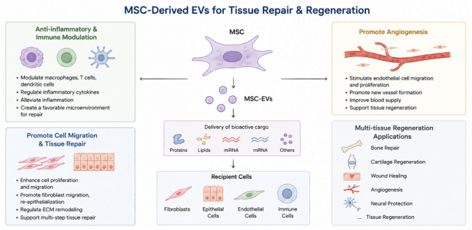
Mesenchymal stem cell-derived extracellular vesicles (MSC-EVs) are utilized for tissue repair and regeneration. Secreted by MSCs, MSC-EVs carry bioactive substances including proteins, lipids and miRNAs. They act on fibroblasts, epithelial cells, endothelial cells and immune cells. Via multiple mechanisms including anti-inflammatory and immunomodulatory effects, promotion of cell migration and tissue repair, and induction of angiogenesis, MSC-EVs enable multi-tissue regenerative applications such as bone repair, cartilage regeneration, wound healing and neuroprotection.
1.5 Representative Validation Model
Using human umbilical cord mesenchymal stem cell-derived extracellular vesicles (hUC-MSC-EVs) as a representative sample, systematic in vitro functional validation models can be established to assess the biological activity of MSC-EVs at multiple levels, including cellular uptake, cell migration and inflammatory regulation. MSC-EVs can be internalized by diverse recipient cells and alter the functional status of recipient cells through delivery of bioactive cargo such as proteins, lipids and RNAs.
In cell migration and tissue repair research, the Scratch Wound Assay and Transwell Migration Assay can be applied to evaluate the effects of hUC-MSC-EVs on the migratory capacity of repair-associated cells including fibroblasts and keratinocytes. Combined with markers of cell proliferation and extracellular matrix, their promotive effects on tissue repair can be analyzed. MSC-EVs have been widely documented to promote cell migration, proliferation, angiogenesis and tissue regeneration.
For anti-inflammatory functional studies, the regulatory effects of hUC-MSC-EVs on inflammatory microenvironments can be evaluated by detecting expression changes of inflammatory factors including TNF-α, IL-1β and IL-6, alongside markers for macrophage polarization and functional indicators of other immune cells. By modulating immune cell function and inflammatory signaling, MSC-EVs create a permissive microenvironment for subsequent repair of damaged tissues.
By integrating EV characterization, cellular uptake detection, cell migration assessment and inflammatory response evaluation, a cellular-level functional validation system for hUC-MSC-EVs can be constructed to further dissect their underlying mechanisms in tissue repair and regeneration. This research framework can be extended to diverse tissue regeneration studies, such as bone repair, cartilage regeneration, cutaneous wound repair and neuroprotection.
Recommended Products:
Product Category | Product Name | Cat.No. | Specification | Core Relevance to This Chapter |
MSC-EV Research Materials | ContExo® High-Purity HuMSC Exosomes | CTE-052 | 2E11 Particles/1 mL | Core product; applied to studies of MSC-EV-mediated tissue repair, immune regulation, angiogenesis, cell migration and regeneration |
EV Isolation and Purification | Exosupur® Rapid Cell Supernatant Purification Kit | ESS0500 / ESS050 | 500 mL / 50 mL | Rapid isolation and purification of EVs from MSC culture supernatant for subsequent functional research on tissue repair |
EV Characterization | ExosuperQC® Exosome 4-Positive-1-Negative WB Characterization Kit | EQC-AP-01 | 5T | Detect CD9, CD63, CD81, ALIX and Calnexin to verify the quality and purity of EV samples |
MSC-EV Identification | ExosuperQC® MSC-Specific Protein Exosome WB Characterization Kit | EQC-SAP-01 | 5T | Combined detection of CD13 and CD73 for identification of MSC-derived EVs |
EV Tracing / Uptake | FluoExo® Exosome Tracing Kit | FE-01 / FE-02 / FE-03 Series | Green / Red / Near-Infrared | For research on MSC-EV cellular uptake, intercellular delivery and interactions with recipient cells |