
info@mail.ucallm.com
```
```
Necroptosis
1 Mechanism Overview
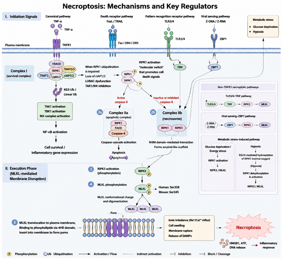
Necroptosis is a caspase‑independent form of programmed cell death regulated by specific molecular mechanisms, with the core regulatory axis consisting of RIPK1–RIPK3–MLKL. This process is typically activated by signaling pathways including TNF‑α/TNFR1, Fas, TRAIL receptors, and TLR3/TLR4. When apoptosis is blocked, particularly due to loss of caspase‑8 activity, the kinase activity of RIPK1 is enhanced and interacts with RIPK3 through their RHIM (RIP homotypic interaction motif) domains, leading to the formation of the necrosome complex. Subsequently, activated RIPK3 phosphorylates MLKL, promoting MLKL oligomerization and translocation to the plasma membrane. Membrane‑associated MLKL forms pores that disrupt plasma membrane integrity, resulting in ionic imbalance, cellular swelling, and membrane rupture, ultimately causing necroptotic cell death. In addition to the classical TNFR1‑mediated pathway, necroptosis can also be triggered by the ZBP1–RIPK3 pathway during viral infection, the TLR3/TLR4–TRIF pathway, as well as metabolic stresses such as hypoxia and glucose deprivation, all of which activate the RIPK1/RIPK3/MLKL signaling axis. By promoting inflammatory responses through the release of intracellular damage‑associated molecular patterns (DAMPs), necroptosis plays important roles in infection, ischemic injury, inflammatory diseases, and tumor development and progression.
Necroptosis pathway schematic. Upon stimulation by TNF‑α/TNFR1, TLR3/4, ZBP1 signaling, and metabolic stress, RIPK1 is activated and interacts with RIPK3 to form the necrosome complex. When caspase‑8 activity is inhibited, RIPK3 further phosphorylates MLKL, promoting MLKL oligomerization and translocation to the plasma membrane, where it forms membrane pores. Ultimately, increased membrane permeability leads to ionic homeostasis disruption, cellular swelling, and plasma membrane rupture, resulting in the release of damage‑associated molecular patterns (DAMPs) and the induction of inflammatory responses.
2 Target List
| Mechanism Module | Target / Detection Marker | Full Name | Biological Function / Mechanistic Significance | Recommended Detection |
|---|---|---|---|---|
| Necroptosis Initiation Recognition | TNFR1 | Tumor necrosis factor receptor 1 | Classical upstream receptor mediating TNF‑α‑induced necroptosis | WB / IF / Flow cytometry |
| TRADD | TNFR1‑associated death domain protein | Component of the TNFR1 death‑inducing signaling complex | WB / IP | |
| FADD | Fas‑associated death domain protein | Regulates the decision between apoptosis and necroptosis | WB / IP | |
| RIPK1 Activation Assessment | RIPK1 | Receptor‑interacting protein kinase 1 | Core initiator protein of necroptosis | WB / IF |
| p‑RIPK1 (Ser166) | Phosphorylated receptor‑interacting protein kinase 1 at Ser166 | Marker of RIPK1 kinase activation and necrosome formation | WB | |
| RIPK1 ubiquitination | RIPK1 ubiquitination modification | Regulates RIPK1 fate determination from survival signaling to death signaling | IP‑WB | |
| Necrosome Formation Detection | RIPK1–RIPK3 interaction | Receptor‑interacting protein kinase 1–RIPK3 interaction | Direct evidence of necrosome formation | Co‑IP / PLA |
| RIPK3 | Receptor‑interacting protein kinase 3 | Core kinase of necroptosis | WB / IF | |
| p‑RIPK3 (Ser227 human / Thr231‑Ser232 mouse) | Phosphorylated RIPK3 activation sites | Activation marker of RIPK3 | WB | |
| MLKL Execution Phase | MLKL | Mixed lineage kinase domain‑like protein | Final execution protein of necroptosis | WB / IF |
| p‑MLKL (Ser358 human / Ser345 mouse) | Phosphorylated mixed lineage kinase domain‑like protein | Most classical and recommended biomarker of necroptosis | WB / IF | |
| MLKL oligomerization | MLKL oligomer formation | Indicates MLKL activation and membrane‑targeting capability | Native PAGE / Cross‑linking | |
| MLKL membrane translocation | MLKL translocation to plasma membrane | Indicates recruitment of MLKL to the plasma membrane for execution | IF / Cell fractionation | |
| Membrane Rupture and Cell Death Validation | LDH release | Lactate dehydrogenase release | Indicator of plasma membrane integrity disruption | ELISA |
| PI uptake / SYTOX Green | Propidium iodide uptake / SYTOX Green staining | Detects increased membrane permeability during late‑stage cell death | Flow cytometry | |
| HMGB1 release | High mobility group box 1 release | Marker of DAMP release and inflammatory cell death | WB / ELISA | |
| ATP depletion | Cellular ATP depletion | Indicates energy depletion associated with necroptotic death | ATP assay | |
| TLR/Virus‑Induced Necroptosis Pathways | TLR3 | Toll‑like receptor 3 | Upstream mediator of TRIF‑dependent necroptosis | WB |
| TLR4 | Toll‑like receptor 4 | Mediates LPS‑induced necroptosis | WB | |
| TRIF | TIR‑domain‑containing adapter‑inducing interferon‑β | Adaptor protein linking TLR3/4 signaling to RIPK3 activation | WB | |
| ZBP1 | Z‑DNA binding protein 1 | Induces necrosome formation during viral infection | WB / IF | |
| Metabolic Stress‑Related Pathways | HIF‑1α | Hypoxia‑inducible factor 1‑alpha | Regulator of hypoxia‑induced necroptosis | WB |
| EGLN/PHD proteins | Egl‑9 family hypoxia‑inducible factor proteins / Prolyl hydroxylase domain proteins | Regulate RIPK1 hydroxylation status | WB | |
| RIPK1 hydroxylation | RIPK1 hydroxylation modification | Mechanistic indicator of RIPK1 activation under hypoxic conditions | IP‑MS / WB | |
| Exclusion of Apoptosis | Cleaved‑Caspase‑8 | Cleaved cysteine‑dependent aspartate‑directed protease 8 | Determines whether signaling is redirected toward apoptosis | WB |
| Cleaved‑Caspase‑3 | Cleaved cysteine‑dependent aspartate‑directed protease 3 | Excludes classical apoptosis activation | WB | |
| Cleaved‑PARP | Cleaved poly(ADP‑ribose) polymerase | Terminal marker of apoptosis | WB | |
| Exclusion of Pyroptosis | GSDMD‑N | Gasdermin D N‑terminal domain | Excludes pyroptotic cell death | WB |
| Caspase‑1 cleavage | Activated Caspase‑1 cleavage | Excludes inflammasome‑mediated pyroptosis activation | WB | |
| Exclusion of Ferroptosis | GPX4 | Glutathione peroxidase 4 | Excludes ferroptosis activation | WB |
| SLC7A11 | Solute carrier family 7 member 11 | Cystine/glutamate antiporter system involved in ferroptosis regulation | WB | |
| ACSL4 | Acyl‑CoA synthetase long‑chain family member 4 | Ferroptosis‑promoting factor involved in PUFA metabolism | WB |
3 Application Scheme
| Category | Marker |
|---|---|
| Core necroptosis signaling axis | RIPK1 + p‑RIPK1 (Ser166) |
| RIPK3 + p‑RIPK3 | |
| MLKL + p‑MLKL (Ser358) | |
| Membrane rupture validation | LDH release / PI uptake |
| Exclusion of apoptosis | Cleaved‑Caspase‑3 + Cleaved‑Caspase‑8 |
| Exclusion of pyroptosis | GSDMD‑N |
4 References
- Yuan J, Ofengeim D.A guide to cell death pathways. Nature Reviews Molecular Cell Biology. 2024;25(6):379‑395. doi: 10.1038/s41580‑023‑00689‑6.
- Zhang T, Xu D, Liu J, et al. Prolonged hypoxia alleviates prolyl hydroxylation‑mediated suppression of RIPK1 to promote necroptosis and inflammation. Nature Cell Biology. 2023;25:950‑962. doi: 10.1038/s41556‑023‑01170‑4.
- Galluzzi L, Vitale I, Aaronson SA, et al. Molecular mechanisms of cell death: recommendations of the Nomenclature Committee on Cell Death 2018. Cell Death & Differentiation. 2018;25:486‑541. doi: 10.1038/s41418‑017‑0012‑4.
- Clucas J, Meier P. Roles of RIPK1 as a stress sentinel coordinating cell survival and immunogenic cell death. Nature Reviews Molecular Cell Biology. 2023;24:835‑852. doi: 10.1038/s41580‑023‑00623‑w.
- Chen X, Zhu R, Zhong J, Ying Y, Wang W, Cao Y, Cai H, Li X, Shuai J, Han J. Mosaic composition of RIP1–RIP3 signalling hub and its role in regulating cell death. Nature Cell Biology. 2022;24(4):471‑482. doi: 10.1038/s41556‑022‑00854‑7.