
info@mail.ucallm.com
1. Multiple Sclerosis
Multiple sclerosis (MS) is a chronic immune-mediated disease of the central nervous system (CNS), characterized by inflammation, demyelination, axonal damage, and progressive neurodegeneration. It is a major cause of neurological disability in young adults and can impose a substantial long-term burden on patients.
The exact causes of MS remain unclear. Genetic susceptibility, environmental factors, and dysregulated immune responses are thought to contribute to disease onset and progression. Abnormal activation of autoreactive T and B cells promotes CNS inflammation and damages neural tissues.
MS can manifest as visual impairment, sensory abnormalities, motor dysfunction, impaired coordination, gait disturbances, fatigue, and cognitive deficits. Pathologically, inflammatory lesions, oligodendrocyte loss, demyelination, and axonal injury occur throughout the brain and spinal cord. Although remyelination and tissue repair can occur, persistent inflammation and progressive neurodegeneration can lead to irreversible neurological disability.
Despite major advances in disease-modifying therapies, current treatments primarily target inflammatory and immune responses and remain insufficient to prevent progressive neurological deterioration in many patients. A deeper understanding of the molecular mechanisms underlying inflammation, demyelination, and neurodegeneration is therefore urgently needed to identify new biomarkers and therapeutic targets for MS.
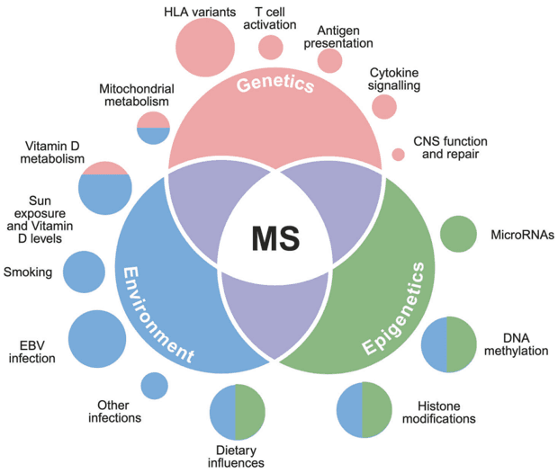
Figure 1. Genetic, Environmental, and Epigenetic Factors Shaping MS Risk (PMID: 41034190).
2. PTMs Studied in MS
Accumulating evidence indicates that diverse PTMs regulate myelin stability, immune response, inflammation, and neurodegeneration in MS. The table below summarizes PTMs that have been investigated in MS and their representative protein targets and biological relevance.
PTM Type | Representative Target Proteins | Biological Processes | Journal Article |
Citrullination | MBP, GFAP, vimentin, PAD2 | Myelin destabilization; antigen generation; autoimmune response; demyelination | PMID: 38425098 PMID: 37506490 PMID: 42139970 |
Phosphorylation | PIM1, Drp1, CSF1R | Myelin stability; cytoskeletal regulation; axonal integrity | PMID: 42331266 PMID: 33097690 |
Acetylation | MBP, histones | Myelin stability; chromatin regulation; Inflammatory responsed | PMID: 29111742 PMID: 42139970 |
Ubiquitination | MBP, RORγt, Nedd4, ABCA1, UBE3A
| Protein degradation; antigen processing; proteostasis | PMID: 39972304 PMID: 40883260 |
Glycosylation | CTLA-4, MOG | Protein stability; immune recognition; cellular signaling | PMID: 41939892 PMID: 21629267 PMID: 16157927 |
3. Representative PTM Research Cases in MS
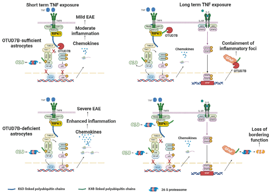
Figure 2. Mechanisms underlying the dual protective roles of astrocytic OTUD7B in EAE (PMID: 41115891).
Title: Astrocytic-OTUD7B ameliorates murine experimental autoimmune encephalomyelitis by stabilizing glial fibrillary acidic protein and preventing inflammation.
Introduction: Reactive astrocytes play complex and context-dependent roles in multiple sclerosis (MS). During CNS inflammation, they produce chemokines that recruit encephalitogenic T cells and amplify inflammatory response, while also forming GFAP-positive astrocytic borders that restrict lesion spread. Ubiquitination and deubiquitination are important regulators of inflammatory signaling; however, how deubiquitination coordinates these distinct astrocyte functions during autoimmune neuroinflammation remains poorly understood.
Biological Questions:
1) Does astrocytic OTUD7B regulate autoimmune neuroinflammation and EAE progression?
2) How does OTUD7B control the pro-inflammatory response of astrocytes and the recruitment of encephalitogenic T cells?
3) Does OTUD7B regulate TNF-induced inflammatory signaling through RIPK1 deubiquitination?
4) How does OTUD7B regulate GFAP stability and the formation of astrocytic borders around inflammatory lesions?
Key Findings:
1) Astrocytic OTUD7B protects against EAE by limiting neuroinflammation in the CNS.
2) OTUD7B suppresses chemokine expression in lesion-associated astrocytes and reduces the recruitment of encephalitogenic CD4+ T cells.
3) OTUD7B sequentially removes K63- and K48- linked ubiquitin chains from PIPK1, thereby limiting NF-κB and MAPK activation and TNF-induced chemokine production.
4) OTUD7B maintains GFAP expression by supporting its transcription and preventing proteasomal degradation through K48- linked deubiquitination.
Research Strategy:
To address these questions, the study combined astrocyte-specific genetic manipulation with EAE models, transcriptomic profiling, spatial analysis, and molecular assay to define the role of OTUD7B in autoimmune neuroinflammation and dissect the underlying deubiquitination mechanisms.
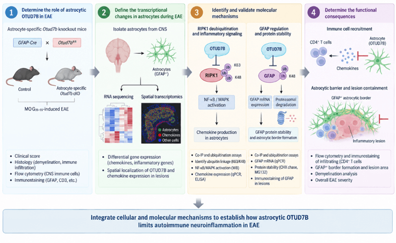
Figure 3. Experimental Strategy for Dissecting the Role of Astrocytic OTUD7B in EAE.
Summary:
This study identifies astrocytic OTUD7B as a key regulator of autoimmune neuroinflammation in EAE. Through deubiquitination of RIPK1 and GFAP, OTUD7B suppresses inflammatory signaling and maintains astrocytic barrier function, thereby limiting immune cell infiltration and disease progression. These findings highlight deubiquitination as an important regulatory mechanism linking astrocyte function to autoimmune neuroinflammation.
5. Key References
1) Boutitah-Benyaich I, Eixarch H, Villacieros-Álvarez J, et al. Multiple sclerosis: molecular pathogenesis and therapeutic intervention. Signal Transduct Target Ther. 2025;10(1):324.
2) Harit K, Yi W, Jeron A, et al. Astrocytic-OTUD7B ameliorates murine experimental autoimmune encephalomyelitis by stabilizing glial fibrillary acidic protein and preventing inflammation. Nat Commun. 2025;16(1):9279.
3) Jakimovski D, Bittner S, Zivadinov R, et al. Multiple sclerosis. Lancet. 2024;403(10422):183-202.