
info@mail.ucallm.com
I. Targeting Advantages of RVG Peptide: From Viral Neurotropism to a Precision Targeting Tool
1.1 Molecular Targeting Mechanism
The RVG (Rabies Virus Glycoprotein) peptide is a 29‑amino‑acid targeting peptide derived from the rabies virus glycoprotein[1]. It specifically binds to the nicotinic acetylcholine receptor (nAChR), which is highly expressed on brain capillary endothelial cells and neurons[2].
1.2 Dual Mechanism for Blood–Brain Barrier Penetration
The RVG peptide crosses the blood–brain barrier (BBB) via receptor‑mediated transcytosis (RMT) through the following four steps:
Step 1: RVG peptide binds to nAChR on the luminal surface of brain microvascular endothelial cells[3];
Step 2: Receptor‑mediated endocytosis is triggered, forming intracellular vesicles;
Step 3: Vesicles are transported across the endothelial cells and released at the basolateral (brain parenchymal) side;
Step 4: The released RVG peptide further targets nAChR on neurons, enabling precise intra‑brain delivery[4].
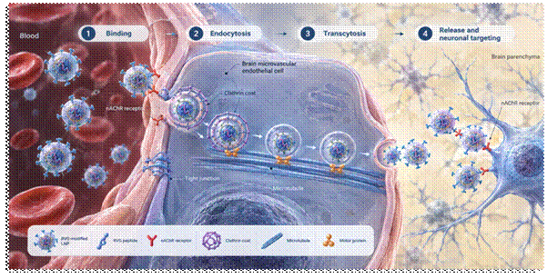
Figure 1. Schematic of the RVG peptide‑mediated BBB penetration mechanism.
II. Product Features: RVG Lipid‑Anchor Kit
Based on the principle of “like dissolves like,” Yaokemei has developed the RVG Lipid‑Anchor Kit as an excellent tool for surface engineering of exosomes. The kit contains a PEG‑cholesterol‑conjugated FAM‑RVG targeting peptide. Cholesterol, being amphiphilic with both lipophilic and hydrophilic moieties (the former being dominant), can insert itself between phospholipid molecules upon interaction with phospholipids, thereby anchoring the cargo molecule onto the outer surface of exosomes. FAM is a green fluorescent dye with excitation at 488 nm and emission at 525 nm. By chemically linking these three components (PEG, cholesterol, and FAM‑RVG), the kit enables the attachment of green‑fluorescent‑labeled RVG targeting peptides onto the exosome surface, making it a superior tool for exosome surface modification.
2.1 Core Components
Table 1. Composition of the RVG Lipid‑Anchor Kit
Component | Specification | Storage Condition |
RVG‑lipid‑anchor targeting peptide | 5 mg/vial | –20 °C |
Co‑incubation Reaction Buffer | 40 mL/bottle | 4 °C |
Wash Buffer (for ultrafiltration) | 250 mL×3 bottles | Room temperature |
2.2 Example Data
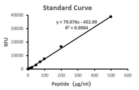
Figure 2. Fluorescence quantitative standard curve for the lipid‑anchored targeting peptide.
Table 2. Example calculation of exosome loading after lipid‑anchoring reaction and purification using the fluorescence quantitative standard curve.
Fluorescence value (10-fold dilution) | Concentration (ug/mL) | Targeted peptide amount (ug) | BCA concentration (ug/mL) | Capacity (nmol/mg) |
357 | 10.24 | 25.61 | 835.83 | 19.16 |
2.3 Precautions
Content | Description |
Form | Liquid/lyophilized powder |
Transport | Ice‑pack shipping |
Storage | –20 °C/room temperature/4 °C (as specified) |
Usage suggestion | Vortex/mix thoroughly before use; protect from light during operation |
Shelf life | Lyophilized powder: 2.5 years; after reconstitution: 1 year at –20 °C |
Important notes | 1. Avoid repeated freeze‑thaw cycles. Due to the inherent properties of the lipid‑anchored peptide, aggregation may occur over time; use promptly after thawing. |
2.4 Applycation
1.Co‑incubation modification: Directly connect targeting peptide molecules via simple co‑incubation to obtain engineered exosomes with targeting capability.
2.Disease directions: Targeting nAChR for functional molecule delivery in mechanistic studies of various brain disorders, including neurodegenerative, infectious, inflammatory, genetic, and neoplastic diseases–such as glioblastoma and other brain tumors, ischemic stroke, major depression, Parkinson’s disease, Alzheimer’s disease, subarachnoid hemorrhage, and cerebral/fetal Zika virus (ZIKV) infection.
[1] Wang Q, Wang D, Liu P, Tang BZ, Li Y, Yin X. Inflammation-targeted nanoplatform: NIR-II imaging-guided encephalitis suppressing by dual antioxidant-ferroptosis action. Biomaterials. 2026 Apr;327:123741.
[2] Izco M, Sola C, Schleef M, Schmeer M, de Toro M, Verona G, Carlos E, Reinares-Sebastian A, Colina S, Marzo-Sola ME, Garcia-Sanmartin J, Fernández-Irigoyen J, Santamaría E, Mugica-Vidal R, Blesa J, Alvarez-Erviti L. Development of human targeted extracellular vesicles loaded with shRNA minicircles to prevent parkinsonian pathology. Transl Neurodegener. 2025 May 26;14(1):26.
[3] Gupta A, Ramanathan R, Raulji CM, I Mahato R. Nanoparticulate delivery and targeting of RNA to the brain. Biochim Biophys Acta Rev Cancer. 2025 Nov;1880(6):189480.
[4] Ding L, Xiao Y, Xu Z, Zhu Z, Gan T, Huang X, Shu H, Liang X, Mo M, Huang X, Zhu X, Huang W, Xu P, Zhang W. RVG-targeted extracellular vesicles loaded with echinatin attenuate dopaminergic neurodegeneration via the IGF-2/PI3K/Akt pathway in Parkinson's disease mice. J Nanobiotechnology. 2026 Jan 7;24(1):117.