
info@mail.ucallm.com
1. Overview
Saint Louis encephalitis virus (SLEV) is a member of the genus Orthoflavivirus within the family Flaviviridae and is the causative agent of St. Louis encephalitis. It is an enveloped, positive-sense, single-stranded RNA virus with a spherical virion approximately 40–50 nm in diameter. SLEV was first isolated during a large encephalitis outbreak in St. Louis, USA, in 1933 and is one of the most important mosquito-borne encephalitis pathogens in the Americas.
SLEV is transmitted primarily by Culex mosquitoes and is maintained in a natural cycle between wild birds and mosquitoes, with humans and horses serving as dead-end hosts. In 2015, SLEV re-emerged in the western United States after more than a decade of apparent absence and has since persisted in California, with its geographic range expanding to previously unaffected areas. In 2025, SLEV cases in the United States increased compared with 2024, and neuroinvasive cases also increased. No licensed SLEV vaccine or specific antiviral drug is currently available, and treatment is supportive.
2. Background Information
2.1 What is Saint Louis Encephalitis Virus?
SLEV is an enveloped, positive-sense, single-stranded RNA virus belonging to the family Flaviviridae and genus Orthoflavivirus. The genome is a positive-sense single-stranded RNA containing a single open reading frame (ORF) encoding a polyprotein precursor that is cleaved by viral and host proteases into three structural proteins—capsid (C), precursor membrane (prM/M), and envelope (E)—and seven non-structural proteins (NS1, NS2A, NS2B, NS3, NS4A, NS4B, NS5). SLEV is closely related to Japanese encephalitis virus, Powassan virus, and West Nile virus.
A 2026 cryo-EM study resolved the structures of mature and immature SLEV particles at 3.3 Å and 4.4 Å resolution, respectively. SLEV is characterised by glycosylated E and prM proteins, the presence of a lipid pocket, and a complex network of inter- and intra-protein interactions between E and (pr)M across maturation stages. Non-lethal single mutations in the conserved histidine residues H285 and H443 of the E protein (H285R and H443R) affect SLEV replication in mammalian and mosquito cell lines and delay death in infected mouse models.
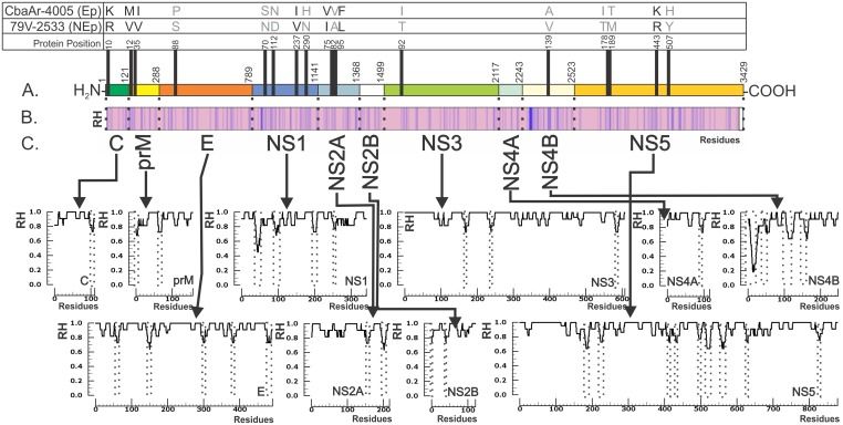
Amino acid differences between epidemic (Ep) and non-epidemic (NEp) SLEV strains and the polyprotein conservation profile. (PMID: 26312485)
2.2 SLEV Genome & Classification
The SLEV genome is a positive-sense single-stranded RNA with a 98-nucleotide 5′ non-coding region, followed by a single continuous open reading frame encoding capsid (C), pre-membrane/membrane (prM/M), and envelope (E) proteins in order, along with seven non-structural proteins (NS1, NS2A, NS2B, NS3, NS4A, NS4B, NS5). SLEV shares amino acid sequence homology of 39%, 66%, 64%, and 43% with yellow fever, Murray Valley encephalitis, West Nile, and dengue viruses, respectively. SLEV belongs to the Japanese encephalitis serocomplex within the genus Orthoflavivirus.
2.3 What Are SLEV Proteins?
SLEV encodes 10 proteins, including 3 structural and 7 non-structural proteins.
2.3.1 Structural Proteins
Protein | Full name | Main Function |
C | Capsid | Encapsidates viral genomic RNA, forming the nucleocapsid core |
prM | pre-Membrane/Membrane | prM is cleaved by furin during maturation to produce M protein; involved in assembly and envelope formation |
E | Envelope | Major surface protein; mediates receptor binding and membrane fusion; primary target of neutralizing antibodies; contains conserved histidine residues H285 and H443 that affect viral fitness |
2.2.2 Non-structural Proteins
The seven non-structural proteins (NS1, NS2A, NS2B, NS3, NS4A, NS4B, NS5) participate in viral RNA replication, virion assembly, and modulation of host immune responses. NS3 possesses both RNA helicase and NTPase activities, is essential for viral genome replication, and is an important target for antiviral drug development. A 2026 study resolved the crystal structure of the SLEV NS3 helicase at 2.5 Å resolution, revealing three subdomains (I–III), of which domains I and II adopt a RecA-like fold critical for NTPase and helicase function. NS5 possesses RNA-dependent RNA polymerase (RdRp) and methyltransferase activities and is the core enzyme for viral replication.
2.4 Mechanism of Cell Entry and Replication
2.4.1 Viral Attachment and Receptor Recognition
SLEV infection begins with the binding of the viral envelope protein E to host cell surface receptors. Like most flaviviruses, SLEV utilises low-density lipoprotein receptor (LDLR) family members and heparan sulfate proteoglycans (HSPGs) as receptors or attachment factors to mediate initial binding and internalisation. In addition, studies suggest that SLEV may enter cells through apoptotic mimicry, using phosphatidylserine (PS) on the viral envelope surface to bind host cell receptors and be taken up by cells.
Host cell molecular chaperones play important auxiliary roles during viral attachment. Heat shock protein 70 (HSP70) and glucose-regulated protein 78 (GRP78) assist in the folding and maturation of viral proteins, especially the E protein, whose correct conformation is essential for viral entry and assembly. In addition, host cell proteases such as furin and signal peptidase participate in processing the viral polyprotein to generate mature structural and non-structural proteins.
2.4.2 Viral Entry and Uncoating
SLEV enters host cells primarily via clathrin-mediated endocytosis. After E protein binding to receptors, viral particles are internalised through clathrin-coated pits, forming clathrin-coated vesicles. After entering the cell, the vesicles fuse with early endosomes.
Acidification of the endosome is a key step triggering viral membrane fusion. As endosomal pH decreases, the E protein undergoes conformational changes, exposing its fusion peptide and mediating fusion of the viral envelope with the endosomal membrane. After fusion, the viral nucleocapsid is released into the host cell cytoplasm and subsequently uncoated, releasing the viral genomic RNA. Studies have shown that agents altering endosomal pH, such as chloroquine, affect the efficiency of SLEV entry, confirming a pH-dependent entry mechanism.
2.4.3 Viral RNA Replication
After release into the cytoplasm, the viral genomic RNA (approximately 11 kb positive-sense RNA) immediately initiates translation on rough endoplasmic reticulum (ER) membranes, producing a polyprotein precursor. This precursor is co- and post-translationally cleaved by viral and host proteases into three structural proteins (C, prM/M, E) and seven non-structural proteins (NS1, NS2A, NS2B, NS3, NS4A, NS4B, NS5).
The non-structural proteins assemble into a replication complex on ER membranes, which associates with ER-derived membrane vesicles. Pulse-labelling studies show that the SLEV replication complex associates with membrane structures in the infected cytoplasm, with a sedimentation coefficient of approximately 250S, and contains viral RNA polymerase, single-stranded viral RNA, and replication intermediates of SLEV RNA.
Within the replication complex, NS5 protein, as the RNA-dependent RNA polymerase (RdRp) and methyltransferase, catalyses viral RNA synthesis and 5′ capping. NS5 first synthesises a negative-sense RNA intermediate using the positive-sense RNA as template, and then synthesises progeny positive-sense genomic RNA using the negative-sense RNA as template. NS1 protein plays a key role in forming the replication complex, participating in recruiting other non-structural proteins to ER-derived membrane structures.
NS3 protein has dual functions in viral replication. Its N-terminal domain acts as a chymotrypsin-like serine protease, forming a complex with the NS2B cofactor to cleave the viral polyprotein; its C-terminal domain exerts RNA helicase and nucleoside triphosphatase (NTPase) activities, driving genome replication by unwinding double-stranded RNA. The 2026 SLEV NS3 helicase crystal structure (2.5 Å resolution) revealed three subdomains (I–III), of which domains I and II adopt a RecA-like fold critical for NTPase and helicase function; domain III is mainly α-helical and partially disordered in the crystal structure. The RNA-binding cleft lies between domains I/II and III, with a strongly positive electrostatic surface, and conserved residues interact with the ribose 2′-hydroxyl and phosphate backbone of single-stranded RNA.
2.4.4 Viral Assembly and Maturation
Newly synthesised progeny positive-sense RNA genomes associate with capsid protein C to form nucleocapsids. Virion assembly occurs on ER membranes, and immature virions bud into the ER lumen. Immature virions display prM-E heterodimers on their surface; prM prevents premature conformational changes in E protein during transport.
Immature virions are then transported through the Golgi apparatus. During this process, furin protease cleaves prM to generate mature M protein, triggering rearrangement of the viral envelope proteins and maturing the virion. The E protein of mature virions adopts a postfusion trimer conformation and is infectious.
2.4.5 Viral Release
Mature virions are released primarily via exocytosis. In late infection, cell lysis can also serve as a release route. In mosquito cells, electron microscopy studies show that virus predominantly accumulates in ER cisternae and is subsequently released from the apical plasma membrane into the glandular lumen. Viral release exhibits directional preference—large amounts of virus are released through the apical plasma membrane, with little association with lateral or basal margins.
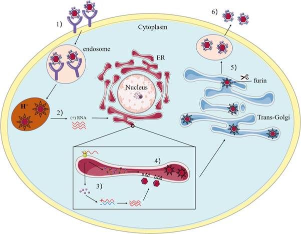
The life cycle of SLEV. (PMID: 33841381)
2.5 Symptoms and Treatment
2.5.1 Clinical Symptoms
The incubation period of SLEV infection is typically 4–14 days. Approximately 99% of infections are asymptomatic. About 1% of infected individuals develop clinical symptoms, ranging from mild febrile illness to severe neuroinvasive disease:
l Fever
l Headache
l Dizziness
l Nausea, vomiting
l Generalised weakness
Symptoms usually begin abruptly and worsen over days to a week. Some patients progress to central nervous system infection, presenting with encephalitis or meningitis, with symptoms including neck stiffness, confusion, disorientation, tremor, and unsteadiness. Severe cases may develop coma.
Case fatality rate: among patients diagnosed with St. Louis encephalitis, 5%–20% die; the risk of death increases with age. US median data from 2014–2018 showed a neuroinvasive rate of 62% and a case fatality rate of 9%. Children and young adults usually have milder disease, while the elderly are more likely to develop severe disease.
2.5.2 Antiviral Treatment
Currently, no specific antiviral therapy is approved for SLEV. Treatment is primarily supportive:
l Antiemetics
l Analgesics
l Hospitalisation: severe patients usually require hospitalisation, supportive care, and/or rehabilitation
l Antiviral drugs: have not been shown to alter the symptoms of SLEV infection
2.6 Targets for Intervention
2.6.1 Viral Targets
Target | Protein | Biological Role |
E | Envelope protein | Receptor binding & membrane fusion |
NS3 | Helicase/NTPase | RNA unwinding, viral genome replication |
NS5 | RNA polymerase (RdRp) | RNA replication |
Methyltransferase | 5′ cap methylation | |
C | Capsid | Nucleocapsid formation |
2.6.2 Host Receptors & Entry Factors
Host Target | Type | Role in Infection |
Not fully characterised | Receptor | Viral entry |
TLR-4 | Pattern recognition receptor | Immune receptor target in vaccine design |
2.6.3 Host Signaling Pathways
Pathway | Key targets | Biological role |
Interferon pathway | IFN-α/β | Antiviral response |
Inflammatory signaling | NF-κB | Neuroinflammatory response |
2.6.4 Immune & Inflammatory Targets
Target | Category | Major role |
IFN-α | Cytokine | Antiviral response |
IFN-β | Cytokine | Antiviral response |
IL-6 | Cytokine | Inflammatory response |
TNF-α | Cytokine | Inflammation |
2.7 Vaccine Types and Development Progress
Currently, no SLEV vaccine is licensed worldwide. However, several vaccine candidates are in development.
2.7.1 Multi-epitope Subunit Vaccine (Reverse Vaccinology Design)
A 2025 study published in Frontiers in Immunology used reverse vaccinology and immunoinformatics to design a multi-epitope subunit vaccine targeting SLEV membrane glycoprotein M, envelope protein E, and anchored capsid protein anchC. The vaccine construct has the following characteristics:
l Structurally stable, antigenic, non-allergenic, non-toxic, and soluble
l Molecular docking showed significant binding affinity to TLR-4, and molecular dynamics simulations supported its stability
l Potential to induce immune responses
l Cloned into pET-28a(+)-6xHis-TEV-ORF9c expression vector for further experimental validation
The authors suggest evaluating the vaccine's therapeutic effect through in vitro and in vivo studies in the near future.
2.7.2 Other Vaccine Directions
No other SLEV vaccine candidates have entered clinical trials. Given the antigenic relatedness of SLEV to Japanese encephalitis virus, West Nile virus, and other flaviviruses, chimeric vaccine platforms or conserved-antigen-based pan-flavivirus vaccine strategies may provide references for SLEV vaccine development.
2.8 Drugs
Currently, no specific small-molecule antiviral is approved for SLEV.
2.8.1 Virus-Targeted Drugs
Target | Drug/candidate | Mechanism | Stage |
NS3 helicase | Carnosine | Molecular docking shows binding to SLEV helicase active site | Computational screening |
Papain inhibitor | Targets SLEV helicase | Computational screening | |
estatin | Targets SLEV helicase | Computational screening | |
THIP hydrochloride | Targets SLEV helicase | Computational screening |
2.8.2 Host-Targeted Drugs
No host-targeted drugs for SLEV have entered clinical research.
2.8.3 Drugs with Unclear or Non-Specific Targets
No drugs with unclear or non-specific mechanisms have entered clinical research for SLEV. The efficacy of broad-spectrum antivirals (e.g., ribavirin, interferon) against SLEV has not been established.
2.9 Epidemiology
l First identified: 1933, St. Louis, USA
l Geographic distribution: the Americas, including the United States, Canada, Mexico, Central America, and South America. SLEV re-emerged in the western United States after 2015 and has continued to circulate
l Vectors: Culex mosquitoes, mainly Culex tarsalis and Culex quinquefasciatus
l Natural cycle: mosquitoes ↔ wild birds; humans and horses are dead-end hosts
l 2025 US outbreak: SLEV cases increased by 200% compared with 2024, and neuroinvasive cases also increased by 200%
l 2025 detection: in September 2025, SLEV was detected for the first time in Midland County, Texas
l Argentina: a 2025 study confirmed continued active circulation of SLEV in multiple mosquito species, indicating broad vector adaptability
l Haemovigilance survey: a 2025 haemovigilance survey provided evidence of silent SLEV circulation, detecting SLEV RNA (strain OR236721) in blood donors, with 97% homology to isolates from Córdoba (2005 epidemic), Santa Fe (1979 non-epidemic), and Buenos Aires (2013 non-epidemic), Argentina
2.10 Diagnosis
Diagnosis of SLEV infection relies on laboratory testing:
l Virus isolation: isolation of SLEV from clinical material
l Nucleic acid testing (NAT) : detection of SLEV RNA in blood or cerebrospinal fluid (RT-PCR), targeting the conserved NS5 gene region
l Serology: detection of anti-SLEV IgM antibodies in serum (ELISA), or detection of seroconversion or four-fold or greater rise in SLEV-specific IgG titres; the most commonly used diagnostic method
Due to serological cross-reactivity with other flaviviruses (e.g., West Nile, Japanese encephalitis, dengue), confirmation should be interpreted alongside clinical and epidemiological information, with neutralisation tests (PRNT) used when necessary.
2.11 Prevention
l Vaccination: no licensed SLEV vaccine is currently available
l Avoid mosquito bites: the most effective way to prevent SLEV
Ø Avoid activities in areas with SLEV outbreaks
Ø Avoid contact with standing water (e.g., ponds, swamps); empty water-holding containers such as bird baths, buckets, watering cans, and trash cans
Ø Use mosquito repellents and wear long-sleeved clothing
Ø Install window and door screens
l Personal protective measures: wear appropriate personal protective equipment (PPE) and use repellents when in mosquito-active areas
l Surveillance and reporting: SLEV is a notifiable disease in the United States; strengthen surveillance of human cases, mosquito vectors, and avian hosts
l Blood safety: implement haemovigilance to effectively detect silent SLEV circulation and prevent transfusion transmission
3. Related Products & Services
l Recombinant Antigens & Receptors
l Antibodies
l Detection Kits
l Recombinant Protein Expression Services
l Antibody Development Services
4. Resources
5. References
1. Ardakani, R., Chauhan, L., Piquet, A. L., et al. (2024). An Overview of Saint Louis Encephalitis. The Neurohospitalist, 14(2), 230–231.
2. Kopp, A., Gillespie, T. R., Hobelsberger, D., et al. (2013). Provenance and geographic spread of St. Louis encephalitis virus. mBio, 4(3), e00322-13.
3. Swetnam, D. M., Stuart, J. B., Young, K., et al. (2020). Movement of St. Louis encephalitis virus in the Western United States, 2014- 2018. PLoS neglected tropical diseases, 14(6), e0008343.
4. Curren, E. J., Lindsey, N. P., Fischer, M., et al. (2018). St. Louis Encephalitis Virus Disease in the United States, 2003-2017. The American journal of tropical medicine and hygiene, 99(4), 1074–1079.
5. Luca, V. C., Nelson, C. A., Fremont, D. H. (2013). Structure of the St. Louis encephalitis virus postfusion envelope trimer. Journal of virology, 87(2), 818–828.
6. Marques, R. E., Del Sarto, J. L., Rocha, R. P., et al. (2017). Development of a model of Saint Louis encephalitis infection and disease in mice. Journal of neuroinflammation, 14(1), 61.
7. Rocha, R. F., Del Sarto, J. L., Gomes, G. F., et al. (2021). Type I interferons are essential while type II interferon is dispensable for protection against St. Louis encephalitis virus infection in the mouse brain. Virulence, 12(1), 244–259.
8. Blaney, J. E., Jr, Speicher, J., Hanson, C. T., et al. (2008). Evaluation of St. Louis encephalitis virus/dengue virus type 4 antigenic chimeric viruses in mice and rhesus monkeys. Vaccine, 26(33), 4150–4159.
9. Renner, M., Dejnirattisai, W., Carrique, L., et al. (2021). Flavivirus maturation leads to the formation of an occupied lipid pocket in the surface glycoproteins. Nature communications, 12(1), 1238.
10. Fischer, C., Jo, W. K., Haage, V., et al. (2021). Challenges towards serologic diagnostics of emerging arboviruses. Clinical microbiology and infection : the official publication of the European Society of Clinical Microbiology and Infectious Diseases, 27(9), 1221–1229.
11. Wang, D. P., Jiang, F. Y., Zeng, X. Y., et al. (2022). Crystal structure of RNA helicase from Saint Louis encephalitis virus and discovery of its inhibitors. Genes & diseases, 10(2), 389–392.
12. Qureshi, A. A., Trent, D. W. (1972). Saint Louis encephalitis viral ribonucleic acid replication complex. Journal of virology, 9(4), 565–573.
13. Trent, D. W., Kinney, R. M., Johnson, B. J., et al. (1987). Partial nucleotide sequence of St. Louis encephalitis virus RNA: structural proteins, NS1, ns2a, and ns2b. Virology, 156(2), 293–304.
14. Trent, D. W., Qureshi, A. A. (1971). Structural and nonstructural proteins of Saint Louis encephalitis virus. Journal of virology, 7(3), 379–388.
15. Ramalingam, P. S., Aranganathan, M., Hussain, M. S., et al. (2025). Unveiling reverse vaccinology and immunoinformatics toward Saint Louis encephalitis virus: a ray of hope for vaccine development. Frontiers in immunology, 16, 1576557.