
info@mail.ucallm.com
1. Overview
Staphylococcus aureus is a Gram-positive coccus of the family Staphylococcaceae and genus Staphylococcus. It is one of the most common and dangerous bacterial pathogens in clinical settings. The bacterium is sphere-shaped and often arranged in grape-like clusters, asymptomatically colonizing the nares, nasopharynx, and skin of approximately 30% of the global human population. When host defense barriers are compromised, S. aureus can transition from asymptomatic colonization to invasive infection, causing diseases ranging from skin abscesses to life-threatening conditions such as sepsis and infective endocarditis.
The emergence and dissemination of methicillin-resistant S. aureus (MRSA) and vancomycin-resistant S. aureus (VRSA) pose significant challenges for treatment. In 2019, MRSA alone caused over 100,000 deaths, making it the second leading pathogen associated with antimicrobial resistance deaths globally. In the 2024 WHO Bacterial Priority Pathogens List, MRSA is categorized as a high-priority pathogen.
2. Background Information
2.1 What is Staphylococcus aureus?
S. aureus is a Gram-positive facultative anaerobic coccus of the family Staphylococcaceae. It is catalase-positive and coagulase-positive, the primary coagulase-positive pathogenic species within the genus Staphylococcus. The S. aureus genome consists of a circular chromosome of approximately 2.8 Mb, containing approximately 2,600–2,900 open reading frames. The genome is composed of two domains: one descended from an ancestral bacterial species containing housekeeping genes, and another containing genes responsible for virulence and drug resistance acquired from other bacterial species via lateral gene transfer.
The S. aureus genome is highly plastic, with genomic islands encoding virulence factors and restriction systems within the core genome, and mobile genetic elements (MGEs) being commonplace. Most strains carry prophages, insertion sequences, transposons, and pathogenicity islands. S. aureus pathogenicity islands (SaPIs) are mobile genetic elements carrying virulence genes that spread upon infection by helper phages that induce their transfer.
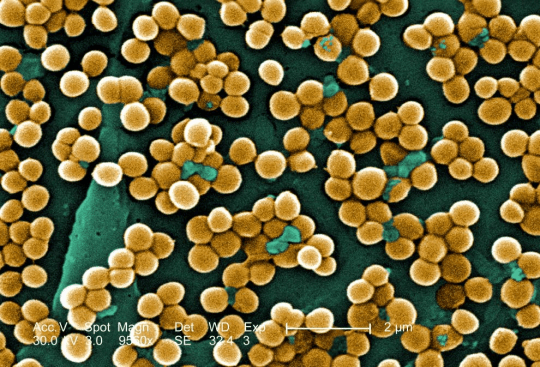
Typical morphology of Staphylococcus aureus. (Source: Wikimedia Commons)
2.2 What Are S. aureus Virulence Factors?
S. aureus virulence factors can be categorized into adhesins, toxins, and immune evasion proteins. The genome carries numerous virulence genes covering various functional categories including adherence, exoenzymes, immune evasion, secretion systems, and toxins.
2.2.1 Adhesins
Protein/Structure | Main Function |
ClfA/ClfB | Fibrinogen-binding proteins, mediate bacterial adhesion to host tissues |
FnBPA/FnBPB | Fibronectin-binding proteins, promote bacterial invasion of host cells |
Cna | Collagen-binding protein, particularly common in mastitis isolates |
Ebh/Ebp/Efb | Extracellular matrix-binding proteins, involved in biofilm formation and tissue colonization |
Atl | Autolysin, involved in cell division and biofilm formation |
2.2.2 Toxins
Toxin | Main Function |
α-Toxin (Hla) | Major pore-forming toxin, lyses erythrocytes, platelets, and leukocytes, promoting bacterial spread |
Panton-Valentine leukocidin (PVL) | Lyses neutrophils, monocytes, and macrophages, evading innate immune responses |
Enterotoxins (SEA-SEE, etc.) | Cause food poisoning; upon translocation into blood, activate T lymphocytes leading to cytokine storm |
Toxic shock syndrome toxin 1 (TSST-1) | Activates T lymphocytes, causing massive cytokine production and leading to toxic shock syndrome |
Exfoliative toxins | Cleave desmosomes in the granular layer of the epidermis, causing staphylococcal scalded skin syndrome |
2.2.3 Immune Evasion Proteins
S. aureus secretes multiple proteins to evade host immune defenses, including:
l CHIPS: Binds FPR1 and C5aR, inhibiting neutrophil chemotaxis
l SCIN: Inhibits complement activation, preventing bacterial lysis and opsonophagocytosis
l SSL-7: Binds the Fc region of IgA and complement C5, blocking antibodies and inhibiting complement activation
l Efb: Binds fibrinogen and complement C3, forming a fibrinogen layer that shields C3b and antibodies from recognition
l Sbi: Forms a tripartite complex with complement C3d and factor H, consuming C3 and blocking the C3d-CR2 association
l Protein A (SpA) : Binds IgG Fcγ via its IgBD domains, blocking antibody-mediated phagocytosis, and exhibits B cell superantigen activity
2.3 Mechanism of Infection and Pathogenesis
2.3.1 Adhesion and Colonization
The first step of S. aureus infection is adhesion to host tissue surfaces. The bacterium binds to host extracellular matrix proteins (fibrinogen, fibronectin, collagen, etc.) through multiple adhesins (ClfA, FnBPs, Cna, etc.), achieving initial colonization. In the nasal cavity, S. aureus can colonize long-term without causing symptoms, forming an asymptomatic carrier state.
2.3.2 Biofilm Formation and Quorum Sensing
S. aureus can establish chronic persistent infections through biofilm formation, which can increase antibiotic tolerance up to 1,000-fold. Biofilm formation is precisely regulated by the accessory gene regulator (agr) quorum-sensing system. The agr system coordinates toxin production and biofilm modulation via the AgrC/AgrA cascade and effector RNA III. When the agr system is downregulated, S. aureus transitions from a highly virulent, toxin-producing phenotype to a persistent, biofilm-forming state. A 2026 study found that cyclic dipeptides (e.g., cyclo(L-Phe, D-Hyp)) significantly inhibited biofilm formation by downregulating the agr-QS system (RNAIII decreased by 75.7%), ica operon, and sarA genes, with low cytotoxicity.
2.3.3 Immune Evasion Mechanisms
S. aureus has evolved multi-layered immune evasion strategies:
l Complement inhibition: SCIN blocks all three complement activation pathways, while Efb and SdrE inhibit the classical and alternative pathways, respectively
l Antibody function interference: SpA binds IgG Fc, and Sbi blocks the C3d-CR2 association, thereby inhibiting B cell antigen recognition
l Phagocyte evasion: Host-induced cell wall thickening leads to reduced exposure of bound antibodies and complement, thereby reducing neutrophil phagocytosis and killing
l Intracellular survival: S. aureus can survive within phagosomes, expressing polysaccharides and proteins that inhibit antibody and complement opsonization
2.3.4 Toxin-Mediated Tissue Damage
Multiple toxins secreted by S. aureus play key roles in pathogenesis. Strains producing high levels of α-toxin and PVL are highly cytotoxic and associated with fatal pneumonia. In necrotizing pneumonia, α-toxin and PVL cause cytotoxicity, inflammation, and necrosis of lung epithelial cells, along with loss of E-cadherin.
2.4 Transmission Routes and Process
Transmission of S. aureus occurs primarily through the following routes:
l Direct contact transmission: Direct contact with an infected person, or contact with contaminated objects (e.g., gym equipment, door knobs, remote controls, elevator buttons)
l Droplet transmission: Inhalation of infected droplets dispersed by sneezing or coughing
l Airborne transmission: Through skin flakes in the air or on towels, sheets, clothing, or sports equipment
l Hand transmission: Carriers can move the bacteria from their nose to other body parts with their hands, sometimes leading to infection
Process: Colonization of the nares or skin → transmission via hand contact or droplets → invasion of compromised skin or mucosa → local infection (e.g., abscess) → if entering the bloodstream, dissemination to distant organs (heart valves, bones, etc.).
2.5 Symptoms and Treatment
2.5.1 Clinical Symptoms
S. aureus infections range from mild to life-threatening:
Skin infections: Most common, causing blisters, abscesses, and redness and swelling in the infected area.
Invasive infections: The bacteria can spread through the bloodstream and infect almost any site, particularly heart valves (endocarditis) and bones (osteomyelitis). The bacteria also tend to accumulate on medical devices in the body, such as artificial heart valves or joints, heart pacemakers, and catheters.
2.5.2 Treatment
Treatment regimens depend on the drug susceptibility of the infecting strain:
l Methicillin-susceptible strains: Penicillinase-resistant β-lactams are typically used
l MRSA infections: Vancomycin, linezolid, and daptomycin remain first-line agents for serious MRSA infections
l Resistance issues: S. aureus has developed resistance to virtually every available antibiotic used to treat it, including MRSA and VRSA
2.6 Prevention
l Hand hygiene: Thorough handwashing with soap and water or use of alcohol-based hand sanitizer is the most important measure to prevent S. aureus transmission
l Wound coverage: Keep wounds clean and covered to prevent bacterial entry
l Avoid sharing personal items: Do not share towels, washcloths, or other personal items
l Nasal decolonization: For MRSA carriers, a 5-day decolonization regimen consisting of 2% mupirocin nasal ointment and 4% chlorhexidine soap is recommended
l Healthcare infection control: Strengthen hand hygiene, isolation measures, and environmental cleaning, especially in high-risk areas such as ICUs
l Novel preventive strategies: Novel preventive strategies are being developed, including S. aureus vaccines, lytic agents, probiotics, microbiota transplants, and phage therapy
2.7 Targets for Intervention
2.7.1 Pathogen Targets
Target | Protein/Pathway | Biological Function |
ClpX | Caseinolytic protease system | Cell division regulation |
HisD | Histidinol dehydrogenase | Links metabolism to virulence |
MgrA | Global transcriptional regulator | Regulates pathogenic programs and resistance traits |
TarGH | Wall teichoic acid transporter | Cell wall synthesis |
TarO | WTA synthesis initiation enzyme | Cell wall synthesis |
2.7.2 Host Targets
Target | Type | Role in Infection |
CR2 | Complement receptor | Sbi blocks C3d-CR2 association, inhibiting B cell antigen recognition |
FPR1/C5aR | Chemokine receptors | CHIPS binding inhibits neutrophil chemotaxis |
ICAM-1 | Adhesion molecule | Eap binding inhibits neutrophil rolling and migration |
2.8 Vaccine Types and Development Progress
Currently, no licensed human S. aureus vaccine is available.
2.8.1 Protein Subunit Vaccines
IMTS8 (SAUSA300_1684) : A 2026 study published in Vaccine identified the novel antigen IMTS8 through machine learning-guided reverse vaccinology. It is highly conserved across major clinical strains and elicited strong antigen-specific IgG responses in mice, conferring near-complete protection in a murine infection model.
2.8.2 Nanoparticle Vaccines
mSEB-mi3 nanoparticle vaccine: A 2026 single-dose nanoparticle vaccine that elicits robust humoral immunity in mice, protecting against SEB intoxication and MRSA infection.
2.8.3 Multi-Epitope Vaccines
"Just the Best Ones" strategy: A 2026 novel multi-epitope vaccine design method using immunoinformatics to induce protective immunity against MRSA infection.
2.8.4 Multi-Epitope Vaccines
AntiPan pipeline: A 2026 genome-informed in silico pipeline that shortlisted 10 candidate antigens (IsdC, EbpS, SspB, EssA, TagH, SirA, EsxA, AmiA, HlgC, HlgB).
2.9 Drugs
2.9.1 Pathogen-Targeted Drugs
Target | Drug/candidate | Mechanism | Stage |
ClpX | Compound 13 | Binds SaClpX, oral bioavailability 37.9%, effective in MRSA skin abscess model | Animal model |
HisD | Pixantrone | Dose-dependent suppression of infection severity and inflammation | Animal model |
MgrA | Negletein | Antivirulence strategy targeting MgrA, improves survival in MRSA pneumonia | Animal model |
TarGH | Targocil-II | Cryo-EM-resolved WTA transporter inhibitor | In vitro |
TarO | 18a | β-lactam sensitizing and anti-biofilm activities | In vitro/ in vivo |
2.9.2 Phage Therapy
Synthetic phage-based CRISPR-Cas delivery systems: 2025–2026 studies developed synthetic phage-based CRISPR-Cas delivery systems for targeted elimination of S. aureus mastitis infections. CRISPR-Cas9-engineered phage K: A 2025 study developed an efficient phage engineering platform using CRISPR-Cas9-assisted counterselection.
2.9.3 Nanomaterials
Silver-based nanomaterials: Exhibit potent antibacterial activity against S. aureus through mechanisms including cell membrane disruption, generation of reactive oxygen species (ROS) , and inhibition of essential cellular processes, offering a novel avenue for overcoming multi-drug resistance.
2.10 Recent Research
2.10.1 Molecular Epidemiology
A 2026 nationwide multicenter study in Brazil showed that MRSA accounted for 25.9% of S. aureus bloodstream infections, with the most common clonal complex being CC5 (72.3%), of which ST105(CC5)-SCCmecII-t002 was the most common strain (56.2%). A 2026 study in Huai'an, China showed that ST398 (18.4%) predominated among all isolates and was strongly associated with MRSA (88.9%).
2.10.2 CRISPR-Cas and Pathogenicity Island Interaction
A 2026 study published in Cell Reports revealed an unexpected synergy between S. aureus pathogenicity islands (SaPIs) and CRISPR-Cas systems: after SaPIs package incomplete helper phage genomes, the defective viral DNA stimulates spacer acquisition in both CRISPR types, enhancing antiphage immunity.
2.10.3 Resistance Evolution
A 2025 study published in Nature tracked the genomic and phenotypic adaptations of MRSA during vancomycin therapy, revealing the evolutionary mechanisms of lipoglycopeptide antibiotic resistance.
2.10.4 Diagnostic Technology Advances
A multiplex real-time PCR method developed in 2025 can simultaneously detect 16S rRNA, nuc, and mecA genes within 30 minutes, with a sensitivity of 1.0×10² CFU/mL, showing high concordance with mass spectrometry.
3. Related Products & Services
l Recombinant Antigens & Receptors
l Antibodies
l Detection Kits
l Recombinant Protein Expression Services
l Antibody Development Services
4. Resources
5. References
1. Tong, S. Y., Davis, J. S., Eichenberger, E., et al. (2015). Staphylococcus aureus infections: epidemiology, pathophysiology, clinical manifestations, and management. Clinical microbiology reviews, 28(3), 603–661.
2. Turner, N. A., Sharma-Kuinkel, B. K., Maskarinec, S. A., et al. (2019). Methicillin-resistant Staphylococcus aureus: an overview of basic and clinical research. Nature reviews. Microbiology, 17(4), 203–218.
3. Lakhundi, S., Zhang, K. (2018). Methicillin-Resistant Staphylococcus aureus: Molecular Characterization, Evolution, and Epidemiology. Clinical microbiology reviews, 31(4), e00020-18.
4. Lee, A., de Lencastre, H., Garau, J., et al. (2018). Methicillin-resistant Staphylococcus aureus. Nat Rev Dis Primers 4, 18033.
5. Cheung, G. Y. C., Bae, J. S., Otto, M. (2021). Pathogenicity and virulence of Staphylococcus aureus. Virulence, 12(1), 547–569.
6. Novick, R. P., Ram, G. (2017). Staphylococcal pathogenicity islands-movers and shakers in the genomic firmament. Current opinion in microbiology, 38, 197–204.
7. Foster T. J. (2005). Immune evasion by staphylococci. Nature reviews. Microbiology, 3(12), 948–958.
8. Otto M. (2014). Staphylococcus aureus toxins. Current opinion in microbiology, 17, 32–37.
9. Spaan, A. N., van Strijp, J. A. G., Torres, V. J. (2017). Leukocidins: staphylococcal bi-component pore-forming toxins find their receptors. Nature reviews. Microbiology, 15(7), 435–447.
10. Archer, N. K., Mazaitis, M. J., Costerton, J. W., et al. (2011). Staphylococcus aureus biofilms: properties, regulation, and roles in human disease. Virulence, 2(5), 445–459.
11. Le, K. Y., Otto, M. (2015). Quorum-sensing regulation in staphylococci-an overview. Frontiers in microbiology, 6, 1174.
12. Wei, J., Li, S., Ma, S., et al. (2026). Cyclic dipeptides inhibit Staphylococcus aureus biofilm formation and virulence via agr-quorum sensing, ica, and sarA pathways. Microbial pathogenesis, 213, 108359.
13. Liu, C., Bayer, A., Cosgrove, S. E., et al. (2011). Clinical practice guidelines by the infectious diseases society of america for the treatment of methicillin-resistant Staphylococcus aureus infections in adults and children. Clinical infectious diseases : an official publication of the Infectious Diseases Society of America, 52(3), e18–e55.
14. Jiang, Y., Wang, Y., Bai, Y., et al. (2025). Genomic and phenotypic adaptations of methicillin resistant Staphylococcus aureus during vancomycin therapy. Scientific reports, 15(1), 15346.
15. Card, K. J., Crozier, D., Durmaz, A., et al. (2025). Evolution under vancomycin selection drives divergent collateral sensitivity patterns in Staphylococcus aureus. Proceedings of the National Academy of Sciences of the United States of America, 122(39), e2507962122.
16. Fatsis-Kavalopoulos, N., Kim, Y. K., Chong, Y. P., et al. (2025). Vancomycin heteroresistance (hVISA) in MRSA links to treatment failure and supports a revised PAP-AUC threshold. Nature communications, 16(1), 11251.
17. Clegg, J., Soldaini, E., McLoughlin, R. M., et al. (2021). Staphylococcus aureus Vaccine Research and Development: The Past, Present and Future, Including Novel Therapeutic Strategies. Frontiers in immunology, 12, 705360.
18. Banh, D. V., Goldberg, G. W., Marraffini, L. A. (2026). Phage induction of Staphylococcus aureus pathogenicity islands promotes the CRISPR-Cas adaptive immune response. Cell reports, 45(1), 116776.