
info@mail.ucallm.com
1. Overview
Tick-borne encephalitis virus (TBEV) is a major member of the genus Orthoflavivirus within the family Flaviviridae and is the causative agent of tick-borne encephalitis (TBE). It is an enveloped, positive-sense, single-stranded RNA virus with a spherical virion approximately 50 nm in diameter. TBEV was first isolated in 1937 in the Russian Far East and is the most important tick-borne viral central nervous system infection in Europe and Asia.
TBEV is transmitted primarily through hard tick bites (mainly Ixodes species), and can also be acquired through consumption of unpasteurised dairy products. It is estimated that over 10,000 cases are reported annually in Europe and Asia, and the endemic range of TBEV continues to expand due to climate change-driven expansion of tick distribution. TBEV exists in three major subtypes: European (TBEV-Eu), Siberian (TBEV-Sib), and Far Eastern (TBEV-FE), which differ in disease severity. The World Health Organization (WHO) has classified TBEV as one of the priority arthropod-borne viruses. Vaccines are licensed globally, but no specific antiviral therapy is currently available.
2. Background Information
2.1 What is Tick-borne Encephalitis Virus?
TBEV is an enveloped, positive-sense, single-stranded RNA virus belonging to the family Flaviviridae and genus Orthoflavivirus. Its genome is a positive-sense single-stranded RNA of approximately 11 kb, containing a single open reading frame (ORF) encoding a polyprotein precursor that is co- and post-translationally cleaved to produce three structural proteins—capsid (C), precursor membrane (prM), and envelope (E)—and seven non-structural proteins (NS1, NS2A, NS2B, NS3, NS4A, NS4B, NS5). The genome possesses a methylated cap at the 5′ end but lacks a poly(A) tail at the 3′ end.
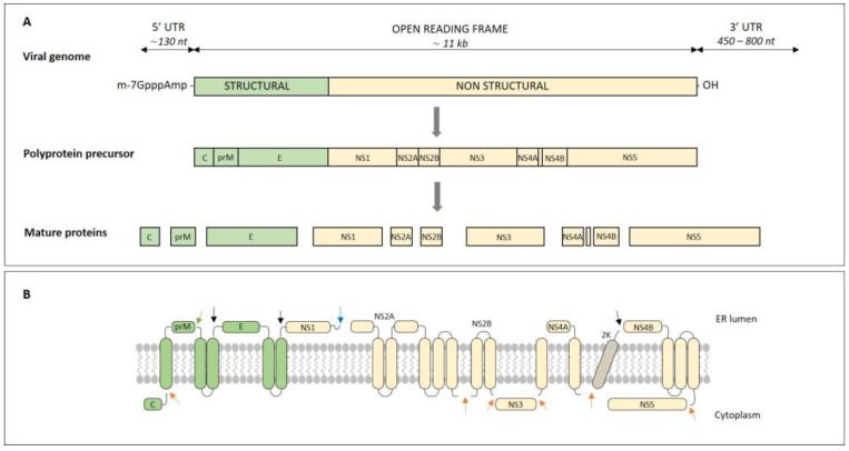
Genome organization of TBE virus (PMID: 37512806)、
2.2 TBEV Genome & Classification
The TBEV genome is approximately 11 kb, encoding 10 proteins. The genome organisation is: 5′-C-prM-E-NS1-NS2A-NS2B-NS3-NS4A-NS4B-NS5-3′. TBEV is traditionally classified into three major subtypes: Far Eastern (TBEV-FE), European (TBEV-Eu), and Siberian (TBEV-Sib). Additional genetic lineages have also been described, such as strain 178-79 (fourth subtype) and the Baikal subtype (TBEV-Baik). In 2026, the Siberian subtype was identified alongside Far Eastern strains in northeastern China.
2.3 What Are Tick-borne Encephalitis Virus Proteins?
TBEV encodes 10 proteins, including 3 structural and 7 non-structural proteins.
2.3.1 Structural Proteins
Protein | Full name | Main Function |
C | Capsid | Encapsidates viral genomic RNA, forming the nucleocapsid core |
prM | pre-Membrane | Cleaved by furin during maturation to produce M protein; involved in assembly and envelope formation |
E | Envelope | Major surface protein; mediates receptor binding and membrane fusion; primary target of neutralizing antibodies |
2.2.2 Non-structural Proteins
The seven non-structural proteins (NS1, NS2A, NS2B, NS3, NS4A, NS4B, NS5) participate in viral RNA replication, virion assembly, and modulation of host immune responses. NS5 possesses both RNA-dependent RNA polymerase (RdRp) and methyltransferase activities and is the core enzyme for viral replication. A 2026 study found that TBEV NS5 interacts with the host protein SIRT1, suppressing the deacetylation of KAP1 and Ku70, thereby impeding DNA damage repair and worsening neurological injury. Additionally, NS5 interacts with P300, inducing G0/G1 cell-cycle arrest and enhancing viral gene expression.
2.4 Mechanism of Cell Entry and Replication
2.4.1 Viral Attachment and Receptor Recognition
TBEV infection begins with the binding of the viral E protein to host cell receptors. In 2025, two independent research teams simultaneously identified low-density lipoprotein receptor-related protein 8 (LRP8) (also known as ApoER2) as a functional entry receptor for TBEV in Nature and PNAS. LRP8 is an apolipoprotein E and reelin receptor with high expression in the brain, and its LA1–2 domains bind with high affinity to the viral E protein, driving viral attachment and internalisation. LRP8 knockdown significantly inhibits TBEV infection in human cells, while ectopic expression enhances infection. LRP8 is used as a receptor by multiple TBEV strains and several closely related tick-borne viruses but not by the mosquito-transmitted orthoflaviviruses tested.
2.4.2 Viral Entry
The virus enters host cells via receptor-mediated endocytosis. Endosomal acidification triggers low-pH-induced conformational changes in the E protein, mediating fusion of the viral envelope with the endosomal membrane and releasing viral RNA. A 2025 cryo-EM study revealed the asymmetric structure of immature TBEV particles—most TBEV particles lack subsets of surface heterodimers, but these irregularities do not impede subsequent maturation.
2.4.3 Viral RNA Replication
Upon release, the viral RNA initiates translation on ER membranes. Non-structural proteins assemble into a replication complex on ER-derived membrane vesicles. NS5 (RdRp) catalyses viral RNA replication, synthesising a negative-sense RNA intermediate, which then serves as template for synthesis of progeny positive-sense genomic RNA. A 2026 study found that the RNA promoter of pathogenic orthoflaviviruses (including TBEV, Japanese encephalitis virus, and West Nile virus) is universal, with its three-dimensional architecture conserved across mosquito- and tick-borne viruses, serving as a target for viral inhibition.
2.4.4 Viral Assembly and Release
Viral assembly occurs in the ER lumen, with immature particles containing prM-E heterodimer spikes. During transport through the Golgi, prM is cleaved by furin protease to produce M protein, triggering rearrangement of the envelope proteins and maturing the virion. Mature virions are finally released via exocytosis. A 2025 study found that irreversible furin cleavage site exposure renders immature tick-borne flaviviruses fully infectious, providing new insights into flavivirus maturation mechanisms.
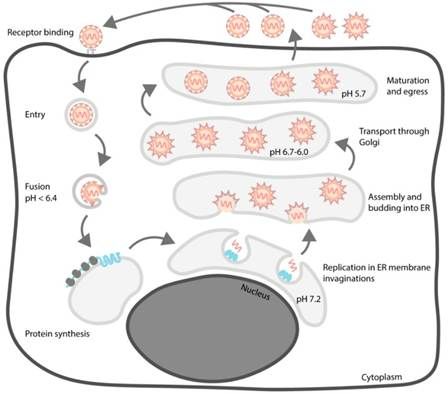
The life cycle of TBEV. (PMID: 29958443)
2.5 Symptoms and Treatment
2.5.1 Clinical Symptoms
TBEV infection presents with a biphasic course. Approximately 20%–30% of infected individuals develop clinical symptoms, with the remainder being asymptomatic. The first phase (viraemic phase) presents with non-specific influenza-like symptoms, including fever, headache, fatigue, and muscle and joint pain, lasting approximately one week. Approximately 20%–30% of symptomatic patients progress to the second phase (neurological phase) , presenting with meningitis, encephalitis, or meningoencephalitis, with symptoms including severe headache, altered consciousness, seizures, and paralysis.
High-risk populations for severe disease: the elderly, immunocompromised individuals, and those infected with the Far Eastern subtype are more likely to develop severe disease. Although the mortality rate is low (<2%), up to 40% of cases may experience post-infectious neurological sequelae.
2.5.2 Antiviral Treatment
Currently, no specific antiviral therapy is approved for TBEV. Treatment is primarily supportive: antipyretics and analgesics (e.g., paracetamol), antiemetics, maintenance of fluid and electrolyte balance, anticonvulsant therapy (benzodiazepines recommended for seizures), respiratory support for patients with neuromuscular paralysis, and intracranial pressure management (osmotherapy may be considered).
2.6 Targets for Intervention
2.6.1 Viral Targets
Target | Protein | Biological Role |
E | Envelope protein | Receptor binding & membrane fusion |
NS3 | Protease/helicase | Polyprotein processing, RNA unwinding |
NS5 | RNA polymerase (RdRp) | RNA replication |
Methyltransferase | 5′ cap methylation | |
Host interactions | SIRT1/P300 interaction, DNA damage repair suppression | |
NS1 | Non-structural protein 1 | Replication complex formation |
2.6.2 Host Receptors & Entry Factors
Host Target | Type | Role in Infection |
LRP8 | Receptor | Functional receptor for TBEV entry |
ER-associated degradation pathway | Host pathway | Required for viral replication |
Lipophagy pathway | Host pathway | Required for viral replication |
2.6.3 Host Signaling Pathways
| Pathway | Key targets | Biological role |
MAPK/ERK pathway | ERK | Viral replication and inflammatory response |
ER stress pathway | PERK, ATF6, IRE1 | Viral replication and inflammatory regulation |
Inflammatory signaling | NF-κB, TNF-α, IL-1β | Neuroinflammation and injury |
2.6.4 Immune & Inflammatory Targets
| Target | Category | Major role |
IFN-α | Cytokine | Antiviral response |
IFN-β | Cytokine | Antiviral response |
IFN-γ | Cytokine | Cellular immunity |
IL-6 | Cytokine | Inflammatory response |
IL-1β | Cytokine | Inflammasome signaling |
TNF-α | Cytokine | Inflammation |
CXCL10 | Cytokine | Immune-cell recruitment |
2.7 Vaccine Types and Development Progress
Vaccination is the most effective means of preventing TBEV infection. Several TBE vaccines are licensed globally, and novel vaccine platforms are rapidly advancing.
2.7.1 Licensed Inactivated Vaccines
TicoVac/TicoVac Junior (Pfizer) is the most widely used TBE vaccine. The standard schedule is: first dose on day 0, second dose 1–3 months later, third dose 5–12 months later. Children aged 1–15 years receive 0.25 mL per dose, and adults aged 16 years and older receive 0.5 mL per dose. A booster dose (fourth dose) may be given at least 3 years after completion of the primary series if ongoing or repeated exposure is expected. In 2026, Pfizer filed for FDA approval of TicoVac based on clinical trial data showing 99–100% of 1–15 year olds and 84–99% of adults produced antibodies following three doses.
2.7.2 mRNA Vaccines
A 2026 study published in Vaccines developed mRNA-PrM/E-LNP vaccine candidates encoding the TBEV prM and E proteins. All tested mRNA candidates conferred 100% survival in mice following lethal challenge with seven distinct TBEV strains (100 LD₅₀). The construct mRNA-PrM/E—Krasny Yar-8 demonstrated the highest immunogenicity, inducing antigen-specific antibodies with a geometric mean titre (GMT) of 1:6625, and the broadest virus-neutralising activity against both homologous and heterologous TBEV strains. The study noted that current inactivated TBEV vaccines offer limited cross-protection against heterologous strains, and the mRNA platform represents a promising strategy for TBEV vaccine development.
2.7.3 Replication-Defective Vaccine (NS1 trans-complementation)
In 2026, researchers used an NS1 trans-complementation platform to generate replication-defective TBE virus particles (TBEV-ΔNS1) with a large in-frame deletion (amino acids 4–298) in the NS1 gene, with the deleted NS1 function provided in trans by a BHK cell line stably expressing heterologous NS1. TBEV-ΔNS1 stably maintained the NS1 deletion during serial passaging, without recombination or reversion to replication-competent wild-type virus. ICR mice inoculated with TBEV-ΔNS1 showed no clinical manifestations or evidence of virulence; a single dose induced neutralising antibodies and robust T-cell responses, with the higher dose providing 100% protection against lethal wild-type virus challenge.
2.7.4 Live-Attenuated Vaccine (Langat Virus Platform)
In 2025, researchers developed Langat virus (LGTV) infectious clones as a live-attenuated TBE vaccine platform. Unlike currently available inactivated TBE vaccines that require repeated boosters, live-attenuated vaccines could potentially offer lifelong immunity with a single dose.
2.7.5 DNA Vaccines
A 2026 study published in Bulletin of Experimental Biology and Medicine showed that the pVAX-prM-E DNA vaccine, administered to mice using a needle-free jet injector, induced high levels of virus-neutralising antibodies, indicating high protective potential.
2.7.6 Virus-Like Particle (VLP) Vaccines
In 2026, researchers developed VLP-based TBEV vaccine candidates. The TBEV VLPs vaccine was able to provide long-term antibody protection against TBEV, making it a promising subunit vaccine candidate.
2.7.7 MVA-Vectored Vaccine
Recombinant Modified Vaccinia Ankara (MVA) expressing TBEV prME (MVA-prME) demonstrated high immunogenicity in mice compared with the licensed vaccine FSME-IMMUN® and afforded full protection against TBEV challenge infection.
2.8 Drugs
Currently, no specific small-molecule antiviral is approved for TBEV. Several antiviral candidates have shown significant progress.
2.8.1 Virus-Targeted Drugs
Target | Drug/candidate | Mechanism | Stage |
NS5 (RdRp) | Remdesivir | RdRp inhibition | In vitro & ex vivo disease models |
Bemnifosbuvir | RdRp inhibition | In vitro & ex vivo disease models | |
E protein | Chelerythrine chloride (CHE) | targets E protein and suppresses MAPK/ERK pathway | In vitro & mouse models |
Multiple mechanisms | Cepharanthine | Modulates ER stress and inflammatory responses, inhibits TBEV RNA replication and virus release | In vitro |
E protein DIII | Cyclic peptides CP2, CP20 | Target E protein receptor-binding sites, neutralise live virus | In vitro |
E protein | Nanobody TA3-dendrimer nanosystem | Crosses BBB, neutralises TBEV | In vitro (BBB model) |
2.8.2 Host-Targeted Drugs
Target | Drug | Mechanism | Stage |
LRP8 receptor | Soluble LRP8-Fc decoy protein | Blocks TBEV infection | In vitro & mouse models |
Multiple host targets | Sunitinib | RIOK1/BRD9 inhibitor | In vitro |
2.8.3 Drugs with Unclear or Non-Specific Targets
No approved drugs with unclear or non-specific targets are currently available for TBEV. The efficacy of broad-spectrum antivirals such as ribavirin and interferon against TBEV remains unclear and is not recommended as standard treatment.
2.9 Epidemiology
l Global disease burden: over 10,000 cases reported annually, with the endemic range continuing to expand
l Geographic distribution: Central Europe, Eastern Europe, Northern Europe, Russia, and parts of East Asia. TBEV is reported as commonly found in at least 27 countries
l Incidence: medically attended TBEV incidence ranged from 0.5/100,000 in Denmark to 21.5/100,000 in Latvia
l Upper Austria: anti-TBE-NS1-IgG seroprevalence was 74/100,000 for the whole population and 594/100,000 in the non-vaccinated population
l Switzerland: 503 cases recorded in 2025
l European trend: TBE cases increased from 2,412 in 2012 to 3,514 in 2022
2.10 Diagnosis
Diagnosis of TBEV infection relies on laboratory testing:
l Virus isolation: isolation of TBEV from clinical material (requires BSL-3 laboratory)
l Nucleic acid testing (NAT) : detection of TBEV RNA in clinical material (blood, cerebrospinal fluid)
l Serology: detection of seroconversion or four-fold or greater rise in TBEV-specific IgM or IgG titres
2.11 Prevention
l Vaccination: TBE vaccines are licensed in multiple European and Asian countries and are the most effective measure for preventing TBEV infection; recommended for residents in endemic areas, travellers, and high-risk occupational groups
l Avoid tick bites: wear long-sleeved clothing and use tick repellents in endemic areas; avoid prolonged stays in tick-active forests and bushes
l Food safety: avoid consuming unpasteurised milk, goat milk, and their products (e.g., cheese) to prevent alimentary transmission
l Personal protective measures: check for and remove attached ticks promptly after entering tick-active areas; veterinarians and field workers should wear appropriate personal protective equipment (PPE)
l Surveillance and reporting: TBE is a notifiable disease in most endemic countries; strengthen surveillance and reporting of human cases, tick vectors, and animal reservoirs
3. Related Products & Services
l Recombinant Antigens & Receptors
l Antibodies
l Detection Kits
l Recombinant Protein Expression Services
l Antibody Development Services
4. Resources
5. References
1. Füzik, T., Formanová, P., Růžek, D., et al. (2018). Structure of tick-borne encephalitis virus and its neutralization by a monoclonal antibody. Nature Communications, 9, 436.
2. Pulkkinen, L. I. A., Barrass, S. V., Domanska, A., et al. (2022). Molecular Organisation of Tick-Borne Encephalitis Virus. Viruses, 14(4), 792.
3. Pichkur, E. B., Vorovitch, M. F., Ivanova, A. L., et al. (2024). The structure of inactivated mature tick-borne encephalitis virus at 3.0 Å resolution. Emerging Microbes & Infections, 13(1), 2313849.
4. Anastasina, M., Füzik, T., Domanska, A.,et al. (2024). The structure of immature tick-borne encephalitis virus supports the collapse model of flavivirus maturation. Science advances, 10(27), eadl1888.
5. Ruzek, D., Avšič Županc, T., Borde, J.,et al. (2019). Tick-borne encephalitis in Europe and Russia: Review of pathogenesis, clinical features, therapy, and vaccines. Antiviral research, 164, 23–51.
6. Ye, R. Z., Li, Y. Y., Wang, X. Y., et al. (2026). Tick-borne viruses: discovery, clinical aspects, influencing factors and control. Nature reviews. Microbiology, 24(9), 654–672.
7. Mittler, E., Tse, A. L., Tran, P. T., et al. (2025). LRP8 is a receptor for tick-borne encephalitis virus. Nature, 646(8086), 945–952.
8. Li, P., Hui, S., Chong, Z., et al. (2025). LRP8 is an entry receptor for tick-borne encephalitis viruses. Proceedings of the National Academy of Sciences of the United States of America, 122(44), e2525771122.
9. Oviedo-Rouco, S., Bertoni, L., Mikkelsen, E., et al. (2026). The RNA promoter for pathogenic orthoflaviviruses replication is universal and serves as target for viral inhibition. PLoS pathogens, 22(5), e1014233.
10. Holoubek, J., Salát, J., Matkovic, M., et al. (2025). Irreversible furin cleavage site exposure renders immature tick-borne flaviviruses fully infectious. Nature communications, 16(1), 7491.
11. Taba, P., Schmutzhard, E., Forsberg, P., et al. (2017). EAN consensus review on prevention, diagnosis and management of tick-borne encephalitis. European journal of neurology, 24(10), 1214–e61.
12. Reusken, C., Boonstra, M., Rugebregt, S., et al. (2019). An evaluation of serological methods to diagnose tick-borne encephalitis from serum and cerebrospinal fluid. Journal of clinical virology : the official publication of the Pan American Society for Clinical Virology, 120, 78–83.
13. Who Publication (2011). Vaccines against tick-borne encephalitis: WHO position paper--recommendations. Vaccine, 29(48), 8769–8770.
14. Domnich, A., Panatto, D., Arbuzova, E. K., et al. (2014). Immunogenicity against Far Eastern and Siberian subtypes of tick-borne encephalitis (TBE) virus elicited by the currently available vaccines based on the European subtype: systematic review and meta-analysis. Human vaccines & immunotherapeutics, 10(10), 2819–2833.
15. Asghar, N., Jaafar, R., Valko, A., et al. (2025). Development of Langat virus infectious clones as a platform for live-attenuated tick-borne encephalitis vaccine. Npj viruses, 3(1), 44.
16. Zhang, J., Zhang, Q. Y., Zhang, Z. R.,et al. (2026). A replication-defective tick-borne encephalitis virus generated by NS1 trans-complementation confers robust protective efficacy. Virologica Sinica, 41(4), 937–946.
17. Kubinski, M., Beicht, J., Zdora, I.,et al. (2023). A recombinant Modified Vaccinia virus Ankara expressing prME of tick-borne encephalitis virus affords mice full protection against TBEV infection. Frontiers in immunology, 14, 1182963.
18. Martello, E., Gillingham, E. L., Phalkey, R., et al. (2022). Systematic review on the non-vectorial transmission of Tick-borne encephalitis virus (TBEv). Ticks and tick-borne diseases, 13(6), 102028.