
info@mail.ucallm.com
1. Overview
Toxoplasma gondii is an obligate intracellular protozoan parasite of the family Sarcocystidae within the phylum Apicomplexa and is the causative agent of toxoplasmosis. T. gondii is considered one of the most successful parasites globally, infecting approximately one-third of the world's human population. Infection is typically asymptomatic or causes only mild flu-like symptoms in healthy individuals, but can cause severe or even fatal disease in immunocompromised patients (e.g., AIDS patients, organ transplant recipients) and congenitally infected fetuses.
T. gondii has a complex two-host life cycle. Felids are the only definitive hosts, and sexual reproduction occurs exclusively in the feline intestine. The intermediate host range is extremely broad, capable of infecting over 200 species of warm-blooded animals, including humans, birds, and rodents. T. gondii has three infectious forms: tachyzoites (causing acute infection), bradyzoites (forming tissue cysts and causing chronic infection), and sporozoites (contained within oocysts).
2. Background Information
2.1 What is Toxoplasma gondii?
T. gondii is an obligate intracellular apicomplexan parasite first discovered by Nicolle and Manceaux in 1908 in a North African rodent (Ctenodactylus gundi). The parasite possesses unique intracellular parasitic capabilities and immune evasion mechanisms that enable it to establish lifelong latent infection in its host.
Major transmission routes of T. gondii include: (1) Foodborne transmission: ingestion of undercooked meat containing tissue cysts with bradyzoites; (2) Environmental transmission: ingestion of food or water contaminated with oocysts from feline feces; (3) Vertical transmission: congenital infection, i.e., transmission from mother to fetus via the placenta after primary maternal infection; (4) Organ transplantation or blood transfusion: rare but possible.
2.2 Genome & Classification
T. gondii belongs to the phylum Apicomplexa, class Conoidasida, order Eucoccidiorida, family Sarcocystidae. Its genome is approximately 70 Mb and consists of 14 chromosomes. The genome is highly plastic, with over 75% of genes containing introns, far exceeding the 5% in yeast.
In 2025, a new T. gondii ME49 reference genome was constructed using Oxford Nanopore long-read sequencing technology, with continuity (NG50 = 6.68 Mb) and structural accuracy significantly exceeding existing reference genomes (RefSeq NG50 = 1.2 Mb). The study identified a 13 kb expansion in the ROP8-ROP2A virulence locus, increasing ROP2A-ROP8 gene copy number from three to six. The T. gondii genome comprises 21 sequence blocks that recombine among themselves, though their order is not entirely random.
T. gondii also carries two important organelles: (1) the apicoplast, a non-photosynthetic plastid essential for its survival; and (2) the mitochondrion, with a unique mitochondrial genome encoding merely three protein-coding genes and 30–40 small RNA genes.
2.3 What Are T. gondii Proteins?
T. gondii secretory organelles (rhoptries, micronemes, and dense granules) secrete numerous effector proteins that are the core weapons for invasion and immune evasion.
Protein | Source Organelle | Main Function |
RON2 | Rhoptry neck | Inserts into host cell membrane, establishes the moving junction |
ROP1 | Rhoptry | Establishes host ER–Toxoplasma membrane contact sites |
ROP6 | Rhoptry | Host cell invasion, parasitophorous vacuole biogenesis; highly immunogenic, vaccine candidate antigen |
ROP21/ROP29 | Rhoptry | Vaccine candidate antigens |
MIC8 | Microneme | Host cell attachment and invasion; vaccine candidate antigen |
AMA1 | Microneme | Moving junction formation; vaccine candidate antigen |
GRA15/GRA60/GRA76 | Dense granule | Host signaling pathway regulation, immune evasion; vaccine candidate antigens |
SAG1 | Surface antigen | High-affinity adhesin; maintains efficient invasion under IFNγ stimulation |
CGP | Conoid complex | Stabilizes conoid structure in mature parasites |
ASAF1 | Conoid complex | Positions and builds conoid structure at an early stage |
2.4 Mechanism of Infection and Pathogenesis
2.4.1 Adhesion and Invasion
T. gondii invasion is a highly complex and precise process:
a. Conoid complex assembly: T. gondii relies on the conoid complex as an "engine" for movement and cell invasion. A 2025 study identified two key proteins: CGP stabilizes the structure in mature parasites, while ASAF1 positions and builds it at an early stage; deletion of either results in failure to properly assemble the invasion machinery.
b. Transient perforation and invasion pore formation: A 2025 study published in EMBO Reports used electrophysiological recordings at sub-200 µs resolution to demonstrate, for the first time, that T. gondii creates a transient perforation in the host cell membrane early in invasion, termed the "invasion pore". This process requires rhoptry exocytosis.
c. Moving junction: T. gondii secretes RON2, which inserts into the host cell membrane to establish the moving junction, a tight connection between the parasite and host cell membrane.
d. Host ER contact: The T. gondii effector TgROP1 establishes membrane contact sites (MCS) between the host ER and the parasite, targeting TOM70 and recruiting host proteins.
2.4.2 Immune Evasion Mechanisms
T. gondii has evolved multiple immune evasion strategies:
l Early immune suppression: A 2025 study published in PLoS Biology found that within 60 minutes of infection, macrophages exhibited upregulated suppressor of cytokine signaling (SOCS) expression, uniquely tied to live infection.
l GRA15-mediated immune suppression: GRA15 expression in dendritic cells suppresses T cell-dependent humoral immune responses.
l Immune pressure during latency: A 2025 study published in Nature Microbiology demonstrated that the latent form (bradyzoites) of T. gondii is under immune pressure, which mitigates infection-induced damage and promotes survival of host and parasite.
2.4.3 Host Immune Response
l γδ T cells and NK cells: A 2025 study found that the early human IFNγ response is driven by Vγ9Vδ2 T-cell sensing of host phosphoantigens and subsequent NK-cell activation.
l Caspase-1/IL-18 axis: Caspase-1 in Cx3cr1-expressing cells drives an IL-18-dependent T cell response that promotes parasite control during acute infection.
l S100A11-RAGE/TLR4 axis: A 2025 study revealed that T. gondii infection induces S100A11 secretion in placental explants, recognized by RAGE and TLR4; inhibition of this pathway prevents parasite-induced placental damage.
2.5 Transmission Routes and Process
Transmission of T. gondii relies on its complex two-host life cycle, involving definitive hosts (felids), intermediate hosts (including humans), and environmental oocysts. Human infection occurs mainly through four routes:
2.5.1 Foodborne Transmission (Tissue Cysts)
This is one of the most common routes of human infection. After infection, intermediate hosts (e.g., pigs, sheep, cattle, poultry) form tissue cysts containing bradyzoites in their muscles and brain. Humans become infected by:
l Eating undercooked meat: especially pork, lamb, and game; tissue cysts survive in meat if cooking temperature is insufficient (core temperature <67°C).
l Cross-contamination: failure to thoroughly wash knives, cutting boards, or hands after handling raw meat before touching ready-to-eat food.
l Consuming unpasteurized dairy products: rarely, raw goat milk may contain tachyzoites.
Process: Ingestion of tissue cysts → gastric acid and digestive enzymes release bradyzoites → bradyzoites invade intestinal epithelial cells → convert to tachyzoites → tachyzoites disseminate via blood and lymph → form new tissue cysts in muscle, brain, and eyes, establishing chronic infection.
2.5.2 Environmental Transmission (Oocysts)
This is another major route, particularly associated with felids. After felids (especially kittens and cats infected for the first time) ingest tissue cysts, sexual reproduction occurs in the intestine, producing oocysts that are shed in feces. Oocysts require 1–5 days in the environment to sporulate and become infectious.
Humans become infected by:
l Ingesting food or water contaminated with oocysts: e.g., unwashed vegetables, fruits, salads, or contaminated drinking water.
l Touching soil or cat litter without washing hands: gardening, cleaning litter boxes, then eating without handwashing.
l Ingesting contaminated soil particles: children playing in sandboxes then putting hands in mouth.
Process: Ingestion of sporulated oocysts → sporozoites released in the intestine → sporozoites invade intestinal epithelium → convert to tachyzoites → disseminate throughout the body → form tissue cysts, establishing chronic infection.
2.5.3 Vertical Transmission (Congenital Infection)
When pregnant women experience primary infection (acute infection) during pregnancy, tachyzoites can cross the placenta and infect the fetus, causing congenital toxoplasmosis.
l Risk: Infection in early pregnancy has lower transmission rate but severe consequences (miscarriage, stillbirth, severe neurological malformations); infection in late pregnancy has higher transmission rate but is often subclinical or mild.
l Process: Maternal tachyzoitemia → tachyzoites cross placental barrier → fetal infection → tissue cysts form in fetal brain and eyes, leading to hydrocephalus, retinochoroiditis, intellectual disability, etc.
2.5.4 Organ Transplantation and Blood Transfusion
Rare but possible through transplantation of organs containing tissue cysts (e.g., heart, kidney) or transfusion of blood products containing tachyzoites. Immunosuppressed recipients often develop severe disease.
2.5.5 Life Cycle Summary
Stage | Host | Form | Event |
Sexual reproduction | Felids (definitive host) | Oocyst | Shed in feces, sporulates in environment |
Asexual reproduction | Intermediate hosts (humans, pigs, sheep, etc.) | Tachyzoite → Bradyzoite | Acute infection → Tissue cyst formation |
Infectious stages | — | Tissue cyst, oocyst, tachyzoite | Transmitted via oral, placental, transplant routes |
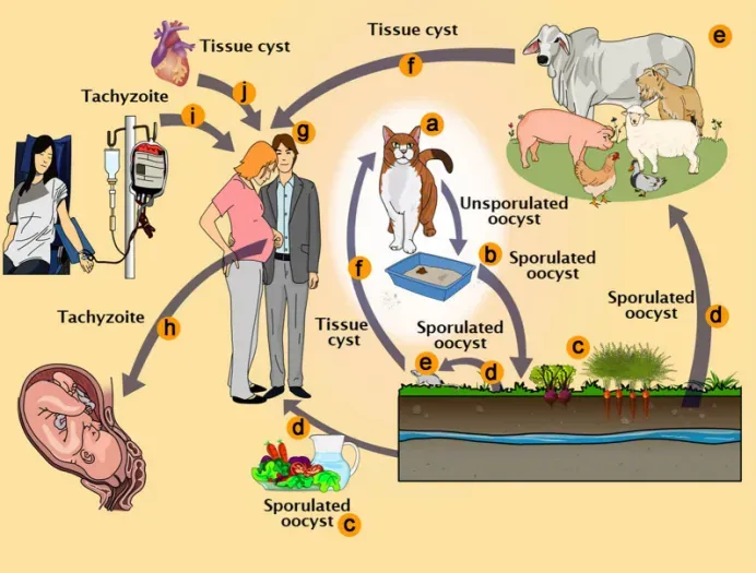
Toxoplasma gondii pathways of transmission. a Feline definitive host (cat). b Unsporulated oocysts in cat feces. c Food contaminated with sporulated oocysts. d Oocysts may be ingested by intermediate hosts via water or raw vegetables. e Intermediate hosts (e.g. cattle, sheep, poultry and swine). f Ingestion of tissue cysts in uncooked meat. g Intermediate hosts (humans). h Tachyzoites transmitted through the placenta to the foetus. i Transmission by blood transfusion and organ transplant (j). (PMID: 33228743)
2.6 Symptoms and Treatment
2.6.1 Clinical Symptoms
Clinical manifestations of toxoplasmosis vary depending on host immune status and route of infection:
Immunocompetent individuals: Usually asymptomatic or present with mild flu-like symptoms including headache, weakness, and fever. Chronic infection is usually asymptomatic, presenting as latent tissue cysts.
Immunocompromised individuals (AIDS patients, organ transplant recipients, cancer patients): Bradyzoites in tissue cysts can reactivate, leading to fatal toxoplasmosis, most commonly presenting as toxoplasmic encephalitis.
Congenital infection: Primary infection during pregnancy can be transmitted to the fetus via the placenta, causing miscarriage, stillbirth, or severe neurological and ocular disease.
Ocular infection: Retinochoroiditis, which can lead to visual impairment.
New associations: Recent studies have linked brain tissue cysts to behavioral disorders.
2.6.2 Treatment
According to the 2025 MSD Manual, treatment regimens for toxoplasmosis are as follows:
Most effective regimen: Pyrimethamine + sulfadiazine + leucovorin (to prevent bone marrow suppression), for 2–4 weeks. For patients with visceral involvement or severe/persistent symptoms, sulfadiazine should be used for at least 6 weeks and continued for 4–6 weeks after resolution of clinical symptoms and signs.
Alternative regimens: For patients allergic to or intolerant of sulfonamides, clindamycin or atovaquone + pyrimethamine may be used.
Ocular toxoplasmosis: Corticosteroids are often given concurrently. The CDC recommends that treatment for ocular toxoplasmosis be continued for 4–6 weeks.
HIV prevention: For HIV-infected patients who are seropositive for T. gondii IgG antibodies and have a CD4 count < 100/mcL, primary chemoprophylaxis is recommended.
Key limitation: Currently, no drugs are available for chronic toxoplasmosis, and existing drugs cannot eliminate bradyzoites within tissue cysts.
2.7 Prevention
Prevention of toxoplasmosis requires comprehensive measures targeting different transmission routes and risk groups.
2.7.1 Food Safety
l Thoroughly cook meat: ensure core temperature reaches above 67°C, or freeze at −20°C for 24 hours to kill tissue cysts.
l Separate raw and cooked: after handling raw meat, thoroughly wash knives, cutting boards, counters, and hands with soap and hot water.
l Avoid tasting raw meat: do not taste meat during cooking before it is fully cooked.
l Wash produce: thoroughly wash or peel fruits and vegetables before eating, especially those eaten raw.
l Avoid unpasteurized dairy: do not consume raw goat milk, raw cow milk, or their products.
2.7.2 Cat-Related Prevention
l Pregnant women and immunosuppressed individuals should avoid handling cat litter: if unavoidable, wear gloves and wash hands immediately with soap and water afterward.
l Clean litter boxes daily: oocysts require 1–5 days to sporulate; daily cleaning reduces risk.
l Keep cats indoors and do not let them hunt: feed commercial cat food or cooked food to prevent feline infection.
l Avoid stray cats: especially kittens and cats of unknown health status.
l Wear gloves when gardening: thoroughly wash hands after contact with soil.
2.7.3 Water Safety
l Drink safe water: in areas with poor sanitation, drink boiled or bottled water.
l Avoid water sources potentially contaminated with cat feces.
2.7.4 Screening and Counseling for Pregnant Women
l Prenatal screening: serological screening for T. gondii IgM/IgG in pregnant women, especially in high-risk areas.
l Management of primary infection: if acute infection is confirmed during pregnancy, promptly assess fetal risk and consider treatment.
l Health education: advise pregnant women to avoid cat litter, raw meat, and unwashed produce.
2.7.5 Prevention in Immunosuppressed Populations
l HIV-infected individuals: if T. gondii IgG positive and CD4 < 100/mcL, primary chemoprophylaxis (e.g., trimethoprim-sulfamethoxazole) should be given.
l Organ transplant recipients: perform donor and recipient serological screening before transplantation; prophylactic medication if necessary.
l Avoid exposure: immunosuppressed individuals should strictly follow the food safety and cat-related prevention measures above.
2.7.6 Prevention in Immunosuppressed Populations Public Health Measures
l Water and wastewater surveillance: regularly test water sources for T. gondii oocysts to prevent large-scale transmission.
l Meat inspection: strengthen T. gondii testing in slaughterhouses and meat products.
l Health education: educate the public about transmission routes and prevention, especially women of childbearing age and immunosuppressed individuals.
2.8 Targets for Intervention
2.8.1 Pathogen Targets
Target | Protein/Pathway | Biological Function |
TgGSK3 | Glycogen synthase kinase 3 | Parasite growth and survival |
TNKS1 | Tankyrase-1 | WNT/β-catenin pathway, parasite proliferation |
TgCDPK1 | Calcium-dependent protein kinase 1 | Host cell invasion |
Protoporphyrinogen oxidase | Heme biosynthesis enzyme | Heme biosynthesis |
C8 peptide | Invasion and autophagy | Inhibits parasite invasion + restores host autophagic defenses |
2.8.2 Host Target
Host Target | Type | Role in Infection |
RAGE/TLR4 | Pattern recognition receptors | S100A11-mediated placental inflammatory response |
Caspase-1 | Inflammasome component | Drives IL-18-dependent T cell response, promoting parasite control |
TOM70 | Mitochondrial outer membrane translocase | Mediates host ER–Toxoplasma membrane contact site formation |
2.9 Vaccine Types and Development Progress
Currently, no licensed human vaccine for toxoplasmosis is available, but several candidates are in preclinical development.
2.9.1 Recombinant Protein Vaccines
rROP6 + Freund's adjuvant: A 2025 study published in Scientific Reports expressed recombinant ROP6 protein (rROP6) in Saccharomyces cerevisiae INVSc1 cells. Immunization of BALB/c mice with rROP6 plus Freund's adjuvant resulted in 100% survival of vaccinated mice, with significantly reduced brain tissue cyst counts and T. gondii DNA loads (P < 0.01; P < 0.0001).
2.9.2 Live-Attenuated Vaccines
PruΔpp2a-c mutant: A 2025 study published in ScienceDirect developed a live-attenuated vaccine with a pp2a-c gene deletion in the T. gondii Pru strain, which induced protective immunity in both mice and cats.
2.9.3 DNA Vaccines
Multivalent DNA vaccine (TgROP6 + TgMIC12): A 2025 study published in Frontiers in Veterinary Science evaluated a bivalent DNA vaccine targeting ROP6 and MIC12, showing protective potential in both acute (RH strain) and chronic (PRU strain) toxoplasmosis mouse models.
ROP21 + ROP29 multi-epitope DNA vaccine: Conferred enhanced protection against both acute and chronic infections.
2.9.4 Viral Vector Vaccines
Recombinant vaccinia virus (rVV) : Recombinant vaccinia viruses expressing MIC8, AMA1, or RON4 induced protection against T. gondii ME49 strain infection.
2.9.5 Immunoinformatics-Designed Vaccines
A 2025 study published in ScienceDirect performed immunoinformatic analysis of six T. gondii dense granule proteins (GRA15, GRA60, GRA76, GRA83, GRA-α, etc.) , predicting global population coverage of up to 93.86%.
2.10 Drugs
2.10.1 Pathogen-Targeted Drugs
Target | Drug/candidate | Mechanism | Stage |
TgGSK3 | LY2090314 | Identified by FDA drug repurposing screen; inhibits Toxoplasma and Cryptosporidium growth | In vitro |
TNKS1 | ZT-5483 | Inhibits TNKS1 activity, suppresses parasite proliferation | In vitro |
TgCDPK1 | QSAR-guided inhibitors | Target calcium-dependent protein kinase 1 | Computational design |
Heme biosynthesis | Oxadiargyl analogs | Inhibit protoporphyrinogen oxidase, blocking heme biosynthesis | In vitro & in vivo |
Invasion and autophagy | C8 peptide | Directly inhibits parasite invasion + restores host autophagic defenses | In vitro |
2.10.2 Host-Targeted Drugs
Current host-targeted drug research for T. gondii focuses primarily on modulation of the RAGE/TLR4 pathway and the Caspase-1 inflammasome pathway, but none have entered clinical research.
2.11 Recent Research
2.11.1 Genomics and Molecular Epidemiology
In 2025, a new T. gondii ME49 reference genome was constructed using Oxford Nanopore long-read sequencing, achieving NG50 of 6.68 Mb and identifying copy number variation in the ROP8-ROP2A virulence locus (increased from three to six). A 2025 systematic review and meta-analysis published in Parasitology International estimated the molecular prevalence of T. gondii in global water and wastewater samples at 11.1% (water samples) and 6.8% (raw and treated wastewater).
2.11.2 New Discoveries in Invasion Mechanisms
A 2025 study published in EMBO Reports used electrophysiological recordings at sub-200 µs resolution to demonstrate, for the first time, that T. gondii creates a transient perforation ("invasion pore") in the host cell membrane early in invasion, a process requiring rhoptry exocytosis.
A 2025 study published in Nature Microbiology found that the T. gondii effector TgROP1 is the major parasite factor required for establishing host ER–Toxoplasma membrane contact sites (MCS) .
A 2025 study published in Nature Communications revealed the functions of two key proteins, CGP and ASAF1, in conoid complex assembly, providing new directions for therapeutic strategies targeting the invasion machinery.
2.11.3 Host-Pathogen Interactions
A 2025 study published in PLoS Biology found that within 60 minutes of T. gondii infection, macrophages exhibited upregulated immunosuppressive factor expression, uniquely tied to live infection. A 2025 study found that the early human IFNγ response is driven by Vγ9Vδ2 T cells sensing host phosphoantigens and activating NK cells. The S100A11-RAGE/TLR4 axis was identified as a significant mediator of placental immune responses.
2.11.4 Diagnostic Technology Advances
RPA-CRISPR/Cas12a (REPORT) : The 2025-developed REPORT method can detect T. gondii in tissue and blood samples within 1 hour, with detection limits of 3.7 copies/μL (target DNA), 3.1 tachyzoites/g (tissue), and 5 tachyzoites/mL (blood), achieving 100% accuracy compared with nested PCR, requiring only a portable heater for on-site detection.
Multi-epitope recombinant protein serological detection: A 2025-developed multi-epitope recombinant protein strategy enabled accurate serological detection of T. gondii infection with no observed cross-reactivity.
Chronic toxoplasmosis biomarkers: A 2025 study identified bradyzoite serological marker (BSM) and cyst-associated BCLA as effective biomarkers for chronic toxoplasmosis, with high sensitivity and specificity.
2.11.5 Anti-Cancer Applications of T. gondii
A 2025 review explored the anti-cancer properties of T. gondii and its therapeutic prospects in cancer immunotherapy. Studies suggest that T. gondii manipulates host cell apoptosis to maintain its intracellular niche and can dampen anti-Toxoplasma immune responses by selectively eliminating immune cells.
3. Related Products & Services
l Recombinant Antigens & Receptors
l Antibodies
l Detection Kits
l Recombinant Protein Expression Services
l Antibody Development Services
4. Resources
5. References
1. Montoya, J. G., Liesenfeld, O. (2004). Toxoplasmosis. Lancet (London, England), 363(9425), 1965–1976.
2. Robert-Gangneux, F., Dardé, M. L. (2012). Epidemiology of and diagnostic strategies for toxoplasmosis. Clinical microbiology reviews, 25(2), 264–296.
3. Attias, M., Teixeira, D. E., Benchimol, M., et al. (2020). The life-cycle of Toxoplasma gondii reviewed using animations. Parasites & vectors, 13(1), 588.
4. Dubey J. P. (2009). History of the discovery of the life cycle of Toxoplasma gondii. International journal for parasitology, 39(8), 877–882.
5. Dubey, J. P. (2010). Toxoplasmosis of Animals and Humans (2nd ed.). CRC Press.
6. Lorenzi, H., Khan, A., Behnke, M. S., et al. (2016). Local admixture of amplified and diversified secreted pathogenesis determinants shapes mosaic Toxoplasma gondii genomes. Nature communications, 7, 10147.
7. Sibley, L. D., Ajioka, J. W. (2008). Population structure of Toxoplasma gondii: clonal expansion driven by infrequent recombination and selective sweeps. Annual review of microbiology, 62, 329–351.
8. van Dooren, G. G., Striepen, B. (2013). The algal past and parasite present of the apicoplast. Annual review of microbiology, 67, 271–289.
9. Dubremetz J. F. (2007). Rhoptries are major players in Toxoplasma gondii invasion and host cell interaction. Cellular microbiology, 9(4), 841–848.
10. Boothroyd, J. C., Dubremetz, J. F. (2008). Kiss and spit: the dual roles of Toxoplasma rhoptries. Nature reviews. Microbiology, 6(1), 79–88.
11. Besteiro, S., Dubremetz, J. F., Lebrun, M. (2011). The moving junction of apicomplexan parasites: a key structure for invasion. Cellular microbiology, 13(6), 797–805.
12. Sibley L. D. (2004). Intracellular parasite invasion strategies. Science (New York, N.Y.), 304(5668), 248–253.
13. The ultrastructure of Toxoplasma gondii. (2020). Toxoplasma gondii: The Model Apicomplexan—Perspectives and Methods, 21–61.
14. Kegawa, Y., Male, F., Jiménez-Munguía, I., et al. (2025). The invasion pore induced by Toxoplasma gondii. EMBO reports, 26(20), 5009–5026.
15. Rosowski, E. E., Lu, D., Julien, L.,et al. (2011). Strain-specific activation of the NF-kappaB pathway by GRA15, a novel Toxoplasma gondii dense granule protein. The Journal of experimental medicine, 208(1), 195–212.
16. Mehra, C., Alvarado Valverde, J., Matias, A. M. N., et al. (2025). Toxoplasma effector TgROP1 establishes membrane contact sites with the endoplasmic reticulum during infection. Nature microbiology, 10(12), 3331–3345.
17. Li, W., Koczy, O., Qin, P., et al. (2025). An apical ring protein essential for conoid complex assembly and daughter cell formation in Toxoplasma gondii. Nature communications, 16(1), 10149.
18. Hunter, C. A., Sibley, L. D. (2012). Modulation of innate immunity by Toxoplasma gondii virulence effectors. Nature reviews. Microbiology, 10(11), 766–778.
19. Hakimi, M. A., Olias, P., Sibley, L. D. (2017). Toxoplasma Effectors Targeting Host Signaling and Transcription. Clinical microbiology reviews, 30(3), 615–645.