
info@mail.ucallm.com
1. Overview
Vesicular stomatitis virus (VSV) is the prototype species of the genus Vesiculovirus within the family Rhabdoviridae. It is an enveloped, negative‑sense, single‑stranded, non‑segmented RNA virus with a bullet‑shaped virion, approximately 70 nm in diameter and 150–180 nm in length.
VSV was first discovered in South America in 1926. The virus is transmitted primarily through sand flies and other insect vectors, as well as through direct contact with infected animals or aerosol routes. VSV has a broad natural host range, including cattle, horses, swine, and various rodents. Human infections are typically self‑limited influenza‑like illnesses with mild or no symptoms.
VSV holds a prominent position in virology as a model virus for studying the replication mechanisms of negative‑strand RNA viruses. Furthermore, VSV has been extensively developed as an oncolytic virus and vaccine vector platform, with multiple recombinant VSV‑based vaccines and oncolytic products having entered clinical trials.
2. Background Information
2.1 What is Vesicular Stomatitis Virus?
Vesicular stomatitis virus is an enveloped, negative‑sense, single‑stranded, non‑segmented RNA virus belonging to the family Rhabdoviridae and genus Vesiculovirus. The virion is bullet‑shaped, approximately 50–90 nm in diameter and 150–180 nm in length. The virion structure consists, from inside to outside, of a nucleocapsid, a matrix protein layer, and an envelope. The envelope is derived from the host cell membrane and is studded with glycoprotein G (G protein) spikes, which are critical for virus recognition and binding to host cell receptors.
The VSV genome is a negative‑sense single‑stranded RNA of approximately 11,161 nucleotides (Indiana serotype). The genome contains five genes in the order 3′ to 5′, encoding five structural proteins.
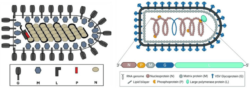
Schematic structure of VSV (PMID: 39772239)
2.2 Vesicular Stomatitis Virus Genome & Classification
VSV belongs to the genus Vesiculovirus in the family Rhabdoviridae. Its genome is a negative‑sense single‑stranded RNA of approximately 11,161 nucleotides. The genome is organized as leader – N – P – M – G – L – trailer from 3′ to 5′. There is a 47‑nucleotide untranslated leader sequence at the 3′ end of the N gene.
The five genes are separated by intergenic sequences, each consisting of two nucleotides. Transcription initiates at the 3′ end of the genome and proceeds obligatorily sequentially in the order N→P→M→G→L. At the end of each gene, a conserved termination‑polyadenylation signal (AUACU₇) causes the polymerase to "stutter," producing a poly(A) tail.
2.3 What Are Chikungunya Virus Structural Proteins?
CHIKV encodes five structural proteins:
Protein | Length (aa) | Main Function |
N (Nucleoprotein) | 422 | Binds tightly to viral RNA, forms the nucleocapsid core |
P (Phosphoprotein) | 265 | Polymerase cofactor, involved in transcription and replication |
M (Matrix protein) | ~25 kDa | Inner envelope protein, mediates assembly and budding |
G (Glycoprotein) | ~65 kDa | Surface glycoprotein, mediates receptor binding and membrane fusion |
L (Large polymerase) | 2109 | RNA‑dependent RNA polymerase (RdRp), catalyzes transcription and replication |
2.4 Mechanism of Cell Entry and Replication
2.4.1 Viral Attachment and Receptor Recognition
VSV initiates infection through the binding of G protein to molecules on the host cell surface. The virus may attach to host cells through non‑specific electrostatic and hydrophobic interactions. Phosphatidylserine has been proposed as a potential receptor for VSV.
2.4.2 Viral Entry
VSV enters host cells via clathrin‑mediated endocytosis (CME) . After internalization, the endosomal pH drops below 6.5, triggering a conformational change in G protein that mediates fusion between the viral envelope and the endosomal membrane, releasing the nucleocapsid into the cytoplasm. M protein dissociates from the nucleocapsid, allowing viral RNA synthesis to commence.
2.4.3 Viral RNA Replication
After nucleocapsid release into the cytoplasm, the L protein (RdRp) together with its cofactor P protein initiates transcription and replication:
l Transcription: initiation at the 3′ end of the genome, first synthesizing a 47‑nucleotide leader RNA
l Subsequent synthesis of five mRNAs in the order N→P→M→G→L
l Each mRNA is capped during transcription and receives a poly(A) tail via "stuttering" at the gene end signal
l Replication: the viral RNA polymerase synthesizes a full‑length positive‑strand RNA (antigenome), which serves as template for progeny genome synthesis
During transcription, attenuation at each gene start results in a gradient of mRNA abundance—N mRNA is the most abundant, and L mRNA the least.
2.4.4 Viral Assembly and Release
Newly synthesized N protein associates with progeny genomic RNA and L protein to form new ribonucleoprotein complexes (RNPs). M protein recruits RNPs to the cell membrane. Newly synthesized G protein is transported from the ER through the Golgi to the plasma membrane via COPII‑ and COPI‑mediated vesicular transport. The virus acquires its envelope by budding from the host cell membrane, releasing bullet‑shaped virions. The VSV replication cycle is extremely short, taking only 2–3 hours from infection to progeny virus production.
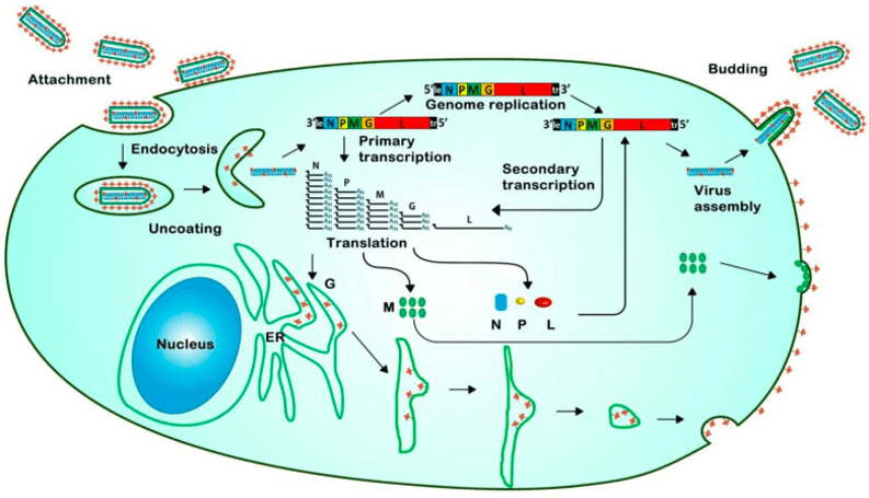
The life cycle of VSV. (PMID: 39772239)
2.5 Symptoms and Treatment
2.5.1 Clinical Symptoms
The incubation period for VSV ranges from 30 hours to 6 days.
Animal symptoms: VSV causes vesicular disease in livestock (cattle, horses, swine, etc.). Vesicles (blisters) and ulcers develop 2–5 days post‑infection in the mouth, tongue, muzzle, ears, genitalia, and above the hooves. Classical signs include excessive salivation, fever, lameness, and teat lesions. Teat lesions are the most disastrous consequence, as mastitis can easily follow incomplete milking.
Human symptoms: Human infection typically presents as influenza‑like illness. Infections with Indiana and New Jersey serotypes can cause high fever (often biphasic) , severe malaise, headaches, myalgia, arthralgia, retrosternal pain, eye aches, and nausea. Chandipura serotype infection can cause fever, sensory disorders, convulsions, vomiting, diarrhea, and encephalitis. The disease typically resolves without complications within 7–10 days.
2.5.2 Treatment
Currently, no specific treatment or vaccine is available for VSV infection. Treatment is primarily symptomatic and supportive:
l Mild antiseptic mouthwashes
l Good sanitation to prevent secondary bacterial infections
l For laboratory exposures, immediate washing of exposed areas with soap and water, and flushing of mucous membranes for 15 minutes
2.6 Targets for Intervention
2.6.1 Viral Targets
Target | Protein | Biological Role |
G | Glycoprotein | Receptor binding & membrane fusion |
L (RdRp) | RNA polymerase | RNA transcription & replication |
M | Matrix protein | Assembly & budding |
N | Nucleoprotein | RNP formation |
2.6.2 Host Receptors & Entry Factors
Host Target | Type | Role in Infection |
Phosphatidylserine | Potential receptor | Viral attachment |
Clathrin‑mediated endocytosis | Endocytic pathway | Viral entry |
COPII/COPI | Vesicular transport | G protein transport to plasma membrane |
2.6.3 Host Signaling Pathways
Pathway | Key Targets | Biological Role |
Type I interferon pathway | IFN‑α/β | Antiviral response; restricts VSV replication in healthy cells |
eIF4F complex | 4E‑BP1, eIF4E | VSV dephosphorylates 4E‑BP1 and eIF4E to prioritize viral mRNA translation |
PI3K pathway | PI3K | PI3K‑IN‑1 effectively inhibits VSV proliferation |
2.7 Vaccine Types and Development Progress
VSV has been extensively developed as a vaccine vector platform, owing to its rapid production, strong immunogenicity, and self‑amplifying RNA properties
2.7.1 rVSV‑Vectored Vaccine Platform
The recombinant vesicular stomatitis virus (rVSV) vector platform has been successfully applied to multiple vaccine candidates:
l rVSV‑RABV: a recombinant VSV expressing the rabies virus glycoprotein
l VSVΔG‑LASV‑GPC: a VSV‑vectored Lassa virus vaccine candidate that has advanced to human clinical trials
l rVSVΔG‑PEDV‑S: a recombinant VSV vaccine expressing the spike protein of porcine epidemic diarrhea virus (PEDV)
2.7.2 Replication‑Deficient VSV Vaccines (rdVSV)
Replication‑deficient vesicular stomatitis virus (rdVSV) vaccines achieve replication deficiency through G gene deletion or other modifications, enhancing safety. Such vaccines can serve as self‑amplifying RNA vaccine platforms.
2.7.3 mini-VSV Platform
In 2025, researchers proposed a "mini‑VSV" platform featuring genomic truncation and UAA‑regulated M protein expression, aiming to develop a safer, higher‑capacity VSV vaccine vector.
2.7.4 Nucleocapsid (NC) Platform
A 2025 study published in Scientific Reports established VSV nucleocapsids (NCs) as a novel, intrinsically safer, self‑amplifying platform for antiviral screening and vaccine development.
2.7.5 Veterinary Vaccines
Currently, no licensed VSV vaccine is available for animals. VSV control relies on quarantine, slaughter, and strict sanitation measures.
2.8 Drugs
Currently, no specific antiviral drug is approved for VSV infection. Treatment relies primarily on supportive care. Several anti‑VSV drug candidates have shown promise:
2.8.1 Virus-Targeted Drugs
Target | Drug/candidate | Biological Role | Stage |
Unknown | SMU‑V18 | Novel pyrimidine/pyridine derivative with optimized anti‑VSV activity | In vitro |
Viral replication | Epipodophyllotoxin acetate | Cyclo‑lignan compound, IC50 = 0.1 μg/mL | In vitro |
3‑Indoleacetonitrile | Broad‑spectrum antiviral | In vitro | |
Ribavirin | Broad‑spectrum nucleoside analog | Research use | |
Homoharringtonine | Protein synthesis inhibitor, broad‑spectrum | In vitro | |
Transcription initiation | Vesiculopolins | Novel anti‑vesiculoviral compounds inhibiting transcription initiation | Research |
2.8.2 Host-Targeted Drugs
Target | Drug | Mechanism | Stage |
Innate immunity | Betrixaban | Activates innate immune responses, broad‑spectrum antiviral | In vitro |
PI3K pathway | PI3K‑IN‑1 | Phosphatidylinositol‑3 kinase inhibitor | In vitro |
Unknown | Doxycycline | Anti‑VSV activity | In vitro |
Unknown | Metformin | Inhibits VSV replication | In vitro |
Pyrimidine synthesis | Brequinar | Host‑targeting antiviral compound | In vitro |
2.9 Oncolytic Virus Therapy
VSV is one of the most prominent candidates in the oncolytic virus field
2.9.1 Mechanism of Oncolysis
VSV can infect almost all cell types, but type I interferon (IFN) ‑mediated antiviral responses prevent productive infection in healthy cells. However, IFN signaling defects frequently coincide with tumorigenesis, allowing VSV to selectively infect and destroy cancer cells with minimal damage to normal cells.
2.9.2 Advantages of VSV as an Oncolytic Virus
l Short replication cycle: only 2–3 hours from infection to progeny virus production
l Broad tissue tropism: G protein‑mediated recognition enables targeting of diverse cancer cells
l Low pre‑existing immunity: natural human infection with VSV is extremely rare; most individuals lack anti‑VSV antibodies
l Hypoxia adaptation: VSV can replicate normally in the hypoxic conditions of the tumor microenvironment
l Easy to engineer: small genome and mature reverse genetics systems facilitate directed modification
2.9.3 Clinical Trial Progress
Recombinant VSV expressing interferon‑β (IFNβ) and sodium‑iodide symporter (NIS) is currently undergoing multiple Phase I clinical trials:
l NCT03017820
l NCT03120624
l NCT02923466
2.9.4 Engineering Strategies
l Reducing neurotoxicity: modification of M and G proteins (e.g., deletion or substitution of methionine 51 in M protein)
l Improving safety: replacing VSV‑G with envelope proteins from other viruses
l Enhancing efficacy: multi‑dose strategies, though neutralizing antibody clearance remains a challenge
2.10 Epidemiology
l First discovered: 1926, South America
l Geographic distribution: primarily endemic in the Americas, including the United States, Mexico, Central America, and South America
l Natural hosts: multiple mammalian species and insects
l Transmission routes:
Ø Vector‑borne: sand flies and mosquitoes
Ø Contact: direct contact with infected livestock
Ø Skin contact: through skin abrasions
Ø Aerosol: inhalation via the nasopharyngeal route
Ø Laboratory‑acquired: accidental autoinoculation or aerosol inhalation
l Laboratory‑acquired infections (LAIs) : as of 1980, 46 cases with New Jersey and Indiana viruses, and 13 cases due to Piry virus (no deaths)
2.11 Diagnosis
Diagnosis of VSV infection relies on laboratory testing:
l Virus isolation: from throat swabs or blood
l PCR (Polymerase Chain Reaction)
l ELISA (Enzyme‑Linked Immunosorbent Assay)
l Neutralization tests
l Complement fixation
l Immunofluorescence
l Electron microscopy
2.11 Prevention
l Good hygiene: recommended for prevention
l Avoid contact: avoid contact with infected animals and their secretions
l Personal protective measures: wear appropriate PPE when handling potentially infected animals
l Laboratory safety:
Ø Indiana, New Jersey, and other serotypes: BSL‑2 recommended
Ø Chandipura and Piry serotypes: BSL‑3 recommended
l Quarantine and sanitation: in animal outbreaks, quarantine, slaughter, and strict sanitation are required
3. Related Products & Services
l Recombinant Antigens & Receptors
l Antibodies
l Detection Kits
l Recombinant Protein Expression Services
l Antibody Development Services
4. Resources
5. References
1. Lyles, D. S., Rupprecht, C. E., Rhabdoviruses Study Group. (2007). Rhabdoviridae: The viruses and their replication. In Fields Virology (5th ed.). Lippincott Williams & Wilkins.
2. Hastie, E., Cataldi, M., Marriott, I., et al. (2013). Understanding and altering cell tropism of vesicular stomatitis virus. Virus Research, 176(1–2), 16–32.
3. Finkelshtein, D., Werman, A., Novick, D., et al. (2013). LDL receptor and its family members serve as the cellular receptors for vesicular stomatitis virus. Proceedings of the National Academy of Sciences of the United States of America, 110(18), 7306–7311.
4. Nikolic, J., Belot, L., Raux, H., et al. (2018). Structural basis for the recognition of the LDL-receptor family members by the vesicular stomatitis virus glycoprotein. Proceedings of the National Academy of Sciences of the United States of America, 115(10), E2346–E2355.
5. Albertini, A. A. V., Baquero, E., Ferlin, A., et al. (2012). Molecular and structural aspects of vesicular stomatitis virus entry. Advances in Virus Research, 83, 109–145.
6. Roche, S., Bressanelli, S., Rey, F. A., et al. (2006). Crystal structure of the low-pH form of the vesicular stomatitis virus glycoprotein G. Science, 313(5784), 187–191.
7. Roche, S., Rey, F. A., Gaudin, Y., et al. (2007). Structure of the prefusion form of the vesicular stomatitis virus glycoprotein G. Science, 315(5813), 843–848.
8. Whelan, S. P. J., Barr, J. N., Wertz, G. W. (2004). Transcription and replication of nonsegmented negative-strand RNA viruses. Current Topics in Microbiology and Immunology, 283, 61–119.
9. Ivanov, I., Lehta, S., Jovičić, A. (2020). The molecular mechanisms of vesicular stomatitis virus replication. Viruses, 12(12), 1407.
10. Harty, R. N., Brown, M. E., McGettigan, J. P., et al. (2001). Rhabdoviruses and the budding process. Advances in Virus Research, 57, 1–31.
11. Harty, R. N., Brown, M. E., McGettigan, J. P., et al. (2000). VSV matrix protein induces apoptosis through a mechanism that is independent of transcription inhibition. Journal of Virology, 74(20), 9802–9809.
12. Finke, S., Conzelmann, K. K. (2003). Dissociation of rabies virus matrix protein functions and virus budding by reverse genetics. Journal of Virology, 77(17), 9633–9643.
13. Lyles, D. S., Rupprecht, C. E. (2007). Rhabdoviridae. In Fields Virology. Lippincott Williams & Wilkins.
14. Clarke, D. K., Nasar, F., Lee, M., et al. (2014). Synergistic attenuation of vesicular stomatitis virus by M protein mutation and glycoprotein replacement. Journal of Virology, 88(14), 8215–8226.
15. Hastie, E., Grdzelishvili, V. Z. (2012). Vesicular stomatitis virus as a flexible platform for oncolytic virotherapy against cancer. Journal of General Virology, 93(12), 2529–2545.
16. Huang, Y., Li, Y., Zhang, Y., et al. (2024). Vesicular stomatitis virus-based oncolytic virotherapy: mechanisms, engineering strategies and clinical applications. Frontiers in Immunology, 15.
17. Hastie, E., Grdzelishvili, V. Z. (2012). Vesicular stomatitis virus as a flexible platform for oncolytic virotherapy against cancer. Journal of General Virology, 93(12), 2529–2545.
18. Garcia, M. N., Hasbun, R., Lednicky, J. A., et al. (2013). Epidemiology and clinical features of vesicular stomatitis virus infection. Clinical Infectious Diseases, 57(9), 1283–1291.
19. Reif, J. S., Webb, P. A., Monath, T. P., et al. (1987). Epizootic vesicular stomatitis in Colorado, 1982: epidemiologic studies along the northern Colorado front range. American Journal of Tropical Medicine and Hygiene, 36(2), 317–326.
20. Hastie, E., Grdzelishvili, V. Z. (2020). Vesicular stomatitis virus as a platform for vaccine development. Expert Review of Vaccines, 19(8), 745–758.